. 2004 Aug;15(8):3829-40.
doi: 10.1091/mbc.e04-02-0085. Epub 2004 Jun 11.

Identification of integrin beta subunit mutations that alter heterodimer function in situ

Affiliations

Identification of integrin beta subunit mutations that alter heterodimer function in situ

Alison L Jannuzi et al. Mol Biol Cell. 2004 Aug.

Abstract

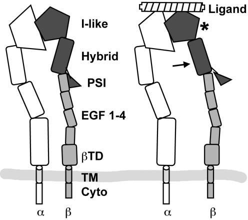
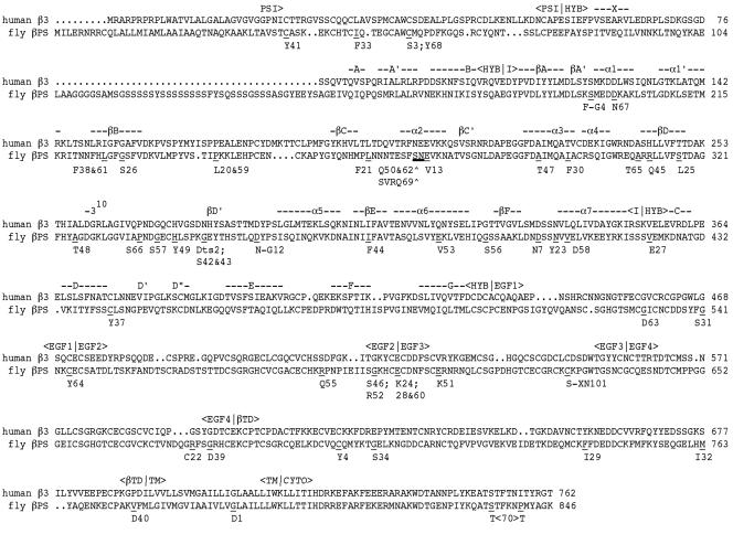
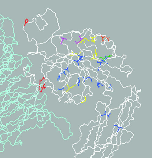
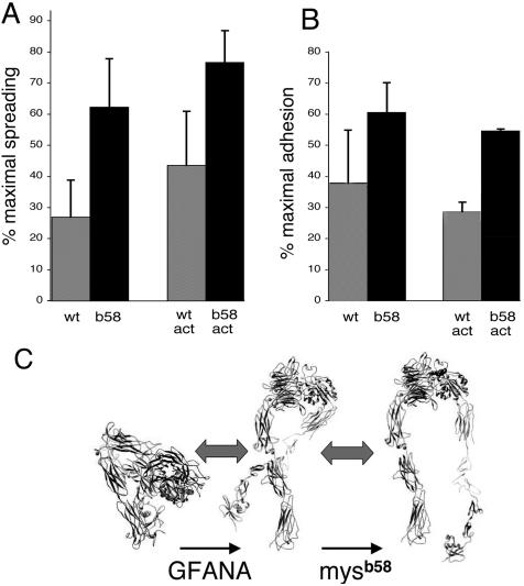
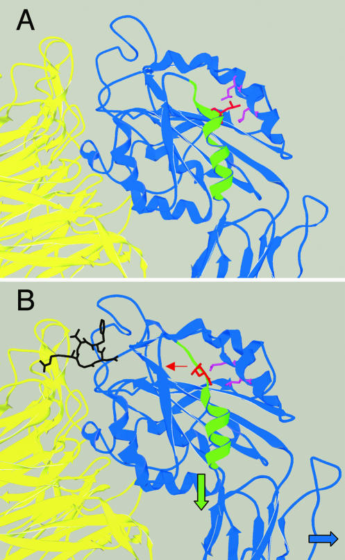
We conducted a genetic screen for mutations in myospheroid, the gene encoding the Drosophila betaPS integrin subunit, and identified point mutants in all of the structural domains of the protein. Surprisingly, we find that mutations in very strongly conserved residues will often allow sufficient integrin function to support the development of adult animals, including mutations in the ADMIDAS site and in a cytoplasmic NPXY motif. Many mutations in the I-like domain reduce integrin expression specifically when betaPS is combined with activating alphaPS2 cytoplasmic mutations, indicating that integrins in the extended conformation are unstable relative to the inactive, bent heterodimers. Interestingly, the screen has identified alleles that show gain-of-function characteristics in cell culture, but have negative effects on animal development or viability. This is illustrated by the allele mys(b58); available structural models suggest that the molecular lesion of mys(b58), V409>D, should promote the "open" conformation of the beta subunit I-like domain. This expectation is supported by the finding that alphaPS2betaPS (V409>D) promotes adhesion and spreading of S2 cells more effectively than does wild-type alphaPS2betaPS, even when betaPS is paired with alphaPS2 containing activating cytoplasmic mutations. Finally, comparisons with the sequence of human beta8 suggest that evolution has targeted the "mys(b58)" residue as a means of affecting integrin activity.


Figures

Figure 1.
Figure 1.
Domain structure of integrin heterodimers, in the extended conformation. α subunit domains are in unshaded outlines. The available data suggest that ligand binding (right) stabilizes an “open” conformation, involving changes in at least two of the three darkly shaded structural domains. In this model, a change in the I-like domain tertiary structure (asterisk) drives a movement of the Hybrid domain; this may also include a dissassociation of the PSI domain from a specific binding site on the stalk. EGF 1–4, EGF-like repeats 1–4; βTD, β terminal domain; TM, transmembrane domain; Cyto, cytoplasmic domain.
Figure 2.
Figure 2.
Locations of βPS point mutants. The Drosophila βPS and human β3 sequences are aligned, with the locations of β3 structural domains and (for the I-like and hybrid domains) secondary structures indicated (from Xiong et al., 2001). Sites of βPS mutants are underlined with the new residue indicated below, followed by the b-series allele number for mutants generated here, or other allele names for preexisting mutants. Note that mysb70 includes two missense changes. Three alleles involve the insertion of a single residue (mysb50 and mysb62) or four amino acids (mysb69) at the splice site indicated by thick underlining between S272 and N273.
Figure 3.
Figure 3.
Expression of αPS2βPS integrins in the posterior margin of third instar larval wing imaginal discs. Typically, αPS2βPS is expressed only in the ventral (lower) half of the epithelium, but these animals are also expressing a transgenic αPS2 subunit with a cytoplasmic activating mutation (deletion of CGFFN) throughout the disk. The upper staining (arrows) is entirely from the transgenic activated αPS2 with βPS, whereas the lower immunostaining is primarily from wildtype αPS2 with βPS, which is much more stable than heterodimers containing the activating αPS2 mutation. For wild-type βPS, the dorsal staining is reduced relative to the ventral staining, and the activated heterodimers are clustered on each cell. For many βPS mutants, expression with the activated αPS2 subunits is reduced much more than for wild-type, or is even nondetectable, as shown here for mysb57. See Table 1 for a summary of the imaginal disk expression data.
Figure 4.
Figure 4.
Examination of integrin surface expression via clonal analysis. A clone of cells homozygous for mysb60 was generated in a heterozygous mysb60/mys+ background, by somatic recombination in a cell of the early wing imaginal disk epithelium. The clone is marked by the loss of nuclear GFP staining (A), which derives from a transgene on the mys+ chromosome. The homozygous mysb60 cells show reduced surface integrin expression, as indicated by staining for βPS (B).
Figure 5.
Figure 5.
Locations of I-like domain and hybrid domain βPS mutants, mapped onto the structure of αv(pale blue)β3(white). Categories of mutations are: α/β interface-red; side chain on surface-yellow; side chain on interior-blue; side chain that coordinates with cation-purple; location of insertions-orange; mysb58-green (see below). There is no obvious hyper-sensitive region for I-like domain mutants.
Figure 6.
Figure 6.
(A and B) Spreading and adhesion of S2 cells expressing αPS2 and wild-type or mysb58 (V409>D) βPS. Cells were allowed to settle on plates coated with an RGD-containing fragment of the Drosophila ECM protein Tiggrin (RBB-Tigg). Spread cells (A) were defined by phase microscopy 3–4 h after plating. Adhesion (B) was defined by the number of cells remaining attached after washing 20 min after settling onto the plate. Ligand concentrations were chosen that give approximately half-maximal spreading or adhesion for each pair of bars (see MATERIALS AND METHODS), and the values are expressed as a percentage of the maximum at high RBB-Tigg concentrations; this adjusts for variations in expression or other factors between experiments. For the right pairs of bars in each histogram (labeled “act”), βPS was combined with αPS2 subunits containing a cytoplasmic-activating mutation (GFFNR>GFANA). In each pairwise comparison, spreading is significantly increased for the mysb58 allele compared with wild-type βPS, and the amount of the increase is approximately the same whether βPS is paired with wild-type or cytoplasmically activated αPS2. (C) These data are most easily explained by a two-stage model of integrin activation, in which the equilibrium between bent and extended heterodimers is regulated primarily by cellular events, and the equilibrium between the open and closed conformations of the integrin head is affected by mysb58 (protein tracings from Shimaoka and Springer, 2003). Data are averages from three experiments, and integrin expression levels in each case were generally similar or slightly higher for the wild-type βPS cells (unpublished data).
Figure 7.
Figure 7.
Structural basis for the activating effects of mysb58. The movement of the β3 residue (L341) homologous to βPS V409, in the unliganded I-like domain (A) and in similar crystals infused with a peptide ligand (B) (structures from Xiong et al., 2001, 2002). In the “liganded” structure, the L341 side chain (red) rotates away from the neighboring hydrophobic side chains of the α1′ helix (purple), toward the solvent. This would facilitate a proposed downward movement of the α7 helix (green) and the rightward rotation of the hybrid domain (blue arrow), although these latter movements are not seen in these crystals, which are still in the “bent” conformation (see Figure 6).
Figure 8.
Figure 8.
Ribbon diagram of the β-terminal domain of β3 (from Xiong et al., 2001), showing structures expected to be missing in β8 (red). The deleted sequences include those that comprise the “deadbolt” that has been hypothesized to interact with the I-like domain and stabilized the bent, inactive conformation (Xiong et al., 2003). Stabilizing disulfides that are retained in β8 are indicated in green.

Similar articles

Cited by

References

    1. Ashburner, M. (1989). Drosophila: A Laboratory Handbook. Cold Spring Harbor, NY: Cold Spring Harbor Laboratory Press.
    1. Baker, S.E., Lorenzen, J.A., Miller, S.W., Bunch, T.A., Jannuzi, A.L., Ginsberg, M.H., Perkins, L.A., and Brower, D.L. (2002). Genetic interaction between integrins and moleskin, a gene encoding a Drosophila homologue of Importin-7 (DIM-7). Genetics 162, 285-296. - PMC - PubMed
    1. Barton, S.J., Travis, M.A., Askari, J.A., Buckley, P.A., Craig, S.E., Humphries, M.J., and Mould, A.P. (2004). Novel activating and inactivating mutations in the integrin β1 subunit A domain. Biochem. J. 380, 401-407. - PMC - PubMed
    1. Beglova, N., Blacklow, S.C., Takagi, J., and Springer, T.A. (2002). Cysteine-rich module structure reveals a fulcrum for integrin rearrangement upon activation. Nat. Struct. Biol. 9, 282-287. - PubMed
    1. Bloor, J.W., and Brown, N.H. (1998). Genetic analysis of the Drosophila αPS2 integrin subunit reveals discrete adhesive, morphogenetic and sarcomeric functions. Genetics 148, 1127-1142. - PMC - PubMed

Publication types

MeSH terms

LinkOut - more resources