. 2004 Aug;15(8):3903-14.
doi: 10.1091/mbc.e04-04-0319. Epub 2004 Jun 11.

Role of the alpha-glucanase Agn1p in fission-yeast cell separation

Affiliations

Role of the alpha-glucanase Agn1p in fission-yeast cell separation

Nick Dekker et al. Mol Biol Cell. 2004 Aug.

Abstract

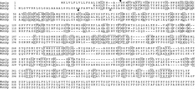
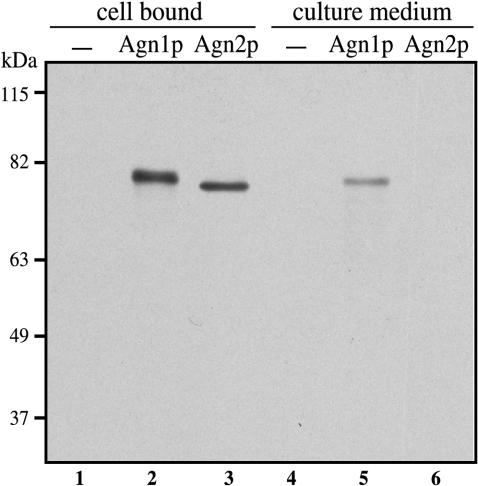
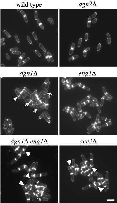
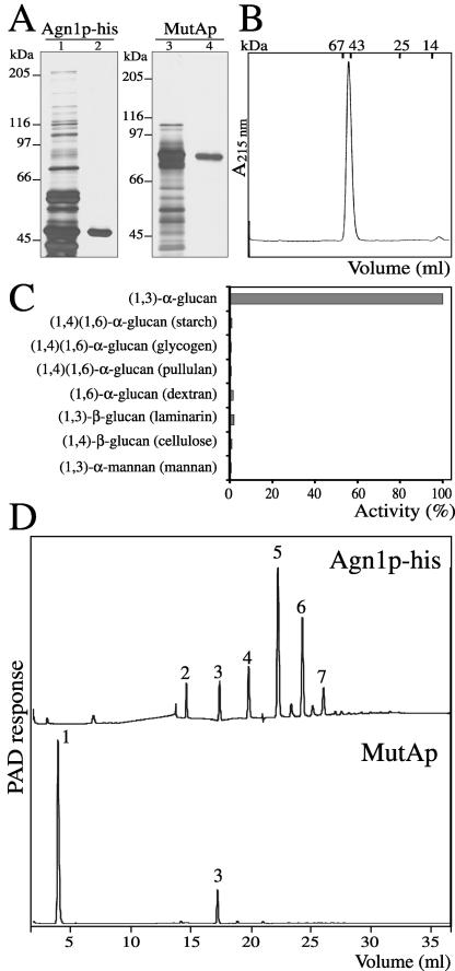
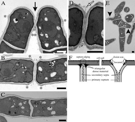
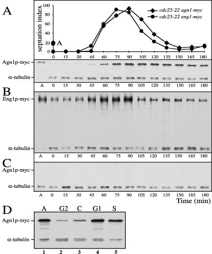
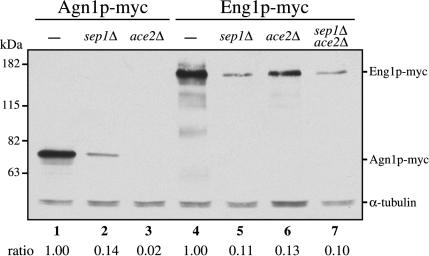
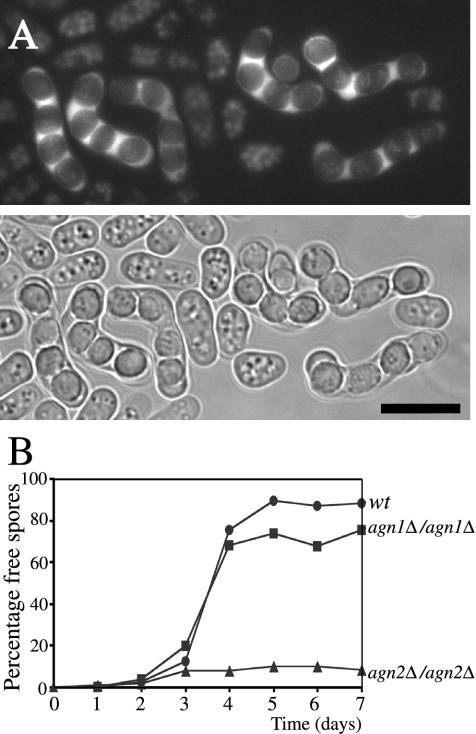
Cell division in the fission yeast Schizosaccharomyces pombe yields two equal-sized daughter cells. Medial fission is achieved by deposition of a primary septum flanked by two secondary septa within the dividing cell. During the final step of cell division, cell separation, the primary septum is hydrolyzed by an endo-(1,3)-beta-glucanase, Eng1p. We reasoned that the cell wall material surrounding the septum, referred to here as the septum edging, also must be hydrolyzed before full separation of the daughter cells can occur. Because the septum edging contains (1,3)-alpha-glucan, we investigated the cellular functions of the putative (1,3)-alpha-glucanases Agn1p and Agn2p. Whereas agn2 deletion results in a defect in endolysis of the ascus wall, deletion of agn1 leads to clumped cells that remained attached to each other by septum-edging material. Purified Agn1p hydrolyzes (1,3)-alpha-glucan predominantly into pentasaccharides, indicating an endo-catalytic mode of hydrolysis. Furthermore, we show that the transcription factors Sep1p and Ace2p regulate both eng1 and agn1 expression in a cell cycle-dependent manner. We propose that Agn1p acts in concert with Eng1p to achieve efficient cell separation, thereby exposing the secondary septa as the new ends of the daughter cells.


Figures

Figure 1.
Figure 1.
S. pombe Agn1 and Agn2 polypeptides show amino acid sequence identities to the catalytic domain of T. harzianum MutAp. cDNA-derived sequences for Agn1p and Agn2p were aligned with that of MutAp (AAF27911). Solid arrowheads indicate cleavage sites in Agn1p and MutAp for signal peptidase predicted by the SIGNALP algorithm, whereas the open arrowhead indicates the start of mature MutA protein observed by Fuglsang et al. (2000). Boxes indicate sequence identities and dashes indicate gaps in the PILEUP alignment.
Figure 2.
Figure 2.
Agn1p is secreted into the culture medium, whereas Agn2p is not. Along with wild-type cells (strain 972; lanes 1 and 4), cells of genotypes agn1-myc (strain ND003; lanes 2 and 5) or agn2-myc (strain ND061; lanes 3 and 6) were grown to midexponential phase in YEA medium. Equivalent amounts of cell-bound fractions were prepared by collecting cells, whereas culture medium fractions were obtained by precipitation on ice with 5% (wt/vol) trichloroacetic acid. Samples were resolved by 8% SDS-PAGE under reducing conditions and visualized by immunoblot analysis using an anti-myc mAb, 9E10. Probing a similar blot with antibodies directed to cytoplasmic protein α-tubulin showed protein in the cell-bound samples, but not in the culture medium samples, from all three strains (our unpublished data).
Figure 3.
Figure 3.
agn1Δ cells clump as a result of inefficient cell separation. Along with wild-type cells (strain 972), cells of genotypes agn2Δ (strain ND028), agn1Δ (strain ND001), eng1Δ (strain ND005), agn1Δ eng1Δ (strain ND019), or ace2Δ (strain ND064) were grown in EMMA medium to midexponential phase and stained with calcofluor white to visualize septa. Note that agn1Δ cells remain attached via a small area near the base of their tips (arrows) and that branching is observed in the agn1Δ eng1Δ and ace2Δ strains (arrowheads). Bar, 10 μm.
Figure 4.
Figure 4.
Agn1p-his is an endo-(1,3)-α-glucanase producing predominantly (1,3)-α-glucan pentasaccharides. (A) Purification of Agn1p-his and T. harzianum MutAp. Agn1p-his was purified from concentrated culture supernatant of strain ND236 by immobilized nickel-nitrilotriacetic acid affinity chromatography (lane 1), followed by anion-exchange chromatography (lane 2). MutAp was purified from a commercial preparation (lane 3) by (1,3)-α-glucan adsorption chromatography (lane 4). Samples were resolved by 8% SDS-PAGE under reducing conditions and visualized by silver staining. (B) Agn1p-his is a monomeric protein. Size-exclusion chromatography of purified Agn1p-his on a Superdex 75 column. Elution volumes of molecular mass markers are indicated. (C) Agn1p-his specifically hydrolyzes (1,3)-α-glucan. Purified Agn1p-his was incubated with indicated substrates at a concentration of 4 mg/ml. To lower backgrounds, substrates were reduced, except for (1,4)-β-glucan (cellulose) and (1,4)(1,6)-α-glucan (starch). To solubilize the (1,3)-α-glucan and (1,4)-β-glucan substrates, they were carboxy-methylated. Data are a percentage of the amount of reducing ends released from reduced and carboxy-methylated (1,3)-α-glucan. (D) Agn1p-his produces mainly (1,3)-α-glucan pentasaccharides. HPAEC analysis with pulsed amperometric detection (PAD) of reaction products released after a 5-h incubation at 37°C from insoluble (1,3)-α-glucan by Agn1p-his (top) or MutAp (bottom). Numbers refer to the degree of polymerization.
Figure 5.
Figure 5.
agn1Δ cells remain attached via remnants of the septum edging. Electron micrographs of thin sections from cells of genotypes: (A) agn1Δ (strain ND001). (B and D) eng1Δ (strain ND005). (C and E) ace2Δ (strain ND064). Cells were grown to midexponential phase in YEA medium. Solid arrow indicates locations of cell attachment, open arrow indicates remnants of the primary septum, solid arrowheads indicate branched outgrowths, and * indicates fission scars. Bar, 1 μm. (F) Schematic representation of the septum and septum edging immediately before (left) and during (right) cell separation. Adapted from Johnson et al. (1973).
Figure 6.
Figure 6.
(A–C) Periodic expression of Agn1p-myc resembles that of Eng1p-myc and depends on ace2+. Cells of genotypes cdc25-22 agn1-myc (strain ND049) (A), cdc25-22 eng1-myc (strain ND051) (B), or cdc25-22 ace2Δ agn1-myc (strain ND111) (C) were arrested at G2-M transition and then released synchronously. Cells were collected at indicated times after start of the release. Degrees of synchrony are indicated by septation index. A indicates asynchronous controls. Quantitation of the blots by normalization to the α-tubulin loading control confirmed the apparent peak in Agn1p levels at about the time of septation and cell separation both in the experiment shown and in a second experiment in which the synchronous population was followed into the second cell cycle (our unpublished data). Note that upon ace2+ deletion Agn1p-myc is not expressed at any stage of the cell cycle. (D) Agn1-myc protein expression is up-regulated during G1 and S phases. Cells of genotypes cdc25-22 agn1-myc (strain ND049; lane 2), cps1-191 agn1-myc (strain ND154; lane 3), and cdc10-129 agn1-myc (strain ND054; lane 4) were grown in YEA medium to midexponential phase at 28°C and then arrested at G2 phase, cytokinesis (C phase), or G1 phase, respectively, by a shift to 36°C for 4 h. For an arrest at S phase, exponentially growing cells of genotype agn1-myc (ND003) were incubated with hydroxyurea at a concentration of 11 mM at 28°C for 4 h (lane 5). Along with a total lysate of asynchronous cells (indicated as A, lane 1), cell lysates were resolved by 8% SDS-PAGE under reducing conditions and visualized by immunoblot analysis by using an anti-myc mAb, 9E10. α-Tubulin served as a loading control.
Figure 7.
Figure 7.
Agn1-myc protein expression depends on ace2+ and sep1+. Cells of genotypes agn1-myc (strain ND003; lane 1), agn1-myc sep1Δ (strain ND140; lane 2), agn1-myc ace2Δ (strain ND132; lane 3), eng1-myc (strain ND007; lane 4), eng1-myc sep1Δ (strain ND144; lane 5), eng1-myc ace2Δ (strain ND136; lane 6), or eng1-myc sep1Δ ace2Δ (strain ND218; lane 7) were grown in YEA medium to midexponential phase. Cell lysates were resolved by 8% SDS-PAGE under reducing conditions and visualized by immunoblot analysis by using an anti-myc mAb, 9E10. α-Tubulin served as a loading control. Ratios indicate normalized Agn1p-myc or Eng1p-myc levels corrected for α-tubulin loading controls.
Figure 8.
Figure 8.
Agn2p localizes to the cytoplasm of the ascus and contributes to hydrolysis of the ascus wall. (A) Haploid cells of genotype agn2-gfp (strain ND172) and matching wild-type cells (strain FYC15) were allowed to mate on a sporulation plate and resulting zygotic asci were examined using fluorescence microscopy. Agn2p-GFP fluorescence is excluded from ascospores, but instead localizes to the cytoplasm of the ascus, the epiplasm. Bar, 10 μm (B). Along with wild-type diploid cells (strain ND080), diploid cells of genotype agn1Δ/agn1Δ (strain ND084) and agn2Δ/agn2Δ (strain ND088) were grown to exponential phase in EMM medium containing 2% ammonium sulfate to inhibit sporulation. Subsequently, sporulation was induced synchronously by a shift to EMM medium containing 0.5% sodium glutamate, a poor nitrogen source. At the indicated time intervals, percentages of free ascospores were determined. Note that in the absence of agn2+ the ascus wall fails to undergo endolysis.

Similar articles

Cited by

References

    1. Akada, R., Kawahata, M., and Nishizawa, Y. (2000). Elevated temperature greatly improves transformation of fresh and frozen competent cells in yeast. Biotechniques 28, 854-856. - PubMed
    1. Bähler, J., Wu, J.-Q., Longtine, M.S., Shah, N.G., McKenzie, A. 3rd, Steever, A.B., Wach, A., Philippsen, P., and Pringle, J.R. (1998). Heterologous modules for efficient and versatile PCR-based gene targeting in Schizosaccharomyces pombe. Yeast 14, 943-951. - PubMed
    1. Baladrón, V., Ufano, S., Dueñas, E., Martín-Cuadrado, A.B., del Rey, F., and Vázquez de Aldana, C.R. (2002). Eng1p, an endo-1,3-β-glucanase localized at the daughter side of the septum, is involved in cell separation in Saccharomyces cerevisiae. Eukaryot. Cell 1, 774-786. - PMC - PubMed
    1. Berlin, A., Paoletti, A., and Chang, F. (2003). Mid2p stabilizes septin rings during cytokinesis in fission yeast. J. Cell Biol. 160, 1083-1092. - PMC - PubMed
    1. Cabib, E., Roh, D.-H., Schmidt, M., Crotti, L.B., and Varma, A. (2001). The yeast cell wall and septum as paradigms of cell growth and morphogenesis. J. Biol. Chem. 276, 19679-19682. - PubMed

Publication types

MeSH terms

Substances

LinkOut - more resources