. 2004 Jul;16(7):1730-40.
doi: 10.1105/tpc.021816. Epub 2004 Jun 11.

MicroRNA-directed cleavage of Nicotiana sylvestris PHAVOLUTA mRNA regulates the vascular cambium and structure of apical meristems

Affiliations

MicroRNA-directed cleavage of Nicotiana sylvestris PHAVOLUTA mRNA regulates the vascular cambium and structure of apical meristems

Neil A McHale et al. Plant Cell. 2004 Jul.

Abstract

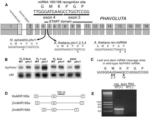
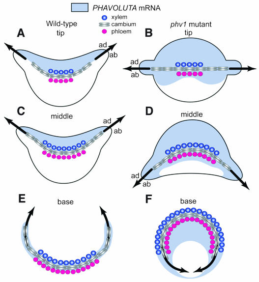
Leaf initiation in the peripheral zone of the shoot apical meristem involves a transition to determinate cell fate, but indeterminacy is maintained in the vascular cambium, a tissue critical to the continuous growth of vascular tissue in leaves and stems. We show that the orientation of cambial growth is regulated by microRNA (miRNA)-directed cleavage of mRNA from the Nicotiana sylvestris ortholog of PHAVOLUTA (NsPHAV). Loss of miRNA regulation in semidominant phv1 mutants misdirects lateral growth of leaf midveins and stem vasculature away from the shoot, disrupting vascular connections in stem nodes. The phv1 mutation also expands the central zone in vegetative and inflorescence meristems, implicating miRNA and NsPHAV in regulation of meristem structure. In flowers, phv1 causes reiteration of carpel initiation, a phenocopy for loss of CARPEL FACTORY/DICER LIKE1, indicating that miRNA is critical to the termination of indeterminacy in floral meristems. Results point to a common role for miRNA in spatial and temporal restriction of HD-ZIPIII mediated indeterminacy in apical and vascular meristems.


Figures

Figure 1.
Figure 1.
Gene Structure and Expression Patterns for NsPHAV and NsMIR166a. (A) PHV gene structure and mutations at the miRNA 165/166 recognition site in Nicotiana and Arabidopsis. (B) NsPHAV mRNA levels in wild-type and mutant phv1 leaves and stems relative to a ubiquitin (UBI) control. (C) The ends of 24 wild-type NsPHAV mRNAs were mapped by 5′ RACE analysis. All were cleaved at the miRNA 165/166 site with a strong preference (n = 19) for a central site flanking the G-to-A base substitution in the phv1 mutant. In heterozygous phv1 mutant plants (data not shown), mutant transcripts terminated at alternative sites 43 and 78 nucleotides upstream. (D) Genomic structure of precursor MIR166 genes in maize and Nicotiana (NsMIR166a), showing the miRNA recognition site (6 box) flanked by two conserved regions. The upstream 5 box represents the predicted foldback region involved in hairpin formation. Downstream conservation in the 7 box suggests that this region is part of the precursor RNA transcript. nt, nucleotides. (E) RT-PCR amplification with (+) and without (−) reverse transcriptase (RT) using primers specific to nonconserved regions internal to the 5 and 7 boxes confirmed transcription of NsMIR166a in RNA samples from the vegetative shoot apex.
Figure 2.
Figure 2.
Leaf Phenotypes and NsPHAV Expression. (A) and (B) Relative positions of adaxial and abaxial ribs domains (adr and abr, respectively) and midveins in wild-type (A) and heterozygous phv1 mutant (B) leaf primordia. Note the enlarged adaxial domain in the mutant. Bars = 160 μm. (C) Cup-shaped leaves in heterozygous phv1 mutant. (D), (E), and (G) Homozygous phv1 mutant rod-like (rdl) leaves (D) with adaxial blade tissue (adb) encircling the primordium (E) and a uniform mesophyll lacking adaxial/abaxial differentiation (G). Bar in (E) = 20 μm; bar in (G) = 80 μm. (F) Elongated epidermal files (ep) marking the adaxial/abaxial boundary in a wild-type leaf. Note the absence of this boundary in the homozygous mutant (E). Bar in (F) = 20 μm. (H) and (I) In situ NsPHAV expression in the wild type (H) and heterozygous phv1 mutant (I) shoot apex showing midveins (mv), leaf blades (lb), and leaf primordia at stages P1 to P4. ad, adaxial; ab, abaxial. Bars = 80 μm.
Figure 3.
Figure 3.
Vascular Phenotypes and NsPHAV Expression. (A) and (B) Wild-type leaf/stem junction. Paraffin section in (A) shows stem pith (sp) and curvilinear growth of interfascicular vascular cambium (ifc) from the flanks of the leaf midvein (mv) connecting with stem vasculature (sv). Wild-type in situ NsPHAV expression is confined to the cambium (B). Bars = 160 μm. (C) to (F) Heterozygous phv1 mutant leaf/stem junctions. Ectopic NsPHAV expression in abaxial domains of leaf petioles (abp) and the stem (abs) and on the lateral flanks of leaf midveins (mvf) (C). Circularized midveins with xylem (x) surrounding phloem (p) (D). Aberrant abaxial curvature of leaf midveins (mv) and stem vasculature (sv) (E). Note that leaf midveins fail to connect with stem vasculature ([C], [D], and [E]). Formation of ectopic axillary meristems (axm) beginning at the flanks of the leaf petiole (lp) and extending down the stem (F). Bar in (C) = 160 μm; bars in (D) and (E) = 200 μm.
Figure 4.
Figure 4.
Meristem and Flower Phenotypes. (A) Wild-type adult vegetative meristem showing central (cz; line = 100 μm) and peripheral zones with P1 to P4 leaf primordia. (B) Wild-type inflorescence meristem showing a terminal flower (tf) and subtending cluster. (C) Wild-type inflorescence. (D) phv1 adult vegetative meristem showing an enlarged central zone (cz; line = 200 μm) with P1 to P4 leaf primordia in the peripheral zone. (E) and (F) phv1 mutant inflorescence meristem showing a terminal flower (tf) (E) and subtending cluster (F) phv1 mutant inflorescence. (G) Transverse section of a wild-type flower showing anthers (a) and carpels (c). (H) Transverse section of a phv1 flower showing anthers (a) and formation of ectopic carpel primordia (ec) emerging on the abaxial side of the primary carpel (c). Lateral growth of petals (p) is often misdirected abaxially, precluding corolla fusion. (I) Scanning micrograph of a phv1 flower showing distorted anthers (a) and inverted curvature in carpels (c), leaving developing ovules (o) on an exposed surface. Bars in (A) and (D) = 40 μm; bars in (B), (E), (I), (G), and (H) = 100 μm.
Figure 5.
Figure 5.
Vascular Development and Leaf Polarity. NsPHAV mRNA accumulates initially throughout wild-type leaf primordia and then localizes to the adaxial domain along a crescent line corresponding to the developing midvein ([A] and [C]). This marks the adaxial/abaxial (ad/ab) boundary, and leaf blades are initiated where this traces to the surface of the primordium. At the base of wild-type petioles (E), NsPHAV expression localizes to the cambium of the midvein, a strip of meristematic tissue proliferating within veins (fascicular) and outward from midveins (interfascicular) along a curvilinear path, which connects to the vascular cylinder of the stem. In heterozygous phv1 mutants ([B], [D], and [F]), loss of miRNA regulation expands the domain of NsPHAV expression, leading to correlated changes in midvein development, leaf polarity, and blade formation, each following a gradient of increasing severity moving from tip to base. Mutant midveins are flattened to straight lines (B), misdirected into abaxial crescents (D), and often circularized at the base (F). Midveins no longer mark the NsPHAV on/off interface ([B] and [D]) but still define the adaxial/abaxial boundary guiding blade formation, suggesting that it is the mutation's effect on the midvein that dictates expansion of the adaxial domain. By positioning negative regulators of HD-ZIPIII expression, such as KANADI and miRNA, the midvein may provide a framework for the onset of adaxial/abaxial polarity in leaves.

References

    1. Bartel, B., and Bartel, D. (2003). MicroRNAs: At the root of plant development? Plant Physiol. 132, 709–717. - PMC - PubMed
    1. Bharathan, G., and Sinha, N. (2001). The regulation of compound leaf development. Plant Physiol. 127, 1533–1538. - PMC - PubMed
    1. Chuck, G., Lincoln, C., and Hake, S. (1996). KNAT1 induces lobed leaves with ectopic meristems when overexpressed in Arabidopsis. Plant Cell 8, 1277–1289. - PMC - PubMed
    1. Clark, S.E., Williams, R.W., and Meyerowitz, E.M. (1997). The CLAVATA1 gene encodes a putative receptor kinase that controls shoot and floral meristem size in Arabidopsis. Cell 89, 575–585. - PubMed
    1. Emery, J.F., Floyd, S.K., Alvarez, J., Eshed, Y., Hawker, N.P., Izhaki, A., Baum, S.F., and Bowman, J.L. (2003). Radial patterning of Arabidopsis shoots by class III HD-ZIP and KANADI genes. Curr. Biol. 13, 1768–1774. - PubMed

Publication types