. 2004 Sep 1;382(Pt 2):463-70.
doi: 10.1042/BJ20040471.

Structural requirements of the glucocorticoid-response unit of the carbamoyl-phosphate synthase gene

Affiliations

Structural requirements of the glucocorticoid-response unit of the carbamoyl-phosphate synthase gene

Onard J L M Schoneveld et al. Biochem J. .

Abstract

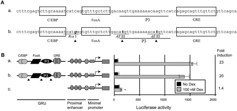
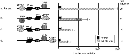
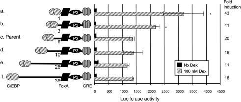
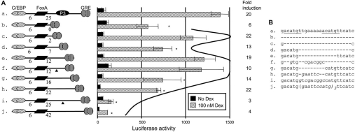
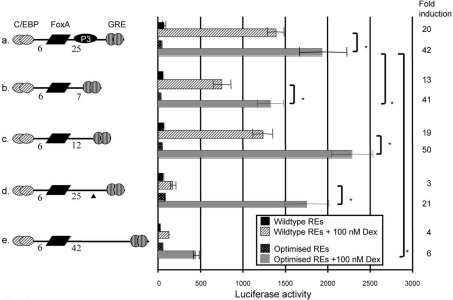
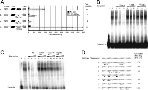
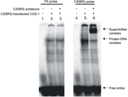
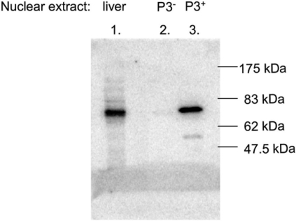
The GRU (glucocorticoid-response unit) within the distal enhancer of the gene encoding carbamoyl-phosphate synthase, which comprises REs (response elements) for the GR (glucocorticoid receptor) and the liver-enriched transcription factors FoxA (forkhead box A) and C/EBP (CCAAT/enhancer-binding protein), and a binding site for an unknown protein denoted P3, is one of the simplest GRUs described. In this study, we have established that the activity of this GRU depends strongly on the positioning and spacing of its REs. Mutation of the P3 site within the 25 bp FoxA-GR spacer eliminated GRU activity, but the requirement for P3 could be overcome by decreasing the length of this spacer to < or =12 bp, by optimizing the sequence of the REs in the GRU, and by replacing the P3 sequence with a C/EBPbeta sequence. With spacers of < or =12 bp, the activity of the GRU depended on the helical orientation of the FoxA and GR REs, with highest activities observed at 2 and 12 bp respectively. Elimination of the 6 bp C/EBP-FoxA spacer also increased GRU activity 2-fold. Together, these results indicate that the spatial positioning of the transcription factors that bind to the GRU determines its activity and that the P3 complex, which binds to the DNA via a 75 kDa protein, functions to facilitate interaction between the FoxA and glucocorticoid response elements when the distance between these transcription factors means that they have difficulties contacting each other.


Figures

Figure 1
Figure 1. Introduction of restriction sites in the GRU does not affect GRU activity
To facilitate cloning, restriction sites were introduced into the GRU. (A) Nucleotide sequence of the CPS GRU and the REs therein (construct a), and the modified GRU upon the introduction of restriction sites (construct b). The triangles indicate the positions of the point mutations. The GRU constructs were cloned upstream of the minimal promoter and proximal enhancer of the CPS gene. These reporter-gene constructs were transiently transfected into FTO-2B hepatoma cells and induced with glucocorticoids for 24 h. The reporter-gene activities, measured in the resulting lysates, are presented as means±S.E.M. luciferase values for at least four experiments and the fold induction. The asterisk indicates significantly different results relative to the wild-type construct a. The data show that the combination of proximal enhancer and minimal promoter does not result in a marked increase in activity when induced by glucocorticoids relative to basal activity (B, construct c). In contrast, both the parent and the modified GRUs exhibited a similarly high level of activity when cultured in presence of glucocorticoids (B, constructs a and b). Dex, dexamethasone.
Figure 2
Figure 2. The specific arrangement of GRU elements is important for the glucocorticoid response
The effects of different GRU arrangements on glucocorticoid-dependent transcription were tested by transient transfections into FTO-2B hepatoma cells. Modified GRUs were placed upstream of the proximal enhancer and minimal promoter. After 24 h of induction with or without glucocorticoids, cell lysates were prepared. Luciferase values are presented as means±S.E.M. for at least four experiments and the fold induction. The asterisk indicates significantly different results relative to parent construct a. The data show that inversion of the GRU (construct b) lowered the activity 2.5-fold, whereas inversion of the FoxA site only (construct c) decreased the activity 3-fold. Displacement of the C/EBP RE downstream of the FoxA- and GR-binding sites (construct d) decreased the activity 3-fold. Dex, dexamethasone.
Figure 3
Figure 3. Effects of variable spacer lengths between the C/EBP- and FoxA-binding sites in the GRU
A series of GRU constructs was made with deletions or insertions between the C/EBP and FoxA REs. These modified GRUs were placed upstream of the proximal enhancer and minimal promoter of the CPS gene, and tested by transient transfection into FTO-2B hepatoma cells. After 24 h of treatment with or without glucocorticoids, cells were lysed and luciferase values were measured. The results are expressed as means±S.E.M. and fold induction from at least four experiments. Results that are significantly different from those with the parent construct c are indicated by asterisks. Shortening of the C/EBP–FoxA spacer (constructs a and b) significantly increased activity relative to the parent construct (c), whereas increasing the distance had no effect (constructs d–f). Dex, dexamethasone.
Figure 4
Figure 4. Both the distance and the sequence of the region between the FoxA and GR REs influence the glucocorticoid response
A series of GRU constructs was made with deletions or insertions between the FoxA and GR REs. Modified GRUs were tested in conjunction with the proximal enhancer and the minimal promoter by transfection into FTO-2B hepatoma cells (A). The results are given as means±S.E.M. for at least four experiments and the fold induction. The asterisks indicate that results are significantly different from those with construct e. The curved line indicates the helical expression profile with respect to the distance between the FoxA and GR REs. The triangles indicate the positions of the point mutations. (B) Sequences of the respective spacers. Italic characters represent nucleotides deviating from the parent sequence. Dex, dexamethasone.
Figure 5
Figure 5. Optimization of the GRU REs increases the glucocorticoid response
GRU variants, differing in the length of the FoxA–GRE spacer, were modified into GRUs with optimized REs. The GRE was replaced by a palindromic GRE (AGAACAnnnTGTTCT) that has a high affinity for GR [25]. The C/EBP REs were replaced by a sequence selected for optimal binding (ATTGCGTAAT) [18], while the FoxA RE was replaced by a consensus FoxA site (TATTGACTTAG) [19]. The triangle indicates the P3-inactivating mutation. The resulting GRUs were placed upstream of the proximal enhancer and minimal promoter of the CPS gene, and were transiently transfected into FTO-2B hepatoma cells. Following 24 h of induction with glucocorticoids, luciferase values were measured in the cell lysates. Shown are mean±S.E.M. luciferase values for at least four experiments and the fold induction. Asterisks indicate significantly different results between the indicated constructs. Dex, dexamethasone.
Figure 6
Figure 6. The P3 RE can be functionally substituted by a C/EBP RE
Specific mutations (indicated by the triangles) were introduced in the P3-binding region of the GRU to give P3 mutants and a construct in which the P3 RE was altered to a consensus C/EBPβ RE (D). These GRUs were placed upstream of the proximal enhancer and minimal promoter of the CPS gene, and the constructs were transfected into FTO-2B hepatoma cells. After 24 h of induction with glucocorticoids, reporter-gene expression was measured in the cell lysates. Results are presented as mean±S.E.M. luciferase values from at least four experiments and the fold induction (A). Asterisks indicate results that are significantly different from those with the parent construct a. (B) To prove specificity, the P3 mutants were also used in a competition assay. A radiolabelled P3-binding region was incubated with 3 μg of rat liver nuclear extract and separated on a 6% (w/v) acrylamide gel (lane 2). Whereas addition of 0.1, 1 or 10 pmol of P3 oligonucleotide efficiently competed with the probe for protein binding (lanes 3–5), the same amounts of mutant P3 oligonucleotides could not (lanes 6–8 and 9–11). To define further the core P3-binding site, a similar competition assay was carried out (C): added to the reaction mixture was 0.1, 1 or 10 pmol of the parent P3 competitor (C, lanes 3–5; D, sequence a), or a competitor in which the left side (C, lanes 6–8; D, sequence e), middle (C, lanes 9–11; D, sequence f), or right side (C, lanes 12–14; D, sequence g) of the P3-binding region had been mutated. Dex, dexamethasone.
Figure 7
Figure 7. The P3-binding region is not a C/EBPβ-binding site
Supershift assays were performed using either the P3-binding region or the consensus C/EBPβ sequence as probe. The probes were incubated with a C/EBPβ-enriched COS-1 cell nuclear extract. To induce a supershift, C/EBPβ-specific antibodies were added to the samples. Both probes gave rise to a protein–DNA complex when incubated with nuclear extract (lanes 2 and 5). Addition of C/EBPβ-specific antibodies supershifted the C/EBPβ protein–DNA complex (lane 6), but not the P3 protein–DNA complex (lane 3).
Figure 8
Figure 8. Identification of the protein that interacts with the P3 site by Southwestern blot analysis
Crude and partially purified rat liver nuclear extracts were resolved on an SDS/9%-PAGE gel and transferred to a PVDF membrane. Following renaturation of the proteins, the blot was incubated with radiolabelled P3 probe. The positions of the molecular size markers are indicated. The liver nuclear extract contained a protein migrating at 75 kDa that was able to bind the P3 sequence (lane 1). A P3-positive MonoS fraction contained the same protein (lane 3), whereas a P3-negative MonoS protein fraction lacked this P3 protein (lane 2).

References

    1. Meijer A. J., Lamers W. H., Chamuleau R. A. Nitrogen metabolism and ornithine cycle function. Physiol. Rev. 1990;70:701–748. - PubMed
    1. Christoffels V. M., van den Hoff M. J., Moorman A. F., Lamers W. H. The far-upstream enhancer of the carbamoyl-phosphate synthetase I gene is responsible for the tissue specificity and hormone inducibility of its expression. J. Biol. Chem. 1995;270:24932–24940. - PubMed
    1. Christoffels V. M., Grange T., Kaestner K. H., Cole T. J., Darlington G. J., Croniger C. M., Lamers W. H. Glucocorticoid receptor, C/EBP, HNF3, and protein kinase A coordinately activate the glucocorticoid response unit of the carbamoylphosphate synthetase I gene. Mol. Cell. Biol. 1998;18:6305–6315. - PMC - PubMed
    1. Mitchell J., Noisin E., Hall R., O'Brien R., Imai E., Granner D. Integration of multiple signals through a complex hormone response unit in the phosphoenolpyruvate carboxykinase gene promoter. Mol. Endocrinol. 1994;8:585–594. - PubMed
    1. Wedel A., Ziegler-Heitbrock H. W. The C/EBP family of transcription factors. Immunobiology. 1995;193:171–185. - PubMed

MeSH terms