. 2004 Jul;112(3):386-96.
doi: 10.1111/j.1365-2567.2004.01896.x.

Presentation of alpha-galactosylceramide by murine CD1d to natural killer T cells is facilitated by plasma membrane glycolipid rafts

Affiliations

Presentation of alpha-galactosylceramide by murine CD1d to natural killer T cells is facilitated by plasma membrane glycolipid rafts

Gillian A Lang et al. Immunology. 2004 Jul.

Abstract

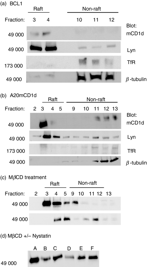
CD1 molecules are non-polymorphic major histocompatibility complex class I-related proteins that bind and present glycolipid antigens to T-cell antigen receptors (TCR) expressed by alphabeta T cells or natural killer-like T cells (NKT). Anti-metastatic properties of NKT cells reactive to the CD1d-binding antigen alpha-galactosylceramide (alpha-GalCer) are now being explored as a contributor to tumour cell killing. In this study, we tested the hypothesis that presentation of alpha-GalCer by murine CD1d (mCD1d) to mCD1d-restricted NKT cells was facilitated by plasma membrane glycolipid rafts. Confocal microscopy of mCD1d-transfected A20 B cells (A20mCD1d) demonstrated that mCD1d was raft-localized. This observation was confirmed by immunoblotting of raft fractions isolated on sucrose density gradients. Raft disruption by the cholesterol-binding agent nystatin, or short-chain ceramides, inhibited presentation of low concentrations of alpha-GalCer to NKT cells. Inhibition of antigen presentation was reversed by treatment of A20mCD1d cells with higher alpha-GalCer concentrations, or removal of raft-disrupting agents. These data indicate that partitioning of mCD1d into membrane rafts increases the capacity of antigen-presenting cells to present limiting quantities of glycolipid antigens, perhaps by stabilizing mCD1d/antigen structures on the plasma membrane and optimizing TCR engagement on NKT cells.


Figures

Figure 1
Figure 1
Plasma membrane distribution of raft and non-raft markers. Cells were incubated at 4° and stained for GM-1 (red) and: mCD1d; BCR; I-Ad; TfR; and CD45 (green). (a) Cells were fixed and analysed by confocal microscopy. Red/green colocalization is shown in yellow. (b) For each marker, images of at least 100 cells were scored for red/green colocalization. Graph shows the percentage of cells in the sample demonstrating GM-1 colocalization of the indicated surface marker.
Figure 2
Figure 2
Nystatin reversibly decreases mCD1d colocalization to rafts. Cells were resuspended in serum-free media and treated with 75 μg/ml nystatin for 15 min and then (a) fixed in 1·0% w/v paraformaldehyde, or (b) allowed to recover in serum-containing media for 30 min, before fixation. Cells were then stained for mCD1d (green) and GM-1 (red). Images shown are representative of at least 100 cells for each condition.
Figure 3
Figure 3
Detection of mCD1d in murine B-cell rafts. (a) BCL1 and (b) A20mCD1d cells were subject to detergent lysis and raft isolation. Equal volumes of gradient fractions (2 × 10⁶ cell equivalents) were resolved by SDS–PAGE before transfer to nitrocellulose and immunoblotting for mCD1d, Lyn, TfR, and β-tubulin. (c) Cells were subject to detergent lysis and raft isolation, or treated with 12·5 mm MβCD for 1 hr at 37° prior to detergent lysis. Equal volumes of gradient fractions were resolved by SDS–PAGE and immunoblotted for mCD1d. (d) Cells were treated as follows: Lanes A, untreated; lane B, 12·5 mm MβCD; lanes C, 12·5 mm MβCD then recovery; lane D, 2·5 mm MβCD + 75 μg/ml nystatin; lanes E, 75 μg/ml nystatin; or lanes F, 75 μg/ml nystatin then recovery, before detergent lysis and raft isolation. Raft fractions were resolved by SDS–PAGE and immunoblotted for mCD1d.
Figure 4
Figure 4
Presentation of α-GalCer to NKT cells is facilitated by membrane rafts. (a, inset) Cells were pulsed with α-GalCer at the concentrations indicated for 4 hr, before fixation in 0·15% w/v paraformaldehyde and washing in NKT culture media. Cells were then cocultured as indicated for 20 hr, before collection of supernatants and measurement of IL-2 concentration by ELISA. Data shows mean ± SD IL-2 concentration in media for triplicate samples. (a, main graph) Cells were pulsed with α-GalCer at the concentrations indicated before fixation and washing (control, closed bars), or nystatin treatment (75 μg/ml), fixation and washing (open bars). Cells were then cocultured with NKT cells as described. (b) After pulsing with α-GalCer cells were untreated, or treated with 75 μg/ml nystatin. Antigen-pulsed, non-fixed A20mCD1d cells were then allowed to recover by washing in NKT culture media, before addition of NKT cells. Data shows the mean ± SD IL-2 production for triplicate samples and is representative of at least three independent experiments.
Figure 5
Figure 5
Recovery from nystatin treatment in fixed cells. (a) A20mCD1d cells were pulsed with α-GalCer at the concentrations indicated before addition of 75 μg/ml nystatin and fixation. (b) Nystatin-containing medium was removed and cells were allowed to recover in serum-containing medium for 30 min, before fixation. Data show the mean ± SD IL-2 production for triplicate samples from a single experiment.
Figure 6
Figure 6
Effects of short-chain ceramides on mCD1d raft colocalization and antigen presentation. (a) A20mCD1d cells were incubated with C2-Cer, or DH-Cer at a 100 μg/ml final concentration, before fixation and staining for mCD1d (green) and GM-1 (red). At least 100 cells were analysed by confocal microscopy. Mid-section images are shown for four representative cells. (b) Cells were treated with C2-Cer or DH-Cer for 1 hr before detergent lysis and raft isolation. Gradient fractions were resolved by SDS–PAGE and immunoblotted for mCD1d. (c) Cells were pulsed with α-GalCer at the concentrations indicated before addition of C2-Cer or DH-Cer. Cells were then fixed, washed, and cocultured with NKT cells before measurement of IL-2 concentrations by ELISA. Data show the mean of triplicate samples ± SEM. (d) The mean of three experiments performed as in (c). Data show mean ± SEM (n = 3) IL-2 production in C2-Cer-treated cells as a percentage of that in DH-Cer-treated cells.

References

    1. Porcelli SA, Modlin RL. The CD1 system. Antigen-presenting molecules for T cell recognition of lipids and glycolipids. Annu Rev Immunol. 1999;17:297–329. - PubMed
    1. Prigozy TI, Sieling PA, Clemens D, et al. The mannose receptor delivers lipoglycan antigens to endosomes for presentation to T cells by CD1b molecules. Immunity. 1997;6:187–97. - PubMed
    1. Moody DB, Ulrichs T, Muhlecker W, et al. CD1c-mediated T-cell recognition of isoprenoid glycolipids in Mycobacterium tuberculosis infection. Nature. 2000;404:884–8. - PubMed
    1. Shamshiev A, Gober HJ, Donda A, et al. Presentation of the same glycolipid by different CD1 molecules. J Exp Med. 2002;195:1013–21. - PMC - PubMed
    1. Burdin N, Brossay L, Koezuka Y, et al. Selective ability of mouse CD1 to present glycolipids: α-galactosylceramide specifically stimulates Vα14+ NK T lymphocytes. J Immunol. 1998;161:3271–81. - PubMed

Publication types

LinkOut - more resources