

Induction of nitric oxide release from the human alveolar epithelial cell line A549: an in vitro correlate of innate immune response to Mycobacterium tuberculosis
- PMID: 15196216
- PMCID: PMC1782514
- DOI: 10.1046/j.1365-2567.2004.01905.x
Induction of nitric oxide release from the human alveolar epithelial cell line A549: an in vitro correlate of innate immune response to Mycobacterium tuberculosis
Abstract
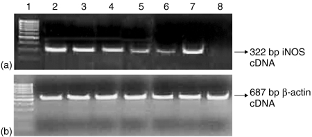
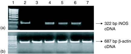
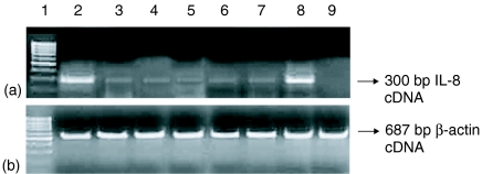
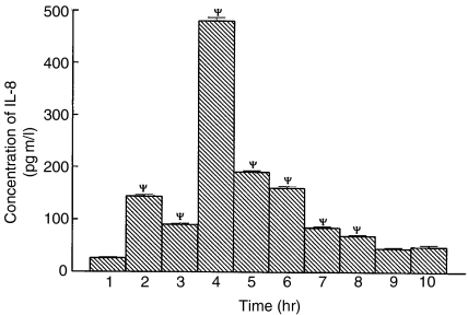
In view of the presence of a large number of epithelial cells in the alveoli of the lung and their ability to produce various cytokines and chemokines, the possible role of alveolar epithelial cells in the innate immune response to tuberculosis was examined. The human alveolar epithelial cell line A549 was used as a model. The ability of A549 cells to induce nitric oxide (NO) in response to Mycobacterium tuberculosis infection was taken as an in vitro correlate of innate immunity. M. tuberculosis infection induced A549 cells to produce significant levels of NO and to express inducible nitric oxide synthase mRNA at 48 hr of infection. However, the amount of NO released at this point was not mycobactericidal. Cytokine stimulation (interferon-gamma, tumour necrosis factor-alpha, interleukin-1beta, alone or in combination) of the infected A549 cells induced a higher concentration of NO. The study of colony-forming units (CFU) as a measure of the mycobactericidal capacity of A549 cells revealed a reduction in CFU of M. tuberculosis by 39.29% (from 10.62 +/- 0.48 - 6.392 +/- 0.54) following cytokine stimulation of the infected cells. Interestingly gamma-irradiated M. tuberculosis H37Rv could also induce higher than basal level of NO. Therefore we examined mycobacterial antigenic components for their possible role in NO production. We observed that A549 cells produced significantly higher amounts of NO at 48 hr when treated with mycobacterial whole cell lysates, cell wall or cell membrane preparations. The release of NO and the resultant mycobactericidal activity could be further enhanced by simultaneously conditioning the M. tuberculosis infected A549 cells with cytokine and mycobacterial components. These results suggest that alveolar epithelial cells respond to their microenvironment, which is constituted of various cytokines and macrophage-processed antigens and may contribute to the innate immune response to tuberculosis.
Figures


Similar articles
-
Pulmonary epithelial cells are a source of interferon-gamma in response to Mycobacterium tuberculosis infection.Immunol Cell Biol. 2007 Apr-May;85(3):229-37. doi: 10.1038/sj.icb.7100037. Epub 2007 Feb 20. Immunol Cell Biol. 2007. PMID: 17310225
-
Differential signaling of inducible nitric oxide synthase induction in Mycobacterium tuberculosis infected alveolar epithelial cell line A549 in response to cytokines IFN-γ, TNF-α and IL-1β.Int J Mycobacteriol. 2014 Mar;3(1):17-24. doi: 10.1016/j.ijmyco.2014.01.008. Epub 2014 Feb 21. Int J Mycobacteriol. 2014. PMID: 26786218
-
Synergistic cytokine-induced nitric oxide production in human alveolar epithelial cells.Nitric Oxide. 1999 Aug;3(4):348-57. doi: 10.1006/niox.1999.0242. Nitric Oxide. 1999. PMID: 10444374
-
What is the role of nitric oxide in murine and human host defense against tuberculosis?Current knowledge.Am J Respir Cell Mol Biol. 2001 Nov;25(5):606-12. doi: 10.1165/ajrcmb.25.5.4487. Am J Respir Cell Mol Biol. 2001. PMID: 11713103 Review.
-
Dynamics of cytokine generation in patients with active pulmonary tuberculosis.Curr Opin Infect Dis. 2003 Jun;16(3):205-10. doi: 10.1097/00001432-200306000-00004. Curr Opin Infect Dis. 2003. PMID: 12821809 Review.
Cited by
-
Ursolic Acid Reduces Mycobacterium tuberculosis-Induced Nitric Oxide Release in Human Alveolar A549 cells.Mol Cells. 2015 Jul;38(7):610-5. doi: 10.14348/molcells.2015.2328. Epub 2015 Jun 18. Mol Cells. 2015. PMID: 26084752 Free PMC article.
-
Water-pipe smoke condensate increases the internalization of Mycobacterium Bovis of type II alveolar epithelial cells (A549).BMC Pulm Med. 2017 Apr 21;17(1):68. doi: 10.1186/s12890-017-0413-7. BMC Pulm Med. 2017. PMID: 28431548 Free PMC article.
-
Effect of mycobacterial secretory proteins on the cellular integrity and cytokine profile of type II alveolar epithelial cells.Lung India. 2012 Oct;29(4):313-8. doi: 10.4103/0970-2113.102796. Lung India. 2012. PMID: 23243342 Free PMC article.
-
The role of lipid raft aggregation in the infection of type II pneumocytes by Mycobacterium tuberculosis.PLoS One. 2012;7(9):e45028. doi: 10.1371/journal.pone.0045028. Epub 2012 Sep 14. PLoS One. 2012. PMID: 23024786 Free PMC article.
-
Control of Francisella tularensis Intracellular Growth by Pulmonary Epithelial Cells.PLoS One. 2015 Sep 17;10(9):e0138565. doi: 10.1371/journal.pone.0138565. eCollection 2015. PLoS One. 2015. PMID: 26379269 Free PMC article.
References
-
- Ellner JJ. Review: The immune response in tuberculosis-implication for tuberculosis control. J Infect Dis. 1997;176:1351–9. - PubMed
-
- Fulton SA, Cross JV, Toossi ZT, Boom WH. Regulation of interleukin-12 by interleukin 10, transforming growth factor-β, tumour necrosis factor-α and interferon-γ in human monocytes infected with Mycobacterium tuberculosis H37Ra. J Infect Dis. 1998;178:1105–14. - PubMed
-
- Rich EA, Torres M, Sada E, Finegan CK, Hamilton BD, Toossi Z. Mycobacterium tuberculosis stimulated production of nitric oxide by human alveolar macrophages and relationship of nitric oxide production to growth inhibition of MTB. Tubercle Lung Dis. 1997;78:247–55. - PubMed
-
- Hernandez-Pando R, Jeyanathan M, Mengistu G, Rook GAW. Persistence of DNA from Mycobacterium tuberculosis in superficially normal lung tissue during latent infection. Lancet. 2000;356:2133–8. - PubMed
Publication types
MeSH terms
Substances
LinkOut - more resources
Full Text Sources