

Polyreactive antigen-binding B (PAB-) cells are widely distributed and the PAB population consists of both B-1+ and B-1- phenotypes
- PMID: 15196248
- PMCID: PMC1809069
- DOI: 10.1111/j.1365-2249.2004.02511.x
Polyreactive antigen-binding B (PAB-) cells are widely distributed and the PAB population consists of both B-1+ and B-1- phenotypes
Abstract
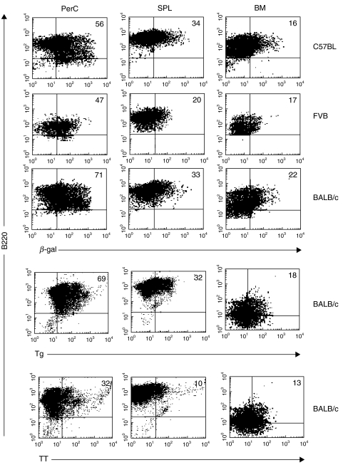
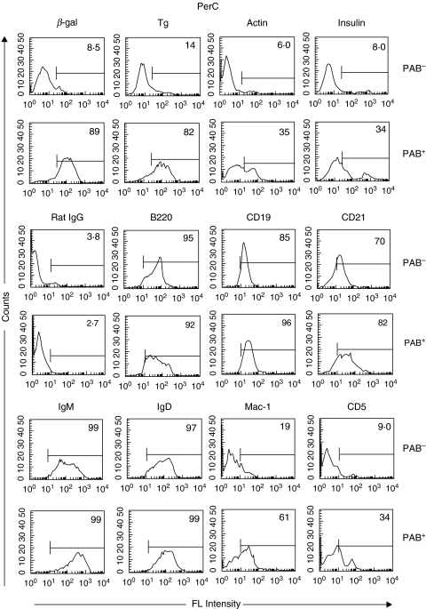
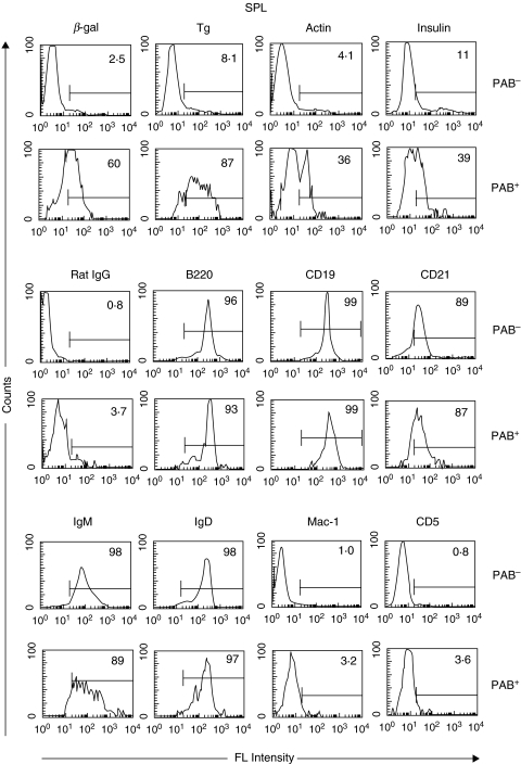
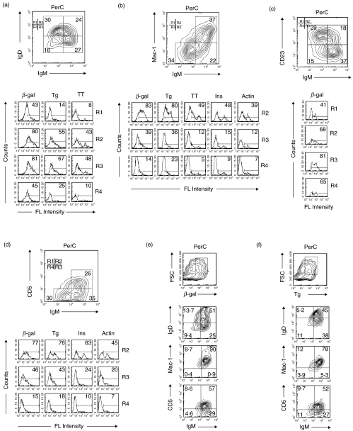
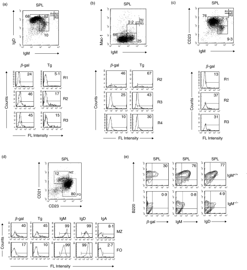
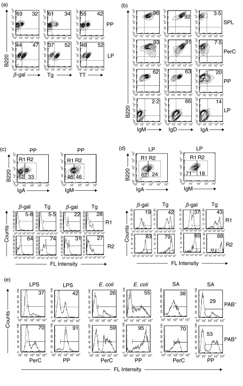
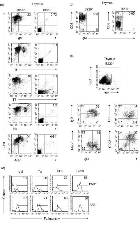
B cells that make polyreactive antibodies (PAB+ cells) express polyreactive Ig receptors on their surface and can bind a variety of different antigens. The present study shows that PAB+ cells are widely distributed, are present in varying numbers in different lymphoid organs and that their phenotype varies depending on the organs from which they are isolated. Up to 10 times more cells in PAB+ enriched populations bind antigens as compared to PAB- populations. Comparison of PAB+ with B-1+ cells showed that a high percentage of PAB+ cells are B-1+, but that many PAB+ cells do not express B-1 cell surface markers and, in fact, are B-1-. It is concluded that the B cell population consists of PAB+/B-1+, PAB+/B-1-, PAB-/B-1+, and PAB-/B-1- cells. The presence of PAB+ cells in the thymus points to the possibility that PAB+ cells may carry endogenous host antigens from peripheral tissues to the thymus where they may contribute to immunological tolerance.
Figures
Similar articles
-
Antigen-binding B cells and polyreactive antibodies.Eur J Immunol. 1995 Feb;25(2):579-86. doi: 10.1002/eji.1830250241. Eur J Immunol. 1995. PMID: 7533091
-
Characterization of murine polyreactive antigen-binding B cells: presentation of antigens to T cells.Eur J Immunol. 2001 Apr;31(4):1106-14. doi: 10.1002/1521-4141(200104)31:4<1106::aid-immu1106>3.0.co;2-5. Eur J Immunol. 2001. PMID: 11298335
-
Polyreactive antigen-binding B cells in the peripheral circulation are IgD+ and B7-.Eur J Immunol. 1996 Dec;26(12):2916-23. doi: 10.1002/eji.1830261217. Eur J Immunol. 1996. PMID: 8977286
-
Function of surface immunoglobulin on murine B cells.Prog Clin Biol Res. 1980;42:46-68. Prog Clin Biol Res. 1980. PMID: 6994118 Review. No abstract available.
-
Xenotransplantation and ABO incompatible transplantation: the similarities they share.Transfus Apher Sci. 2006 Aug;35(1):45-58. doi: 10.1016/j.transci.2006.05.007. Epub 2006 Aug 14. Transfus Apher Sci. 2006. PMID: 16905361 Review.
Cited by
-
Stimulation of Toll-Like Receptors profoundly influences the titer of polyreactive antibodies in the circulation.Sci Rep. 2015 Oct 14;5:15066. doi: 10.1038/srep15066. Sci Rep. 2015. PMID: 26463758 Free PMC article.
-
Evidence to Support a Contribution of Polyreactive Antibodies to HLA Serum Reactivity.Transplantation. 2016 Jan;100(1):217-26. doi: 10.1097/TP.0000000000000840. Transplantation. 2016. PMID: 26285015 Free PMC article.
-
Autoantibodies in COVID-19: implications for disease severity and clinical outcomes.Front Immunol. 2025 Jan 6;15:1509289. doi: 10.3389/fimmu.2024.1509289. eCollection 2024. Front Immunol. 2025. PMID: 39835117 Free PMC article. Review.
-
The broad antibacterial activity of the natural antibody repertoire is due to polyreactive antibodies.Cell Host Microbe. 2007 Mar 15;1(1):51-61. doi: 10.1016/j.chom.2007.01.002. Cell Host Microbe. 2007. PMID: 18005681 Free PMC article.
-
Autoantibodies targeting neuronal proteins as biomarkers for neurodegenerative diseases.Theranostics. 2022 Mar 28;12(7):3045-3056. doi: 10.7150/thno.72126. eCollection 2022. Theranostics. 2022. PMID: 35547759 Free PMC article. Review.
References
-
- Casali P, Notkins AL. Probing the human B-cell repertoire with EBV. polyreactive antibodies and CD5+ B lymphocytes. Annu Rev Immunol. 1989;7:513–35. - PubMed
-
- Satoh J, Prabhakar BS, Haspel MV, Ginsberg-Fellner F, Notkins AL. Human monoclonal autoantibodies that react with multiple endocrine organs. N Engl J Med. 1983;309:217–20. - PubMed
-
- Dighiero G, Lymberi P, Mazie JC, et al. Murine hybridomas secreting natural monoclonal antibodies reacting with self antigens. J Immunol. 1983;131:2267–72. - PubMed
-
- Prabhakar BS, Saegusa J, Onodera T, Notkins AL. Lymphocytes capable of making monoclonal autoantibodies that react with multiple organs are a common feature of the normal B cell repertoire. J Immunol. 1984;133:2815–7. - PubMed
-
- Casali P, Burastero SE, Nakamura M, Inghirami G, Notkins AL. Human lymphocytes making rheumatoid factor and antibody to ssDNA belong to Leu-1+ B-cell subset. Science. 1987;236:77–81. - PubMed
MeSH terms
Substances
LinkOut - more resources
Full Text Sources
