

Jejunal microvilli atrophy and reduced nutrient transport in rats with advanced liver cirrhosis: improvement by Insulin-like Growth Factor I
- PMID: 15196310
- PMCID: PMC434503
- DOI: 10.1186/1471-230X-4-12
Jejunal microvilli atrophy and reduced nutrient transport in rats with advanced liver cirrhosis: improvement by Insulin-like Growth Factor I
Abstract
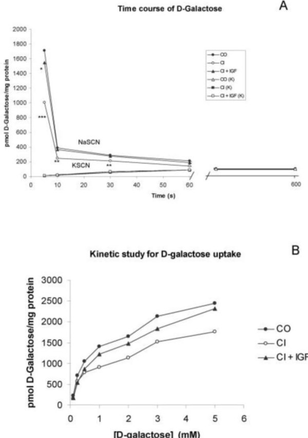
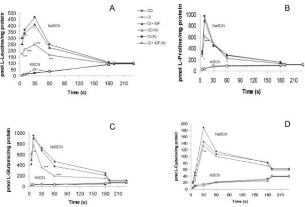
Background: Previous results have shown that in rats with non-ascitic cirrhosis there is an altered transport of sugars and amino acids associated with elongated microvilli. These alterations returned to normal with the administration of Insulin-Like Growth Factor-I (IGF-I). The aims of this study were to explore the evolution of these alterations and analyse the effect of IGF-I in rats with advanced cirrhosis and ascites. Thus, jejunal structure and nutrient transport (D-galactose, L-leucine, L-proline, L-glutamic acid and L-cystine) were studied in rats with ascitic cirrhosis.
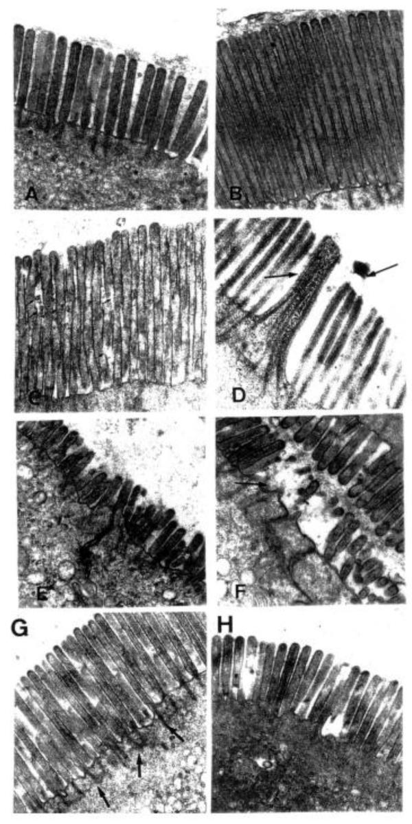
Methods: Advanced cirrhosis was induced by CCl4 inhalation and Phenobarbital administration for 30 weeks. Cirrhotic animals were divided into two groups which received IGF-I or saline during two weeks. Control group was studied in parallel. Jejunal microvilli were studied by electron microscopy. Nutrient transport was assessed in brush border membrane vesicles using 14C or 35S-labelled subtracts in the three experimental groups.
Results: Intestinal active Na+-dependent transport was significantly reduced in untreated cirrhotic rats. Kinetic studies showed a decreased Vmax and a reduced affinity for sugar and four amino acids transporters (expressed as an increased Kt) in the brush border membrane vesicles from untreated cirrhotic rats as compared with controls. Both parameters were normalised in the IGF-I-treated cirrhotic group. Electron microscopy showed elongation and fusion of microvilli with degenerative membrane lesions and/or notable atrophy.
Conclusions: The initial microvilli elongation reported in non ascitic cirrhosis develops into atrophy in rats with advanced cirrhosis and nutrient transports (monosaccharides and amino acids) are progressively reduced. Both morphological and functional alterations improved significantly with low doses of IGF-I.
Figures
Similar articles
-
Altered intestinal transport of amino acids in cirrhotic rats: the effect of insulin-like growth factor-I.Am J Physiol Gastrointest Liver Physiol. 2000 Aug;279(2):G319-24. doi: 10.1152/ajpgi.2000.279.2.G319. Am J Physiol Gastrointest Liver Physiol. 2000. PMID: 10915640
-
Impaired intestinal sugar transport in cirrhotic rats: correction by low doses of insulin-like growth factor I.Gastroenterology. 1997 Oct;113(4):1180-7. doi: 10.1053/gast.1997.v113.pm9322513. Gastroenterology. 1997. PMID: 9322513
-
Effect of insulin-like growth factor I on in vivo intestinal absorption of D-galactose in cirrhotic rats.Am J Physiol. 1999 Jan;276(1):G37-42. doi: 10.1152/ajpgi.1999.276.1.G37. Am J Physiol. 1999. PMID: 9886976
-
Effect of insulin-like growth factor-I on nitrogen balance and intestinal galactose transport in rats with moderate liver cirrhosis.Br J Nutr. 2003 Nov;90(5):929-37. doi: 10.1079/bjn2003974. Br J Nutr. 2003. PMID: 14667186
-
[Insulin-like growth factor I (IGF-I) and liver cirrhosis].Rev Esp Enferm Dig. 2007 Mar;99(3):156-64. doi: 10.4321/s1130-01082007000300007. Rev Esp Enferm Dig. 2007. PMID: 17516829 Review. Spanish.
Cited by
-
IGF-I increases markers of osteoblastic activity and reduces bone resorption via osteoprotegerin and RANK-ligand.J Transl Med. 2013 Oct 25;11:271. doi: 10.1186/1479-5876-11-271. J Transl Med. 2013. PMID: 24161214 Free PMC article.
-
Comparative Proteomic Analysis of Two Differently Extracted Coptis chinensis in the Treatment of Type 2 Diabetic Rats.Evid Based Complement Alternat Med. 2018 Sep 13;2018:3248521. doi: 10.1155/2018/3248521. eCollection 2018. Evid Based Complement Alternat Med. 2018. PMID: 30302116 Free PMC article.
-
IGF-1 modulates gene expression of proteins involved in inflammation, cytoskeleton, and liver architecture.J Physiol Biochem. 2017 May;73(2):245-258. doi: 10.1007/s13105-016-0545-x. Epub 2017 Jan 26. J Physiol Biochem. 2017. PMID: 28124277 Free PMC article.
-
Contribution of gut bacteria to liver pathobiology.Gastroenterol Res Pract. 2010;2010:453563. doi: 10.1155/2010/453563. Epub 2010 Jul 28. Gastroenterol Res Pract. 2010. PMID: 20706692 Free PMC article.
-
Is insulin-like growth factor-1 involved in Parkinson's disease development?J Transl Med. 2020 Feb 11;18(1):70. doi: 10.1186/s12967-020-02223-0. J Transl Med. 2020. PMID: 32046737 Free PMC article. Review.
References
-
- Crawford DHG, Shepherd RW, Halliday JW. Body composition in non-alcoholic cirrhosis: the effect of aetiology and severity on nutritional compartments. Gastroenterology. 1994;106:1611–1617. - PubMed
-
- Kondrup J, Nielsen K, Hamberg O. Nutritional therapy in patients with liver cirrhosis. Eur J Clin Nutr. 1992;46:239–246. - PubMed
-
- Morgan MY. Oxford Textbook of Clinical Hepatology. Oxford: Medical Publications, New York; 1999. pp. 1923–1981.
-
- Caufriez A, Reding P, Urbain DJ. Insulin-like growth factor I: a good indicator of functional hepatocellular capacity in alcoholic liver cirrhosis. Endocrinol Invest. 1991;14:317–321. - PubMed
-
- Donaghy A, Ross R, Gimson A. Growth hormone, insulin-like growth factor-I, and insulin-like growth factor binding proteins 1 and 3 in chronic liver disease. Hepatology. 1995;21:680–688. - PubMed
