. 2004 Jun 21;199(12):1671-7.
doi: 10.1084/jem.20032247. Epub 2004 Jun 14.

Phosphorylation of histone H2B at DNA double-strand breaks

Affiliations

Phosphorylation of histone H2B at DNA double-strand breaks

Oscar Fernandez-Capetillo et al. J Exp Med. .

Abstract

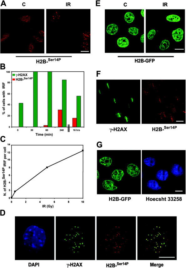
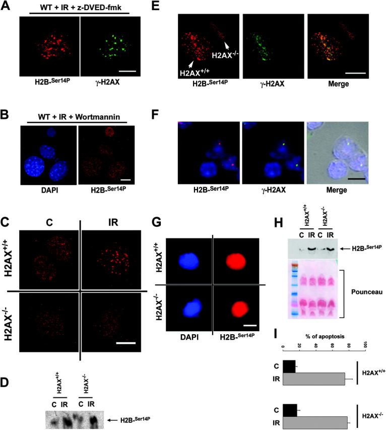
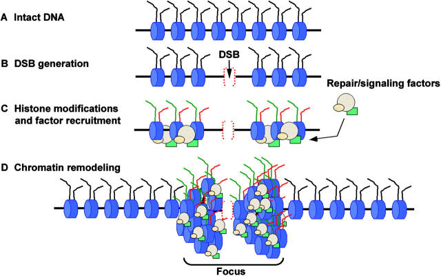
Posttranslational modifications of histone tails regulate numerous biological processes including transcription, DNA repair, and apoptosis. Although recent studies suggest that structural alterations in chromatin are critical for triggering the DNA damage response, very little is known about the nature of DNA damage-induced chromatin perturbations. Here we show that the serine 14 residue in the NH(2)-terminal tail of histone H2B is rapidly phosphorylated at sites of DNA double-strand breaks. At late time points after irradiation, the phosphorylated form of H2B, H2B-(Ser14P), accumulates into irradiation-induced foci. H2B-(Ser14P) foci formation is not associated with the apoptotic phosphorylation of H2B but is strictly dependent on the phosphorylated isoform of H2AX. Our results broaden the spectrum of histone modifications that constitute the DNA damage "histone code" and suggest a model for the underlying chromatin structure within damage-induced foci.


Figures

Figure 1.
Figure 1.
H2B Ser 14 is phosphorylated at DSBs. (A) Immunostaining of H2B-Ser14P (red) in MEFs 4 h after treatment with 10 Gy of IR together with the corresponding nonirradiated control (C). Bar, 10 μm. (B) Percentage of cells with H2B-Ser14P or γ-H2AX foci at various times after a single exposure to 10 Gy of IR. (C) Average number of H2B-Ser14P IRIF per cell after exposure to IR (0, 0.5, 5, and 10 Gy). (D) Immunostaining of H2B-Ser14P (red) and γ-H2AX (green) in MEFs 4 h after treatment with 10 Gy. DNA was counterstained with DAPI. Bar, 5 μm. (E) H2B–GFP–expressing CHO cells 5 h after treatment with 10 Gy of IR (IR) (and nonirradiated control [C]). No foci-like H2B–GFP distribution was observable in irradiated cells. Bar, 10 μm. (F) Immunostaining of H2B-Ser14P (red) and γ-H2AX (green) in MEFs that had been exposed to laser damage. Bar, 20 μm. (G) H2B–GFP expressing CHO cells 5 h after damage by laser scissors. No significant alteration in the distribution of H2B was noted by live cell imaging of these cells during the entire observation period. Note that the path of the laser can be deduced from the bleached nuclear region of the Hoecsht 33258 image. Bar, 10 μm.
Figure 2.
Figure 2.
H2B-Ser14P IRIF formation is not associated with the apoptotic phosphorylation of H2B but is dependent on γ-H2AX. (A) Immunostaining of H2B-Ser14P in wild-type MEFs 4 h after treatment with 10 Gy of IR. The caspase inhibitor z-DEVD-fmk (200 μm) was added to the cells 1 h before the exposure to IR and was maintained in the medium throughout the length of the experiment. Bar, 10 μm. (B) Immunostaining of H2B-Ser14P in wild-type MEF 4 h after treatment with 10 Gy of IR. The PIKK inhibitor wortmannin (200 μm) was added to the cells 15 min before irradiation and was maintained in the medium throughout the length of the experiment. Bar, 10 μm. (C) Immunostaining of H2B-Ser14P (red) in H2AX+/+ and H2AX−/− MEFs 4 h after mock treatment (C) or exposure to 10 Gy of IR. Bar, 5 μm. (D) Western blot analysis of H2B-Ser14P levels in H2AX+/+ and H2AX−/− MEF 4 h after exposure to 100 Gy. (E) Immunostaining of H2B-Ser14P (red) and γ-H2AX (green) in a mixed population of H2AX+/+ and H2AX−/− MEFs that had been exposed to laser damage. Bar, 5 μm. (F) Immunostaining of H2B-Ser14P (red) and γ-H2AX (green) in freshly isolated thymocytes. Bar, 5 μm. (G) Example of the massive H2B-Ser14P (red) staining in H2AX+/+ and H2AX−/− apoptotic thymocytes (see H and I). Bar, 5 μm. (H) Western blot detection of H2B-Ser14P in histone extracts that were prepared by acid extraction from H2AX+/+ and H2AX−/− thymocytes 8 h after exposure to 5 Gy of IR. The Ponceau staining of the nitrocellulose membrane is shown as a loading control. (I) Apoptosis in irradiated thymocytes (IR) measured by flow cytometric analysis of the percentage of cells with a sub-G1 DNA content. Nonirradiated cells (C) that were kept in culture media during the 8-h period were used as a control. When shown (A, B, E, and F), DNA was counterstained with DAPI (blue).
Figure 3.
Figure 3.
The chromatin compaction model. (A) Intact DNA molecule showing the nucleosomes (blue) with protruding tail motifs (black). (B) Generation of a DSB. (C) Histone tails become specifically modified in the chromatin surrounding the lesion (i.e., γ-H2AX [green], H2B-Ser14P [red]). At the same time, DNA repair/signaling factors accumulate in the region. (D) The DSB-induced pattern of histone modifications promotes the compaction of the chromatin and associated factors cytologically detectable as foci.

References

    1. Bakkenist, C.J., and M.B. Kastan. 2003. DNA damage activates ATM through intermolecular autophosphorylation and dimer dissociation. Nature. 421:499–506. - PubMed
    1. Jenuwein, T., and C.D. Allis. 2001. Translating the histone code. Science. 293:1074–1080. - PubMed
    1. Rogakou, E.P., D.R. Pilch, A.H. Orr, V.S. Ivanova, and W.M. Bonner. 1998. DNA double-stranded breaks induce histone H2AX phosphorylation on serine 139. J. Biol. Chem. 273:5858–5868. - PubMed
    1. Chen, H.T., A. Bhandoola, M.J. Difilippantonio, J. Zhu, M.J. Brown, X. Tai, E.P. Rogakou, T.M. Brotz, W.M. Bonner, T. Ried, and A. Nussenzweig. 2000. Response to RAG-mediated VDJ cleavage by NBS1 and gamma-H2AX. Science. 290:1962–1965. - PMC - PubMed
    1. Petersen, S., R. Casellas, B. Reina-San-Martin, H.T. Chen, M.J. Difilippantonio, P.C. Wilson, L. Hanitsch, A. Celeste, M. Muramatsu, D.R. Pilch, et al. 2001. AID is required to initiate Nbs1/gamma-H2AX focus formation and mutations at sites of class switching. Nature. 414:660–665. - PMC - PubMed

MeSH terms