. 2004 Jun 22;101(25):9282-5.
doi: 10.1073/pnas.0401558101. Epub 2004 Jun 14.

Rat adult stem cells (marrow stromal cells) engraft and differentiate in chick embryos without evidence of cell fusion

Affiliations

Rat adult stem cells (marrow stromal cells) engraft and differentiate in chick embryos without evidence of cell fusion

Radhika R Pochampally et al. Proc Natl Acad Sci U S A. .

Abstract

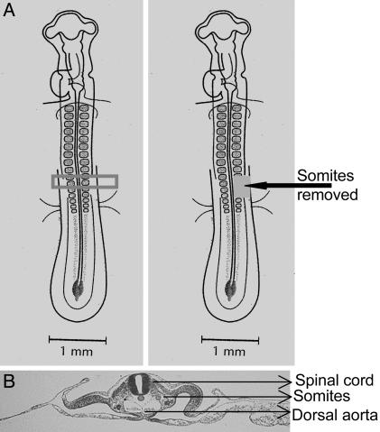
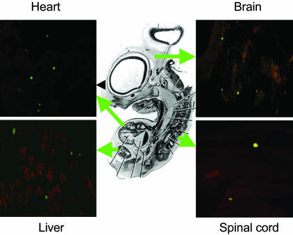
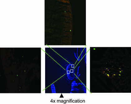
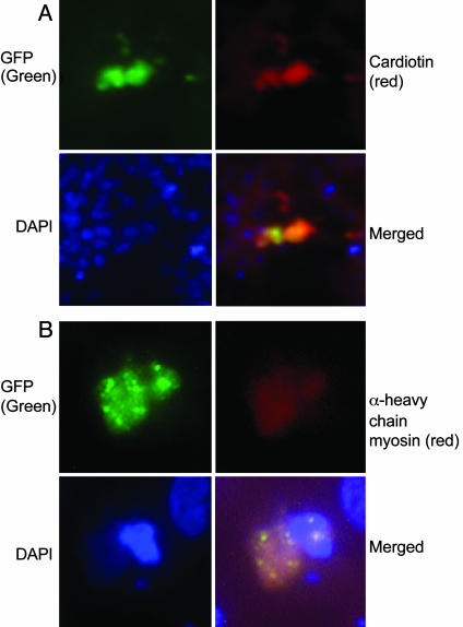
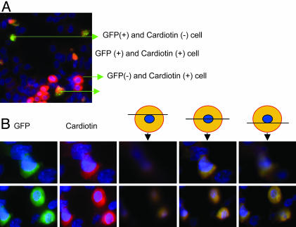
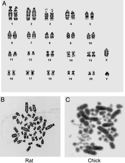
Cell fusion was recently reported to account for the plasticity of adult stem cells in vivo. Adult stem cells, referred to as mesenchymal stem cells or marrow stromal cells, from rat marrow, were infused into 1.5- to 2-day-old chick embryos. After 4 days, the rat cells had expanded 1.3- to 33-fold in one-third of surviving embryos. The cells engrafted into many tissues, and no multinuclear cells were detected. The most common site of engraftment was the heart, apparently because the cells were infused just above the dorsal aorta. Some of the cells in the heart expressed cardiotin, and alpha-heavy-chain myosin. GFP(+) cells reisolated from the embryos had a rat karyotype. Therefore, the cells engrafted and partially differentiated without evidence of cell fusion.


Figures

Fig. 1.
Fig. 1.
Schematic representation of the surgery performed on chick embryos. (A) The two or three most recently formed somites at 48 h were crushed or removed. From 5,000 to 50,000 male rat GFP+ MSCs were injected into the space generated with a glass capillary pipet. (B) Transverse section of stage 12 embryo. [Diagrams reprinted with permission from ref. (Copyright 1997, Elsevier).]
Fig. 2.
Fig. 2.
Rat MSCs expressing GFP detected in organs of 6-day-old chicken embryo by epifluorescence microscopy (×10).
Fig. 3.
Fig. 3.
Rat GFP+ MSCs engrafted and partially differentiated in heart 4 days postinfusion. Five of six embryos assayed had GFP+ cells in heart; section of 20 μm shown at ×4 and ×10.
Fig. 4.
Fig. 4.
Epifluorescent immunohistology of sections from chick embryos infused with rat GFP+ MSCs. (A) Cells positive for both GFP (green) and cardiotin (red) or (B) cells positive for both GFP (green) and α heavy-chain myosin (red). 4′,6-Diamidino-2-phenylindole stain was used to identify nuclei (×63).
Fig. 5.
Fig. 5.
Photomicrograph by 3D deconvolution microscopy of immunohistology sections from chick embryos infused with rat GFP+ MSCs. (A) 3D deconvolution microscopy of section with GFP+ (green) cells and stained for cardiotin (red). The GFP+ cells were mononuclear. (B) 3D deconvolution microscopy to demonstrate that cells that were GFP+ and cardiotin+ were mononuclear (×63).
Fig. 6.
Fig. 6.
Karyotyping of cells expressing GFP. (A) Ideogram of GFP-expressing cells isolated by fluorescence-activated cell sorting from embryos 4 days postinjection. Thirty-five metaphase spreads of GFP-expressing cells were analyzed, and all of the cells contained 42 chromosomes characteristic of rat cells. (B) Metaphase spread of rat chromosomes. (C) Metaphase spread of chick chromosomes. The image was kindly provided by M. E. Delany, University of California, Davis.

References

    1. Friedenstein, A. J., Petrakova, K. V., Kurolesova, A. I. & Frolova, G. P. (1968) Transplantation 6, 230-247. - PubMed
    1. Owen, M. & Friedenstein, A. J. (1988) Ciba Found. Symp. 136, 42-60. - PubMed
    1. Caplan, A. I. (1990) Biomaterials 11, 44-46. - PubMed
    1. Prockop, D. J. (1997) Science 276, 71-74. - PubMed
    1. Eglitis, M. A. & Mezey, E. (1997) Proc. Natl. Acad. Sci. USA 94, 4080-4085. - PMC - PubMed

Publication types

Substances