. 2004 Jun 22;101(25):9405-10.
doi: 10.1073/pnas.0403060101. Epub 2004 Jun 14.

Rabies virus nucleoprotein as a carrier for foreign antigens

Affiliations

Rabies virus nucleoprotein as a carrier for foreign antigens

Martin L Koser et al. Proc Natl Acad Sci U S A. .

Abstract

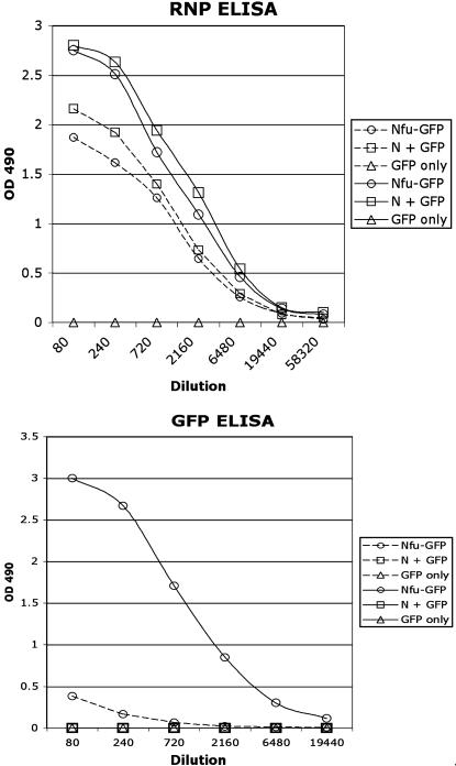
Rabies virus (RV) nucleoprotein (N) tightly encapsidates the genomic and antigenomic RNA of RV to form the viral ribonucleoprotein (RNP) complex. Antigens, such as N, presented in a highly organized structure are sufficient and even desirable to activate B cells to proliferate and produce antibodies. In addition to activating B cells to proliferate, it has been shown that RV N in the RNP complex induces potent T helper cell responses resulting in long-lasting and strong humoral immune responses against RV. The possibility to systematically incorporate foreign genes into the genome of RV and produce a recombinant virus allows us to examine whether the immunogenicity of foreign antigens can be enhanced by incorporation into the RV RNP structure. To test this hypothesis we constructed a recombinant RV expressing a RV N-GFP fusion protein. The chimeric N-GFP fusion protein was efficiently expressed and incorporated into RV RNP and virions. Moreover, the recombinant RNP induces a strong humoral immune response against GFP in mice. In contrast, mice inoculated with GFP alone or a combination of wild-type RV RNPs and GFP did not trigger any GFP-specific humoral responses using the same immunization schedule. These data indicate the usefulness of RV-based vectors as killed vaccines against other infectious diseases.


Figures

Fig. 1.
Fig. 1.
Recombinant RVs expressing N fusion proteins. An RV vector, which allows the expression of an RV N fusion protein, was constructed by deleting the RV N stop codon and introducing the two single restriction sites BsiWI and NheI (BNSP-Nfu). This vector site was the target for introducing the coding sequence of GFP (BNSP-Nfu-GFP). Another RV-based vaccine vector expressing an N-GFP fusion protein from an extra gene in the RV genome was constructed by introducing the gene encoding Nfu-GFP as a new RV transcription unit in pSPBN (pSPBN-Nfu-GFP), resulting in the virus vector SPBN-Nfu-GFP. The SPBN-GFP vector schematic shows an RV vector expressing GFP alone from the same site.
Fig. 2.
Fig. 2.
Recombinant N-GFP fusion protein colocalizes with RV N. BSR cells were infected with virus vector SPBN-GFP (A and A′) or SPBN-Nfu-GFP (B and B′). Cells were fixed 2 days later and immunostained with a monoclonal antibody directed against GFP (A′ and B′) or an antibody specific for RV N protein (A and B). The results indicated that wild-type GFP has a diffuse distribution within the infected cell, whereas the Nfu-GFP fusion protein localizes similar to RV N protein.
Fig. 3.
Fig. 3.
Characterization of recombinant RV expressing Nfu-GFP. (A) To analyze whether the recombinant Nfu-GFP protein is associated with RV RNPs, RNPs from SPBN-Nfu-GFP infected cells were purified by CsCl density centrifugation. A sharp band, which fluoresces green during exposure to light, can be seen. (B) Twelve fractions were collected from the CsCl gradient, dialyzed, and resolved by SDS/PAGE. Probing a Western blot with an RV N-specific antibody detected the highest concentration of RNPs in fraction 9, similar to the results seen for wild-type RNPs (not shown). In addition, a GFP-specific antibody detected equal amounts of Nfu-GFP and N in all fractions, further indicating incorporation of Nfu-GFP into RV RNPs. (C) Virions from BSR cells infected with SPBN or SPBN-Nfu-GFP were purified over 20% sucrose, separated by SDS/PAGE, and analyzed by Western blotting. The results indicate that the Nfu-GFP protein is incorporated into RV virions.
Fig. 4.
Fig. 4.
One-step growth curve of RV vectors. BSR cells were infected with SPBN or SPBN-Nfu-GFP at a multiplicity of infection of 5. Aliquots of culture supernatants were collected at the indicated time points postinfection, and viral titers were determined in duplicate on BSR cells.
Fig. 5.
Fig. 5.
Comparison of GFP immunogenicity using different approaches. Groups of five mice were primed with RNPs containing the RV N-GFP fusion protein (Nfu-GFP, ○), a mixture of recombinant GFP protein and wild-type RV RNPs (N + GFP, □), or GFP protein only (GFP only, ▵). Because no seroconversion was detected after priming, the data points for the prime (first immunization) are not shown in this graph. The same antigens were applied in a second (1st boost, dotted lines) and third (2nd boost, solid lines) immunization. (Upper) RNP-specific ELISA. (Lower) GFP-specific ELISA.

References

    1. Wagner, R. R. & Rose, J. K. (1996) in Fields Virology, ed. Knipe, D. M. (Lippincott, New York), Vol. 1, pp. 1121-1136.
    1. Conzelmann, K. K., Cox, J. H., Schneider, L. G. & Thiel, H. J. (1990) Virology 175, 485-499. - PubMed
    1. Tordo, N. & Kouknetzoff, A. (1993) Onderstepoort J. Vet. Res. 60, 263-269. - PubMed
    1. Tordo, N., Poch, O., Ermine, A., Keith, G. & Rougeon, F. (1988) Virology 165, 565-576. - PubMed
    1. Mebatsion, T., Weiland, F. & Conzelmann, K. K. (1999) J. Virol. 73, 242-250. - PMC - PubMed

Publication types

LinkOut - more resources