Clinical Trial
. 2004 Jun 22;101(25):9363-8.
doi: 10.1073/pnas.0403271101. Epub 2004 Jun 14.

Immunodominant CD4+ responses identified in a patient vaccinated with full-length NY-ESO-1 formulated with ISCOMATRIX adjuvant

Affiliations
Clinical Trial

Immunodominant CD4+ responses identified in a patient vaccinated with full-length NY-ESO-1 formulated with ISCOMATRIX adjuvant

Qiyuan Chen et al. Proc Natl Acad Sci U S A. .

Abstract

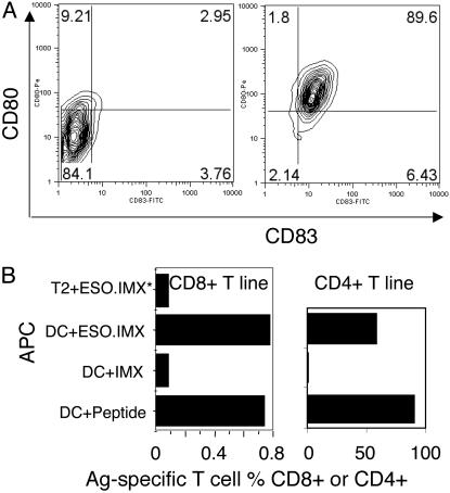
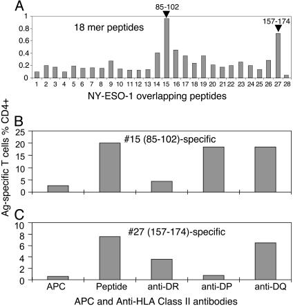
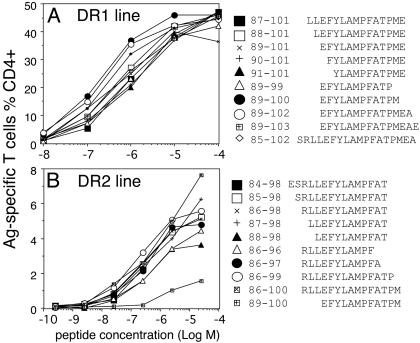
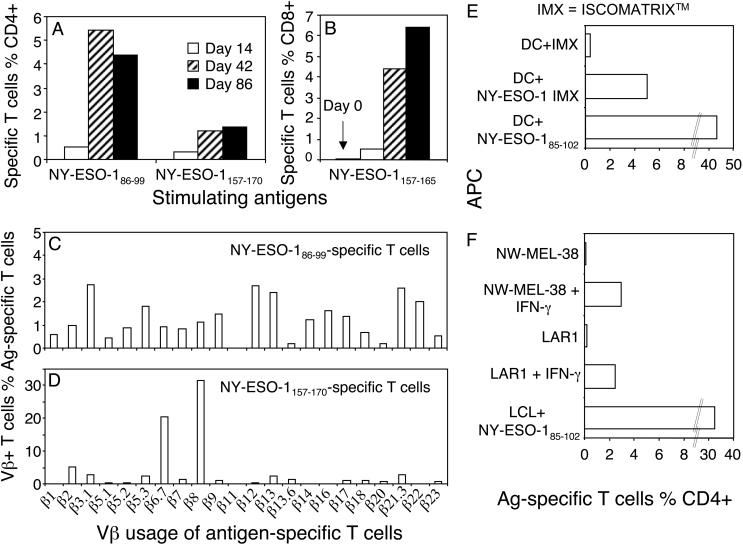
There is increasing evidence showing the involvement of CD4(+) T cells in initiating and maintaining antitumor immune responses. NY-ESO-1 is expressed by various tumors but not normal tissues except testis. We conducted a cancer clinical trial by using full-length NY-ESO-1 protein formulated with ISCOMATRIX adjuvant and injected into patients intramuscularly. Autologous dendritic cells pulsed with NY-ESO-1 ISCOMATRIX in combination with overlapping synthetic peptides were used to identify immunodominant T cells from a vaccinated patient. We show here the identification and characterization of two novel CD4(+) T cell epitopes. T cells specific to these epitopes not only recognized autologous dendritic cells loaded with NY-ESO-1 but also NY-ESO-1-expressing tumor cell lines treated with IFN-gamma. One of the two responses identified was greater than the previously identified immunodominant HLA-DP4-restricted response and correlated with NY-ESO-1-specific CD8(+) T cell induction after vaccination. This T cell response was vaccinated in most patients who expressed HLA-DR2. This study has systematically surveyed patients vaccinated with full-length tumor antigen for a vaccinated CD4 helper T cell response.


Figures

Fig. 1.
Fig. 1.
MoDCs process T cell epitopes from NY-ESO-1 IMX. (A) The MoDCs from patient 7 were loaded with NY-ESO-1 IMX, matured, and stained for CD80 and CD83 (and also for CD86 and HLA-DR; data not shown). (B) Preestablished T cell lines specific to either NY-ESO-1157–165 or NY-ESO-1157–170 were used to detect antigen presentation after NY-ESO-1 IMX loading onto DC or T2 cells.
Fig. 2.
Fig. 2.
Identification of previously uncharacterized CD4+ HLA-DR-restricted NY-ESO-1-specific T cells. (A) T cells stimulated as per Fig. 1 were assessed for their specificities against NY-ESO-1 18-mer overlapping peptides by using autologous BLCLs as APCs in the presence of FCS. (B and C) T cells used in A were stimulated one more time in vitro with either peptide 15 (NY-ESO-185–102)(B) or peptide 27 (NY-ESO-1157–174)(C). The cells were then assayed in the presence or absence of anti-class II antibodies in intracellular cytokine staining.
Fig. 3.
Fig. 3.
Two DR-restricted CD4 determinants within peptide NY-ESO-185–102. BLCLs expressing homozygous HLA-DR molecule and a short-term T cell line (used in Fig. 2 A) and a long-term subline were used in these experiments. (A) The short-term line showed that the majority of T cells were restricted by DR2 and the minority of T cells were restricted by DR1. (B) Long-term subline derived from peptide 15-specific T cells showed exclusive DR1 restriction.
Fig. 4.
Fig. 4.
Fine mapping of the minimum T cell determinants. By using T cell lines [DR1-restricted (A) and DR2-restricted (B)], truncated or extended peptides surrounding the core sequences were tested by titration. Autologous PBMCs were pulsed with the peptides in the absence of FCS for 60 min. Excess peptides were washed out before addition of antigen-specific T cells.
Fig. 5.
Fig. 5.
The previously uncharacterized DR2-restricted T cells are more abundant and more polyclonal than the DP4-restricted T cells and recognize melanoma cell lines. (A) Multiple PBMC samples from patient 7 were thawed and stimulated with NY-ESO-186–99 or NY-ESO-1157–170. The T cell responses were analyzed on day 11. Note that the day-0 sample was not available for this assay. (B) An earlier analysis for NY-ESO-1157–165-specific CD8+ T cell response was performed including the day-0 sample. (C and D) T cells from day 86 after vaccination were stimulated with either NY-ESO-1157–170 or NY-ESO-186–99 and assessed with intracellular cytokine staining plus single Vβ antibodies. Vβ-positive and antigen-specific T cells were displayed as the percent of total antigen-specific T cells. (E and F) The NY-ESO-185–102-specific T cell line was used to read out antigen presentation of autologous MoDCs pulsed with NY-ESO-1 IMX (10 μg/ml, E) and DR1+ (NW-MEL-38) or DR2+ (LAR1) melanoma cell lines with or without IFN-γ treatment (F).

References

    1. Cella, M., Scheidegger, D., Palmer-Lehmann, K., Lane, P., Lanzavecchia, A. & Alber, G. (1996) J. Exp. Med. 184, 747-752. - PMC - PubMed
    1. Bennett, S. R., Carbone, F. R., Karamalis, F., Flavell, R. A., Miller, J. F. & Heath, W. R. (1998) Nature 393, 478-480. - PubMed
    1. Fearon, E. R., Pardoll, D. M., Itaya, T., Golumbek, P., Levitsky, H. I., Simons, J. W., Karasuyama, H., Vogelstein, B. & Frost, P. (1990) Cell 60, 397-403. - PubMed
    1. Gao, F. G., Khammanivong, V., Liu, W. J., Leggatt, G. R., Frazer, I. H. & Fernando, G. J. (2002) Cancer Res. 62, 6438-6441. - PubMed
    1. Cardin, R. D., Brooks, J. W., Sarawar, S. R. & Doherty, P. C. (1996) J. Exp. Med. 184, 863-871. - PMC - PubMed

Publication types