

Review
doi: 10.1038/nrmicro928.
Developing animal models for polymicrobial diseases
Affiliations
- PMID: 15197391
- PMCID: PMC7097426
- DOI: 10.1038/nrmicro928
Item in Clipboard
Review
Developing animal models for polymicrobial diseases
Nat Rev Microbiol.
2004 Jul.
Abstract
Polymicrobial diseases involve two or more microorganisms that act synergistically, or in succession, to mediate complex disease processes. Although polymicrobial diseases in animals and humans can be caused by similar organisms, these diseases are often also caused by organisms from different kingdoms, genera, species, strains, substrains and even by phenotypic variants of a single species. Animal models are often required to understand the mechanisms of pathogenesis, and to develop therapies and prevention regimes. However, reproducing polymicrobial diseases of humans in animal hosts presents significant challenges.
Conflict of interest statement
The author declares no competing financial interests.
Figures

Polymicrobial diseases and suspected polymicrobial diseases (indicated by an asterisk) are listed in the anatomical niche in which the disease pathology is mainly observed.
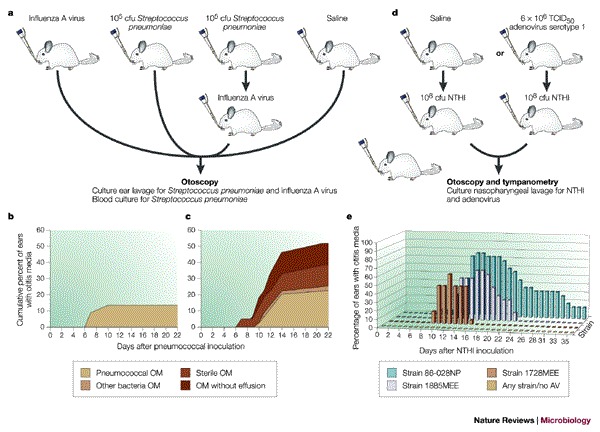
a | Chinchillas were infected intranasally using a pipettor with influenza A virus, 105 colony-forming units (cfu) of Streptococcus pneumoniae or sterile saline. After 2 days, one set of animals inoculated with S. pneumoniae was inoculated intranasally with influenza A virus. All animals were assessed for 22 days by nasopharyngeal lavage every 2–3 days to detect cultures of S. pneumoniae or influenza A virus, by otoscopy to detect symptoms of otitis media, by aspirates from middle-ear effusions to culture S. pneumoniae and influenza A virus and by cardiac puncture to culture S. pneumoniae. The graphs in panels b and c show the incidence of ears with otitis media (OM) that were observed following each infection regime. Panels b and c are reproduced with permission from Ref. © (1980) American Society for Micobiology. d | Chinchillas were infected intranasally using a standard pipettor with either saline (control) or 6 × 10⁶ TCID50 adenovirus serotype I. Seven days later, the same animals were inoculated intranasally with 108 cfu of one of three clinical isolates of nontypeable Haemophilus influenzae (NTHI, strains #86-028NP, #1885MEE or #1728MEE). e | All animals were assessed for 35 days by otoscopy and tympanometry (every 2 days after adenovirus inoculation) for OM and by nasopharyngeal lavage on days 1,4,7,10,14,18,21,28 and 35 to assess NTHI by culturing. TCID50, tissue culture infectious dose 50%, where 50% of aliquots initiate infecton.
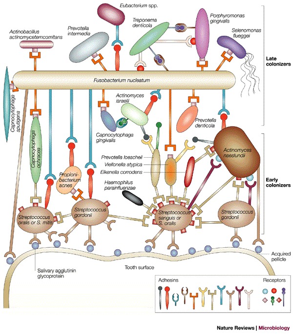
The tooth surface is covered by an acquired pellicle comprised of lipids and proteins, including salivary agglutinin glycoprotein. The pellicle is recognized by primary colonizing bacteria (Streptococcus oralis, Streptococcus mitis, Streptococcus gordonii and Streptococcus sanguis) that express receptors for salivary agglutinin glycoprotein. Other bacteria then colonize in a spatial and temporal manner as shown, using receptors and adhesins to eventually form dental plaque. Reproduced with permission from Ref. . © (2002) American Society for Microbiology.

a | In the mouse model the molar is trimmed to expose the dental pulp. Bacterial suspensions that are being tested for the ability to cause periodontal disease are injected into the dental pulp and the mouse is monitored for signs of periodontal disease by methods that include examination of osseous lesions, tissue necrosis, inflammatory cell recruitment, bacterial tissue penetration and osteoclastogenesis. b | In the rat model bacteria are grown using standard laboratory procedures and washed 3 times with phosphate-buffered saline (PBS) supplemented with 3% sucrose. The rats are pretreated with antibiotics and the mouth is swabbed with chlorhexidine to deplete the oral flora. The bacterial suspension is mixed into the rat's food so that this animal model replicates, as far as possible, the natural route of infection for periodontal disease. After daily inoculation the rats are assessed for bacterial colonization and bone loss. Using this model, different strains of bacteria and the contribution of different virulence loci can be tested. cfu, colony-forming units.
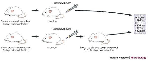
Using an engineered strain of Candida albicans the switch from the yeast to the filamentous forms can be modulated by growth on doxycycline. After pretreatment with this antibiotic, the mice are infected by injection into the tail vein with C. albicans and the mice can be analysed to determine the virulence of the yeast and filamentous forms — under the strict control of antibiotic treatment. In this model, candidiasis can be assessed by measuring the fungal burden in different parts of the anatomy and through histopathological examination.

These diseases share a tick vector, Ixodes scapularis, and to analyse whether Ehrlichia sp. and Borrelia burgorferi (the causative agents of HGE and Lyme disease, respectively) co-infection leads to increased severity of spirochaete-induced Lyme arthritis a mouse model has been developed. Mice are infected intradermally with either spirochaetes (B. burgdorferi cultured in vitro) or HGE (blood culture from a SCID mouse, see inset panel). Arthritis and presence of the two pathogens can then be determined through histopathology, PCR to detect bacterial DNA and by assessing immune responses. Ticks were allowed to feed on all groups of mice to assess transmission of the pathogens. After feeding, PCR (HGE) and immunofluorescence (B. burgdorferi) were used for pathogen detection.
References
-
- Brogden KA, Guthmiller JM. Polymicrobial diseases, a concept whose time has come. ASM News. 2003;69:69–73.
-
- Smith H. The role of microbial interactions in infectious disease. Philos. Trans. R. Soc. Lond. B Biol. Sci. 1982;297:551–561. - PubMed
-
- Jakab GJ. Mechanisms of bacterial superinfections in viral pneumonias. Schweiz Med. Wochenschr. 1985;115:75–86. - PubMed
-
- Hament JM, et al. Respiratory viral infection predisposing for bacterial disease: a concise review. FEMS Immunol. Med. Microbiol. 1999;26:189–195. - PubMed
-
- McCullers JA, Tuomanen EI. Molecular pathogenesis of pneumococcal pneumonia. Front. Biosci. 2001;6:D877–D889. - PubMed
Publication types
MeSH terms
LinkOut - more resources
Full Text Sources
Other Literature Sources
Medical
