. 2004 Oct 1;383(Pt 1):129-38.
doi: 10.1042/BJ20040585.

Heparan sulphate proteoglycans interact with neurocan and promote neurite outgrowth from cerebellar granule cells

Affiliations

Heparan sulphate proteoglycans interact with neurocan and promote neurite outgrowth from cerebellar granule cells

Kaoru Akita et al. Biochem J. .

Abstract

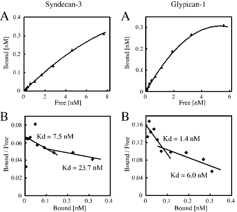
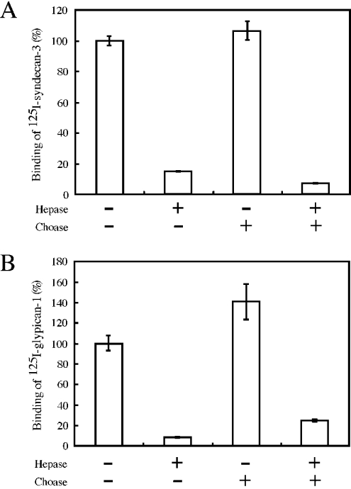
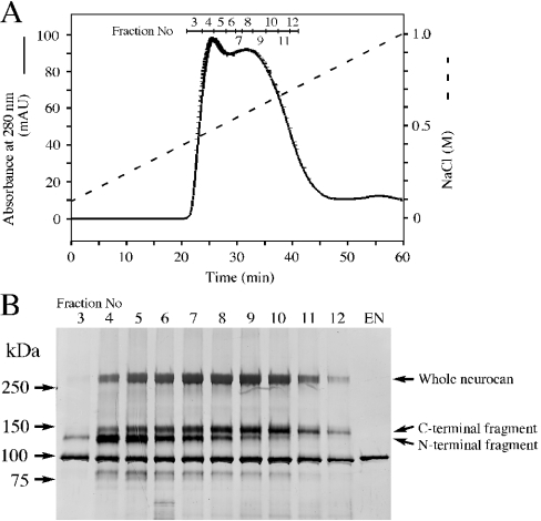
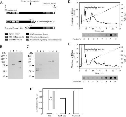
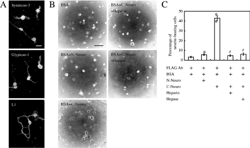
We found that neurocan, a major brain chondroitin sulphate proteoglycan, interacts with HSPGs (heparan sulphate proteoglycans) such as syndecan-3 and glypican-1. Binding of these HSPGs to neurocan was prevented by treatment of the HSPGs with heparitinases I and II, but not by treatment of neurocan with chondroitinase ABC. Scatchard plot analysis indicated that neurocan has two binding sites for these HSPGs with different affinities. It is known that neurocan in the rodent brain is proteolytically processed with aging into N- and C-terminal fragments. When a mixture of whole neurocan and N- and C-terminal fragments prepared from neonatal mouse brains or recombinant N- and C-terminal fragments was applied to a heparin column, the whole molecule and both the N- and C-terminal fragments bound to heparin. A centrifugation cell adhesion assay indicated that both the N- and C-terminal neurocan fragments could interact with these HSPGs expressed on the cell surface. To examine the biological significance of the HSPG-neurocan interaction, cerebellar granule cells expressing these HSPGs were cultured on the recombinant neurocan substrate. A significant increase in the rate of neurite outgrowth was observed on the wells coated with the C-terminal neurocan fragment, but not with the N-terminal one. Neurite outgrowth-promoting activity was inhibited by pretreatment of neurocan substrate with heparin or the addition of heparitinase I to culture medium. These results suggest that HSPGs such as syndecan-3 and glypican-1 serve as the cell-surface receptor of neurocan, and that the interaction of these HSPGs with neurocan through its C-terminal domain is involved in the promotion of neurite outgrowth.


Figures

Figure 1
Figure 1. Purification and identification of syndecan-3-binding protein
(A) Syndecan-3-binding protein was fractionated by chromatography on Sepharose CL-4B and the elution profile and elution volume Ve were analysed. The void volume Vo and the maximum elution volume Vt were measured with respect to the elution positions of Blue Dextran and p-nitrophenol respectively. (B) Aliquots of each fraction were subjected to ligand overlay assay using biotinylated soluble syndecan-3. (C, D) Positive fractions [Kav=(VeVo)/(VtVo)=0.27–0.44] were collected, treated with (+) or without (–) chondroitinase ABC and then separated by SDS/PAGE (6% gel), followed by either visualization by CBB staining (C) or immunochemical detection using anti-neurocan monoclonal antibody 3A11 (D). EN, the same amount of chondroitinase ABC used in (C, D) was subjected to SDS/PAGE followed by the same procedure as described above.
Figure 2
Figure 2. Identification of neurocan-binding proteins
An extract of neonatal mouse brains was fractionated by ammonium sulphate precipitation and ion-exchange chromatography on a column of DEAE-Sephacel as described in the Materials and methods section. The fractions eluted with 0.3–1.5 M NaCl from DEAE-Sephacel were applied to a column of heparin–Sepharose. (A) The pass-through fraction from the heparin column was applied to a column of neurocan–Sepharose. After washing, the bound components were eluted with 10 mM EDTA and then with 1.0 M NaCl. (B) After treatment with (lanes 2, 4 and 6) or without (lanes 1, 3 and 5) heparitinase I, aliquots of each fraction were subjected to SDS/PAGE (2–15% gel), followed by immunoblot analysis with F69-3G10 monoclonal antibodies. Lanes 1 and 2, pass-through fraction from the heparin column; lanes 3 and 4, 10 mM EDTA eluate; lanes 5 and 6, 1 M NaCl eluate. (C) The eluate obtained with 1.0 M NaCl, before and after treatment with heparitinase I, was subjected to SDS/PAGE (7% gel) followed by immunoblot analysis with anti-syndecan-3 antibodies (lane 1) and anti-glypican-1 (core protein) antibodies (lane 2) respectively.
Figure 3
Figure 3. Binding of neurocan to syndecan-3 or glypican-1
(A) Neurocan–Sepharose was incubated with increasing amounts of 125I-syndecan-3 (left panel) or 125I-glypican-1 (right panel) as described in the Materials and methods section. Specific binding was calculated by subtracting the non-specific binding from the total binding, where total binding represents the binding with increasing concentrations of 125I-syndecan-3 or 125I-glypican-1 and non-specific binding represents the binding in the presence of a 200-fold excess of unlabelled syndecan-3 or glypican-1. The bound radioactivity was measured with a gamma counter. Results are expressed as the means for duplicate analyses. (B) Results of Scatchard plot analysis.
Figure 4
Figure 4. Involvement of glycosaminoglycans in the interaction between neurocan and syndecan-3 or glypican-1
(A) 125I-syndecan-3 (10000 c.p.m.) was treated with (+) or without (–) heparitinases I and II (Hepase) and then mixed with neurocan–Sepharose treated with (+) or without (–) chondroitinase ABC (Choase). As a control, Sepharose CL-4B was used instead of neurocan–Sepharose CL-4B. After washing, the bound radioactivity was measured with a gamma counter. (B) Binding of neurocan to glypican-1 was examined similarly using 125I-glypican-1 (10000 c.p.m.). Results are expressed as the averages for duplicate determinations. Error bars represent the S.E.M.
Figure 5
Figure 5. Elution profile of neurocan and its fragments on TSKgel Heparin-5PW
An extract of neonatal mouse brains was fractionated by ammonium sulphate precipitation and DEAE-Sepharose and heparin-affinity chromatographies as described in the Materials and methods section. (A) The fractions eluted from heparin–Sepharose with 0.1–0.7 M NaCl were refractionated with a linear gradient of NaCl from 0.1 to 1.0 M on TSKgel Heparin-5PW. (B) After digestion with chondroitinase ABC, aliquots of each fraction were subjected to SDS/PAGE (2–15% gel), with detection by silver staining. EN, chondroitinase ABC was used for the same procedure.
Figure 6
Figure 6. Binding of recombinant N- and C-terminal neurocan fragments to heparin or HSPGs
(A) FLAG-tagged recombinant neurocan fragments, as represented schematically, were prepared as described in the Materials and methods section. EGF, epidermal growth factor; GAG, glycosaminoglycan; HA, hyaluronic acid; Ig, immunoglobulin. (B) BL21 competent cells were transformed using plasmids constructed with cDNA encoding the N-terminal (lanes 1 and 2) or C-terminal fragment (lanes 3 and 4) of mouse neurocan. Before (lanes 1 and 3) and after (lanes 2 and 4) induction with isopropyl β-D-thiogalactoside, lysates of each type of cell were subjected to SDS/PAGE (7% gel) followed by immunoblot analysis. FLAG-tagged recombinant neurocan fragments were detected with HRP-conjugated anti-FLAG monoclonal antibodies. (C) Cell lysates containing either the FLAG-tagged recombinant N-terminal (lanes 1–3) or C-terminal (lanes 4–6) recombinant neurocan fragment were mixed with anti-FLAG monoclonal antibody-conjugated agarose (lanes 1 and 4), heparin–Sepharose (lanes 2 and 5) or Sepharose CL-4B (lanes 3 and 6). The bound proteins were subjected to SDS/PAGE and immunoblot analysis as described above. Additional bands corresponding to lower molecular masses, which were detected in (B) (lane 4) and (C) (lanes 1 and 2), were probably produced by proteolysis. (D, E) Cell lysates containing the FLAG-tagged recombinant N- (D) or C-terminal (E) neurocan fragment were applied to a column of TSKgel Heparin-5PW. The bound proteins were eluted with a linear gradient of NaCl from 0.1 to 1.0 M. FLAG-tagged recombinant neurocan fragments were detected by dot-blot analysis with HRP-conjugated anti-FLAG monoclonal antibodies. (F) Purified N-terminal neurocan fragment was subjected to SDS/PAGE and stained with Coomassie Brilliant Blue (inset). This fragment was added to the wells coated with syndecan-3 or glypican-1. Bound fragment was detected as described in the Materials and methods section.
Figure 7
Figure 7. Neurite outgrowth from mouse cerebellar granule cells on recombinant neurocan substrate
(A) Cerebellar granule cells from neonatal mice aged 6 days were cultured on poly-D-lysine-coated glass slides for 48 h. The cells were immunostained with anti-syndecan-3 antibodies or anti-glypican-1 antibodies. The cells were also immunostained with antibodies against cell adhesion molecule L1, as a marker for cerebellar granule cells. As a control, the cells treated with normal goat IgG did not exhibit any positive reactivity (results not shown). Scale bar, 20 μm. (B) For the neurite outgrowth assay, the wells were coated with anti-FLAG antibodies only or coated with FLAG-tagged recombinant N- or C-terminal neurocan fragment via anti-FLAG antibodies. To examine further the effect of the heparan sulphate chains, the granule cells were cultured on the C-terminal neurocan fragment-substrate treated with heparin (10 μg/ml) or on the same substrate in the presence of heparitinase I (0.1 unit/ml). The granule cells from neonatal mice aged 6 days were cultured on these substrates for 48 h. These experiments were repeated three times and images from a typical experiment are shown. Scale bar, 100 μm. (C) The percentage of neurite-bearing cells is presented as the means for triplicate determinations. Error bars represent the S.E.M. *P<0.05, **P<0.001, compared with the neurocan-uncoated wells. #P<0.001, compared with recombinant C-terminal neurocan fragment-coated wells. FLAG Ab, anti-FLAG monoclonal antibody; N-Neuro, FLAG-tagged recombinant N-terminal neurocan fragment; C-Neuro, FLAG-tagged recombinant C-terminal neurocan fragment; Hepase, heparitinase I.

References

    1. Carey D. J. N-syndecan: structure and function of a transmembrane heparan sulfate proteoglycan. Perspect. Dev. Neurobiol. 1996;3:331–346. - PubMed
    1. Chernousov M. A., Stahl R. C., Carey D. J. Schwann cells secrete a novel collagen-like adhesive protein that binds N-syndecan. J. Biol. Chem. 1996;271:13844–13853. - PubMed
    1. Kinnunen T., Kaksonen M., Saarinen J., Kalkkinen N., Peng H. B., Rauvala H. Cortactin-Src kinase signaling pathway is involved in N-syndecan-dependent neurite outgrowth. J. Biol. Chem. 1998;273:10702–10708. - PubMed
    1. Lauri S. E., Kaukinen S., Kinnunen T., Ylinen A., Imai S., Kaila K., Taira T., Rauvala H. Regulatory role and molecular interactions of a cell-surface heparan sulfate proteoglycan (N-syndecan) in hippocampal long-term potentiation. J. Neurosci. 1999;19:1226–1235. - PMC - PubMed
    1. Erdman R., Stahl R. C., Rothblum K., Chernousov M. A., Carey D. J. Schwann cell adhesion to a novel heparan sulfate binding site in the N-terminal domain of alpha 4 type V collagen is mediated by syndecan-3. J. Biol. Chem. 2002;277:7619–7625. - PubMed

Publication types

MeSH terms