. 2004 Jun 15:2:14.
doi: 10.1186/1741-7007-2-14.

Segmental identity and cerebellar granule cell induction in rhombomere 1

Affiliations

Segmental identity and cerebellar granule cell induction in rhombomere 1

Mark Eddison et al. BMC Biol. .

Abstract

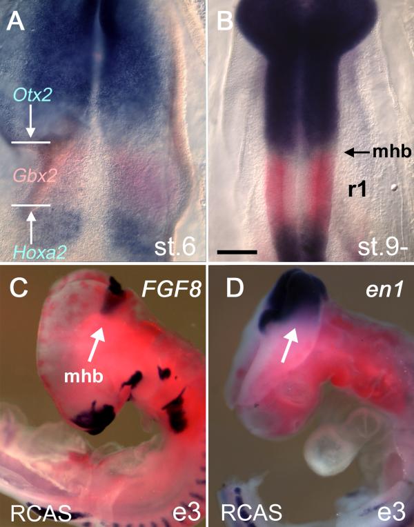
Background: Cerebellar granule cell precursors are specifically generated within the hindbrain segment, rhombomere 1, which is bounded rostrally by the midbrain/hindbrain isthmus and caudally by the boundary of the Hoxa2 expression domain. While graded signals from the isthmus have a demonstrable patterning role within this region, the significance of segmental identity for neuronal specification within rhombomere 1 is unexplored. We examined the response of granule cell precursors to the overexpression of Hoxa2, which normally determines patterns of development specific to the hindbrain. How much does the development of the cerebellum, a midbrain/hindbrain structure, reflect its neuromeric origin as a hindbrain segment?

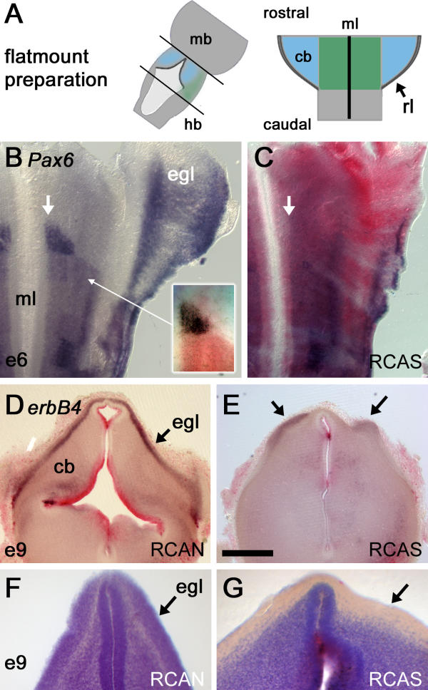
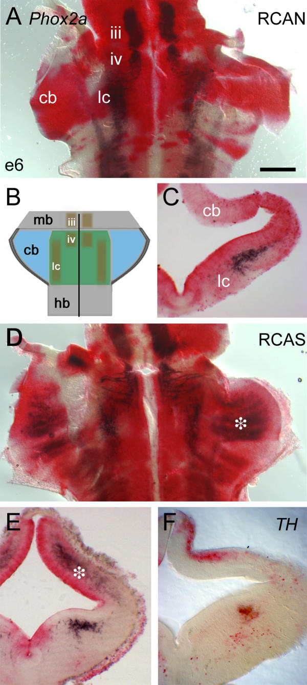
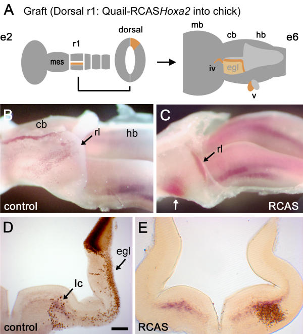
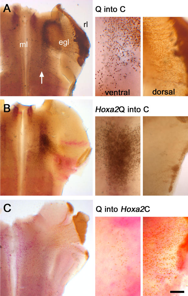
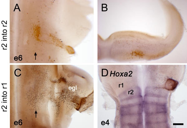
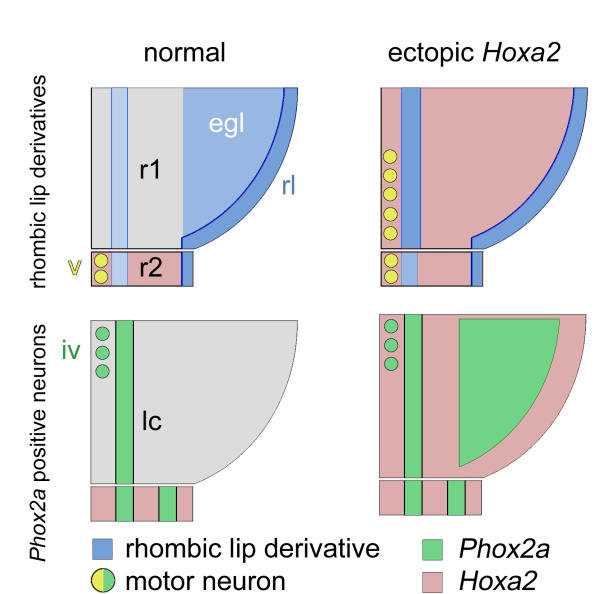
Results: We show that a Gbx2-positive, Otx2-/Hoxa2-negative territory corresponding to rhombomere 1 forms prior to an identifiable isthmic organiser. Early global overexpression of Hoxa2 at embryonic day 0 has no effect on the expression of isthmic signalling molecules or the allocation of rhombomere 1 territory, but selectively results in the loss of granule cell markers at embryonic day 6 and the depletion of cell bodies from the external granule cell layer. By comparison the trochlear nucleus and locus coeruleus form normally in ventral rhombomere 1 under these conditions. Microsurgery, coupled with electroporation, to target Hoxa2 overexpression to rhombic lip precursors, reveals a profound, autonomous respecification of migration. Rhombic lip derivatives, normally destined to occupy the external granule cell layer, violate the cerebellar boundary to form a ventrolateral nucleus in a position comparable to that occupied by rhombic lip derived neurons in rhombomere 2.

Conclusions: Different overexpression strategies reveal that the recognition of migration cues by granule cell precursors is dependent on their identity as rhombomere 1 derivatives. Segmental patterning cues operate autonomously within the rhombic lip precursor pool. By contrast, a subset of coextensive nuclei is refractory to ectopic Hoxa2 and is presumably induced solely by isthmic organiser activity. Thus, graded (isthmic) and segmental mechanisms may operate exclusively of one another in the specification of different neuronal populations within rhombomere 1. The early designation of an Otx2-negative, Hoxa2-negative region, prior to the appearance of the isthmic organiser, is a key initial step in the specification of the cerebellum.


Figures

Figure 1
Figure 1
Establishment of rhombomere 1: Isthmic gene expression is refractory to Hoxa2 overexpression (A) At stage [st.] 6, Otx2 and Hoxa2 expression (both in blue) are separated by a Gbx2-positve domain (red) that defines rhombomere 1 (r1). The domain of Gbx2 expression encompasses r1–3. (B) At st.9-, the boundaries of the Otx2 and Hoxa2 expression domains define the segmental boundaries of r1 as determined by fate-map experiments [2, 17]. (C) Viral overexpression of RCASBP(B)Hoxa2 (red) at embryonic day (E) 0 fails to suppress the activation of either FGF8 (blue) or (D) en1 at E3 (e3). Scale bar (A,B) = 200 μm. mhb – midbrain/hindbrain isthmus.
Figure 2
Figure 2
Global overexpression of Hoxa2 depletes rhombomere 1 of granule cell precursors (A) Flatmounts of brains up to embryonic day (E) 6 were prepared by opening the neural tube along the dorsal midline: cb – cerebellum, mb – midbrain, hb – hindbrain, rl – rhombic lip, ml – ventral midline. (B) The expression of Pax6 (blue) at E6 in one half of the anterior hindbrain viewed as a flatmount (rostral top, mediolateral axis runs left to right). Granule cell precursors born at the rhombic lip express Pax6 as they migrate over dorsal rhombomere 1 (r1) to condense as the external granule cell layer (EGL [egl]). Pax6 is also expressed in a ventral wedge of r1 precursors (arrow) abutting the anterior r2 boundary and ventral midline. The relationship of this Pax6 positive precursor pool (blue) to the anterior boundary of Hoxa2 (red) is shown inset. (C) Overexpression of Hoxa2 (red) leads to a loss of Pax6 in the EGL and prominent ventral wedge (arrow). This is replaced by a weaker, broadened, ventral Pax6 domain similar to that in the hindbrain. (D) At E9, in transverse section through a control embryo, the neuregulin receptor erbB4 labels the EGL. Red label dorsally reflects high levels of expression of the control virus, RCANBP(B)Hoxa2 (red), within the ventricular zone of the cerebellum (cb). (E) Injection of the active RCASBP(B)Hoxa2 virus leads to a reduction in erbB4. Patches of expression (arrows) correspond to residual EGL overlying a locally thickened cerebellum. (F) Cresyl violet stains in control and (G) RCASBP(B)Hoxa2-infected cerebella reveal an absence of cell bodies within a superficial, subpial layer (arrows), normally occupied by the EGL, following Hoxa2 overexpression. Scale bar (D,E) = 500 μm.
Figure 3
Figure 3
Locus coeruleus and trochlear nucleus formation is normal following global Hoxa2 overexpression (A) Flatmounted control embryo at embryonic day (E) 6, infected with RCANBP(B)Hoxa2 virus (red); Phox2a expression (black) marks the oculomotor nucleus (iii) in the midbrain, and trochlear motor nucleus (iv) and locus coeruleus (lc) in rhombomere 1 (r1). There is no Phox2a expression within the cerebellar anlage (cb). (B) Schematic diagram showing the arrangement of Phox2a-positive nuclei within r1, as shown in (A). (C) This lack of dorsal expression is confirmed in transverse section, while the locus coeruleus is clearly identified by Phox2a. (D) Hoxa2 up-regulation with RCASBP(B)Hoxa2 (red) leads to ectopic Phox2a expression in the dorsal r1/cerebellum (asterisk). (E) In a transverse section slightly caudal to that in C, ectopic expression of Phox2a is confined to the deeper cerebellar layers (asterisk), while ventral expression of Phox2a in the locus coeruleus is unaltered. (F) Hoxa2 up-regulation does not result in increased tyrosine hydroxylase (TH) staining within dorsal r1. Scale bar (A,D) = 400 μm. mb – midbrain, hb – hindbrain.
Figure 4
Figure 4
Targeted Hoxa2 overexpression respecifies the migration of rhombic lip derivatives (A) Schematic diagram showing the location of donor tissue grafted from Hoxa2 electroporated quail embryos at embryonic day (E) 2 (e2; left). A graft of dorsal rhombomere 1 (r1) maps to the cerebellar rhombic lip and roofplate at E6 (e6; right) and contributes to both the external granule cell layer (EGL) and neural crest derivatives in the trochlear (iv) and trigeminal (v) nerves [2]. (B) Lateral view of a wholemount dissection of the cerebellum (cb) and hindbrain (hb) of a control E6 chimaeric embryo stained with a quail-specific antibody, Q¢PN (brown), and stained for Phox2a (purple). The rostrocaudal axis runs left to right. A stream of ventrally migrating quail cells originating from caudal rhombic lip (rl) lies over the presumptive cerebellum (point of origin indicated by a black arrow). A thin strip of quail cells also lies in the roofplate separating the bilateral anlage. (C) Migration patterns are substantially altered when grafted donor cells (black arrow) are electroporated with RCASBP(B)Hoxa2. Rhombic lip derivatives fail to condense in the EGL but rather migrate into a single ventrolateral nucleus (white arrow), which expresses high levels of ectopic Hoxa2 (red). (D) Transverse section through the cerebellum of a control chimaera showing the donor progenitors in the dorsal neural tube. The majority of quail cell derivatives lie within the EGL (egl), with a scattering of cells ventrally overlapping the Phoxa2 expression in the locus coeruleus (light blue). (E) By contrast, the majority of cells from grafts electroporated with Hoxa2 condense ventrally (where quail cell nuclear diaminobenzidine label obscures the red in situ product marking Hoxa2). Scale bar (D,E) = 200 μm. mb – midbrain; mes – mesencephalon.
Figure 5
Figure 5
Effects on tangential migration of ectopic Hoxa2 are rhombic-lip-autonomous A comparison of E6 quail-chick chimaeras with different combinations of targeted Hoxa2 overexpression (red), in which the relative proportions of dorsal and ventral rhombic lip derivatives are compared (right). (A) Uninfected quail (Q) donor tissue in a normal chick (C) host (Q into C) gives rise to EGL cells and a small number of neurons in ventral r1 (white arrow). (B) Quail cells expressing Hoxa2 in a normal host (Q-Hoxa2) bypass the EGL and condense ventrally. (C) Normal quail cells migrate in a host expressing ectopic Hoxa2 (Q into Hoxa2 C) migrate as controls. Scale bar for high-power insets = 100 μm. egl – external granule cell layer; ml – midline.
Figure 6
Figure 6
Derivatives of r2 dorsal neural tube have a characteristic migration pattern correlated to maintenance of endogenous Hoxa2 expression (A) Flatmount of a fate-map chimaera at embryonic day (E) 6 (e6), in which quail r2 was orthotopically grafted into rhombomere 2 (r2) of a chick host. At E6, in flatmount, quail derivatives (brown) condense primarily in a single ventrolateral nucleus. (B) In transverse section, r2 migrants can be seen to settle in a subpial location similar to that of ventral derivatives in r1. (C) When transplanted into r1, quail r2 cells contribute to the EGL while maintaining a sparse ventral contribution. (D) When analysed by in situ hybridisation at E4 (e4), r2 grafted into r1 has lost its endogenous Hoxa2 expression (blue). Scale bar = 100 μm.
Figure 7
Figure 7
Summary of the effects of Hoxa2 overexpression on r1 derivatives The arrangement of rhombic lip derivatives in rhombomere 1 (r1) (top) and Phox2a-positive neurons (bottom) shown on a schematised flatmount of a half neural tube from normal (left) and Hoxa2-infected (right) embryos. (Top) Hoxa2 overexpression in r1 results in the loss of the EGL and an expansion of ventral rhombic lip derivatives. Ectopic trigeminal-like motor neurons are generated in caudal r1 [14]. (Bottom) The trochlear nucleus and locus coeruleus form normally when Hoxa2 is overexpressed; however, dorsal r1 expresses high levels of ectopic Phox2a. The cells of this latter population are neither TH-positive locus coeruleus precursors that have stalled during migration nor rhombic lip derivatives that have altered their fate. egl – external granule cell layer; rl – rhombic lip.

Similar articles

Cited by

References

    1. Lumsden A, Krumlauf R. Patterning the vertebrate neuraxis. Science. 1996;274:1109–1115. doi: 10.1126/science.274.5290.1109. - DOI - PubMed
    1. Wingate RJT, Hatten ME. The role of the rhombic lip in avian cerebellum development. Development. 1999;126:4395–4404. - PubMed
    1. Joyner AL. Engrailed, Wnt and Pax genes regulate midbrain – hindbrain development. Trends Genet. 1996;12:15–20. doi: 10.1016/0168-9525(96)81383-7. - DOI - PubMed
    1. Rhinn M, Brand M. The midbrain – hindbrain boundary organizer. Curr Opin Neurobiol. 2001;11:34–42. doi: 10.1016/S0959-4388(00)00171-9. - DOI - PubMed
    1. Jaszai J, Reifers F, Picker A, Langenberg T, Brand M. Isthmus-to-midbrain transformation in the absence of midbrain-hindbrain organizer activity. Development. 2003;130:6611–6623. doi: 10.1242/dev.00899. - DOI - PubMed

Publication types

MeSH terms

Substances