. 2004 Jun 15;18(12):1439-51.
doi: 10.1101/gad.1184204.

Spindle checkpoint regulates Cdc20p stability in Saccharomyces cerevisiae

Affiliations

Spindle checkpoint regulates Cdc20p stability in Saccharomyces cerevisiae

Jing Pan et al. Genes Dev. .

Abstract

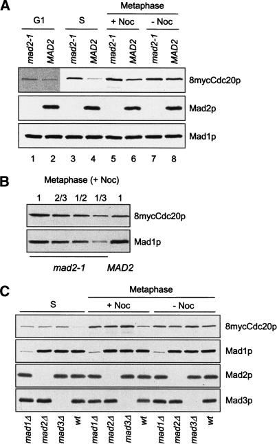
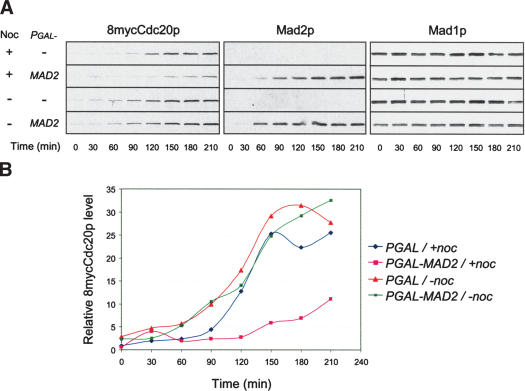
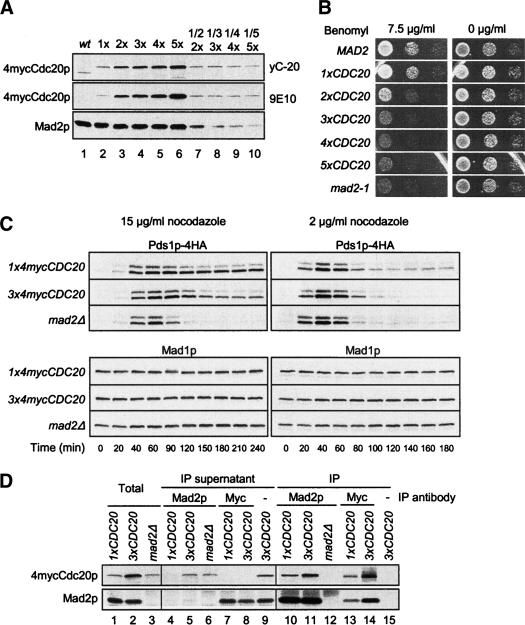
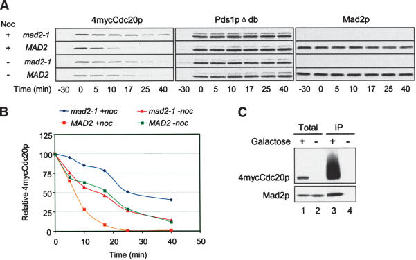
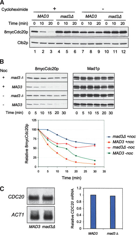
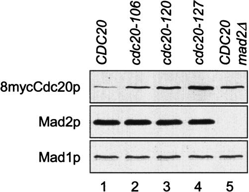
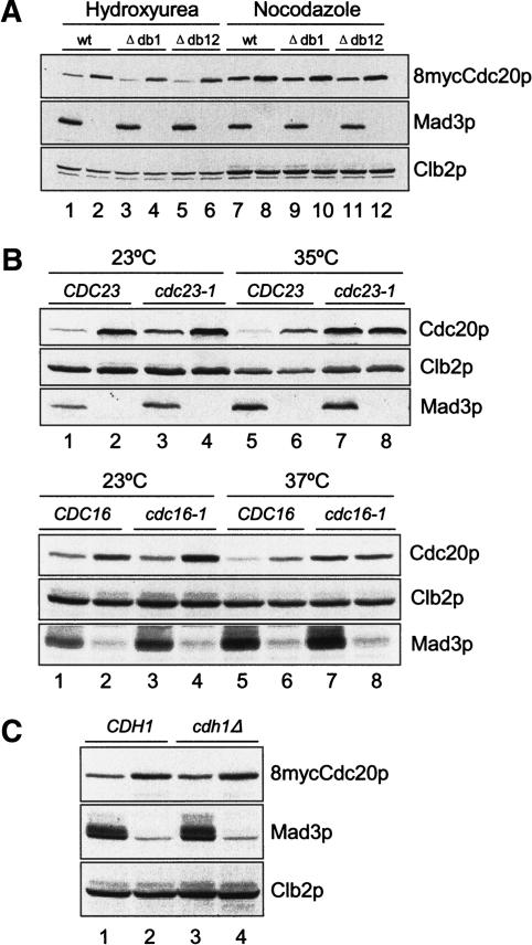
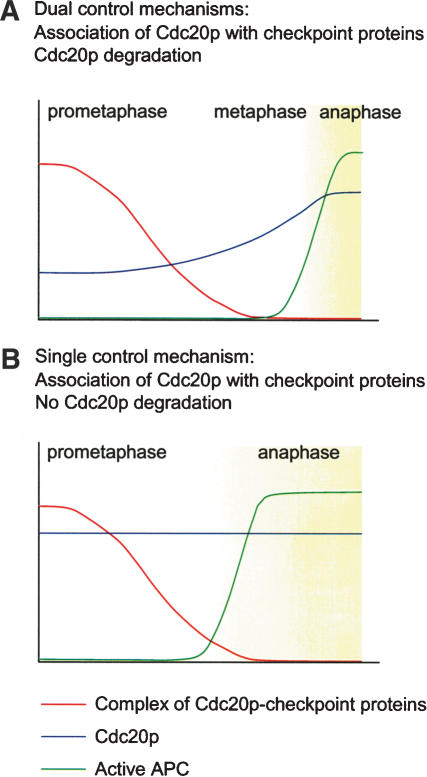
The spindle checkpoint arrests cells at the metaphase-to-anaphase transition until all chromosomes have properly attached to the mitotic spindle. Checkpoint proteins Mad2p and Mad3p/BubR1p bind and inhibit Cdc20p, an activator for the anaphase-promoting complex (APC). We find that upon spindle checkpoint activation by microtubule inhibitors benomyl or nocodazole, wild-type Saccharomyces cerevisiae contains less Cdc20p than spindle checkpoint mutants do, whereas their CDC20 mRNA levels are similar. The difference in Cdc20p levels correlates with their difference in the half-lives of Cdc20p, indicating that the spindle checkpoint destabilizes Cdc20p. This process requires the association between Cdc20p and Mad2p, and functional APC, but is independent of the known destruction boxes in Cdc20p and the other APC activator Cdh1p. Importantly, destabilization of Cdc20p is important for the spindle checkpoint, because a modest overexpression of Cdc20p causes benomyl sensitivity and premature Pds1p degradation in cells treated with nocodazole. Our study suggests that the spindle checkpoint reduces Cdc20p to below a certain threshold level to ensure a complete inhibition of Cdc20p before anaphase.


Figures

Figure 1.
Figure 1.
Cdc20p levels are elevated in spindle checkpoint mutants. (A) mad2-1 and MAD2 cells containing 8mycCDC20 and a galactose-inducible nondegradable Pds1pΔdb (PGAL-pds1Δdb; RHC435 and RHC436) were arrested at G1 with α-factor (lanes 1,2), or at S phase with hydroxyurea (lanes 3,4) in YEPD at 30°C. For metaphase arrest, cells were first synchronized at G1 with α-factor in YEP containing raffinose (YEPR), followed by induction of Pds1pΔdb with 2% galactose for 1 h at 30°C during the G1 arrest. Cells were then washed and released into YEPD in the presence (lanes 5,6) or absence (lanes 7,8) of nocodazole. Pds1pΔdb ensures mitotic arrest of mad2-1, which lacks the spindle checkpoint and cannot be blocked at metaphase by nocodazole. Cell lysates were prepared at 100 min after the release for immunoblots of 8mycCdc20p, Mad2p, and Mad1p, using 9E10, anti-Mad2p, and anti-Mad1p antibodies, respectively. The Mad1p blot serves as a loading control. Because of the low level of Cdc20p at G1, a long exposure of the Cdc20p blot is presented for G1 samples (lanes 1,2). (B) The lysate prepared from mad2-1 cells arrested at metaphase by Pds1pΔdb in the presence of nocodazole was loaded in serial dilutions to compare its 8mycCdc20p level with that in the MAD2 cell lysate. (C) mad1Δ, mad2Δ, mad3Δ, or wild-type cells containing 8myc-CDC20 and PGAL-pds1Δdb (RHC472, RHC473, RHC474, and RHC476) were arrested at S phase with hydroxyurea, or at metaphase by Pds1pΔdb in the presence or absence of nocodazole, as described in A. Immunoblot analysis was performed as indicated. The weak signals present in mad1Δ lanes are from a cross-reacting protein recognized by anti-Mad1p antibody.
Figure 2.
Figure 2.
Overexpression of Mad2p reduces Cdc20p levels in the presence of nocodazole. (A) 8mycCDC20, PGAL-pds1Δdb cells containing PGAL vector alone or PGAL-MAD2 (RHC401 and RHC402) were first arrested at G1 with α-factor, then washed and released into YEPR in the presence or absence of nocodazole at 30°C. When ∼10% of the cells began to bud, 2% galactose was added to the medium. Samples were taken at 30-min intervals after galactose addition for immunoblot analysis. Mad1p blots serve as loading controls. (B) Signals of 8mycCdc20p in A were quantified and plotted against time.
Figure 3.
Figure 3.
Increased Cdc20p expression impairs the spindle checkpoint. (A) Cdc20p levels increase with additional copies of integrated 4mycCDC20. Cell lysates were prepared from wild-type and cdc20Δ cells containing one to five copies of 4mycCDC20 (1×, 2×, 3×, 4×, and 5×; RHC542–RHC546) temporarily arrested at metaphase with nocodazole. Immunoblots were performed with 40 μg of total protein (lanes 16), 20 μg of protein from the 2× strain (lane 7), 16.7 μg of protein from the 3× strain (lane 8), 10 μg of protein from the 4× strain (lane 9), and 8 μg of protein from the 5× strain (lane 10). 4mycCdc20p was detected with either anti-Cdc20p antibody yC-20 (top) or anti-myc antibody 9E10 (middle). The Mad2p blot serves as a loading control. (B) Increased Cdc20p expression leads to benomyl sensitivity. MAD2, mad2-1, and cdc20Δ cells containing one to five copies of integrated 4mycCDC20 (1×, 2×, 3×, 4×, and 5× CDC20) were spotted onto YEPD plates with or without 7.5 μg/mL benomyl. The cells were in 10-fold serial dilutions from left to right. (C) Cells overexpressing 4mycCDC20 by threefold are defective in maintaining Pds1p levels in the presence of nocodazole. PDS1-4HA cells containing one (RHC639) or three copies of 4mycCDC20 (RHC641), or one copy of 4mycCDC20 in mad2Δ (RHC644), were first arrested in G1 with α-factor. Cells were then released at time 0 into YEPD containing 15 μg/mL nocodazole at 25°C (left panel) or 2 μg/mL nocodazole at 30°C (right panel). α-Factor was added back at 1 h 20 min (left panel) or 1 h (right panel) to prevent re-entry into the next cell cycle. Samples were taken at the indicated time points for immunoblot analysis. Mad1p blots serve as loading controls. (D) A fraction of Cdc20p is not bound with Mad2p when Cdc20p is overexpressed. 1xCDC20 (RHC639), 3xCDC20 (RHC641), and mad2Δ (RHC644) cells were first arrested in G1 and then released into YEPD containing 15 μg/mL nocodazole at 25°C. Cell lysates were prepared 2 h after the release, followed by immunoprecipitation of Mad2p and 4mycCdc20p with anti-Mad2p and 9E10 antibodies, respectively. Mock immunoprecipitation was performed with the 3xCDC20 cell lysate as a control for 9E10 immunoprecipitation. The cell lysates (lanes 13), the supernatants from immunoprecipitations (lanes 49), and the immunoprecipitates (lanes 1015) were immunoblotted for 4mycCdc20p and Mad2p as indicated.
Figure 4.
Figure 4.
Spindle checkpoint activation reduces the stability of galactose-induced Cdc20p. (A) Galactose-induced Cdc20p has a longer half-life in mad2-1 than in MAD2 cells in the presence of nocodazole. mad2-1 and MAD2 cells containing PGAL-4mycCDC20 and PGAL-pds1Δdb (RHC421 and RHC422) were arrested at G1 with α-factor in YEPR, followed by a 40-min induction of Pds1pΔdb with 2% galactose at 30°C. Cells were then released into YEPR at 25°C in the presence or absence of nocodazole. The short induction of Pds1pΔdb was sufficient to arrest cells at metaphase. When >95% of the cells were arrested at metaphase (-30 min), expression of 4mycCdc20p was induced with 2% galactose for 30 min and then shut off with 2% glucose and 1 mg/mL cycloheximide (0 min). Samples were taken at the indicated time points for immunoblot analysis. Pds1pΔdb was detected with 16B12 as loading controls. (B) 4mycCdc20p levels were quantified and plotted against time. (C) 4mycCDC20 induced from the GAL promoter binds Mad2p. MAD2 cells containing PGAL-4mycCDC20 and PGAL-pds1Δdb (RHC422) were arrested at G1, briefly induced for Pds1pΔdb expression, and released into YEPR containing nocodazole as described in A. One-half of the culture was then induced for 4mycCdc20p expression with 2% galactose for 30 min (lanes 1,3), and the other half was kept uninduced (lanes 2,4). Cell lysates were prepared for anti-myc immunoprecipitation, followed by immunoblot analysis for 4mycCdc20p and Mad2p.
Figure 5.
Figure 5.
Spindle checkpoint activation reduces the stability of endogenous Cdc20p. (A) Inhibition of protein synthesis rapidly depletes Cdc20p in spindle checkpoint-active cells. MAD3 (RHC476) and mad3Δ cells (RHC474) were arrested in mitosis by Pds1pΔdb in the presence of nocodazole at 30°C, as described for Figure 1A. Cycloheximide was then added at 1 mg/mL to half of the cultures, and samples were taken at the indicated time points for immunoblot analysis of 8mycCdc20p and Clb2p. The latter serves as controls for mitotic arrest and equal loading. (B) Endogenous Cdc20p has a longer half-life in mad3Δ cells than in MAD3 cells. MAD3 (RHC476) and mad3Δ cells (RHC474) were arrested in mitosis by Pds1pΔdb in the presence or absence of nocodazole at 25°C. Cycloheximide was then added at 1 mg/mL, and samples were taken at the indicated time points for immunoblot analysis. The Mad1p blots serve as loading controls. 8mycCdc20p levels were quantified and plotted against time. (C) Wild-type and mad3Δ cells contain similar levels of CDC20 mRNA in mitosis. Wild-type MAD3 (RHC292) and mad3Δ cells (RHC482) were arrested in mitosis by Pds1pΔdb in the presence of nocodazole at 30°C. Total RNA was prepared for Northern blot analysis of CDC20 and ACT1 mRNAs. The plot shows the relative CDC20 mRNA levels normalized with ACT1 signals.
Figure 6.
Figure 6.
Mutant Cdc20p that cannot bind Mad2p is present at elevated levels. Cells containing 8myc-tagged CDC20, cdc20-106, cdc20-120, or cdc20-127 on centromeric plasmids (lanes 14, RHC554–RHC557), or mad2Δ cells containing 8mycCDC20 (lane 5, RHC558) were arrested at metaphase by Pds1pΔdb in the presence of nocodazole, as described for Figure 1A. Cell lysates were prepared for immunoblot analysis. The Mad1p blot serves as a loading control.
Figure 7.
Figure 7.
The spindle checkpoint regulates Cdc20p levels through APC. (A) The destruction-box sequences in Cdc20p are not important for spindle checkpoint-induced instability. MAD3 (odd lanes) or mad3Δ cells (even lanes) containing 8mycCDC20 (lanes 1,2,7,8), 8mycCDC20Δdb1 (lanes 3,4,9,10), or 8mycCDC20Δdb12 (lanes 5,6,11,12) on centromeric plasmids (RHC582–RHC587) were arrested at S phase with hydroxyurea (lanes 16) or at metaphase with Pds1pΔdb in the presence of nocodazole (lanes 712). Cell lysates were prepared for immunoblot analysis. (B) Regulation of Cdc20p levels by the spindle checkpoint is dependent on functional APC. (Upper panel) CDC23 (lanes 1,5, RHC292), CDC23 mad3Δ (lanes 2,6, RHC482), cdc23-1 (lanes 3,7, RHC576), and cdc23-1 mad3Δ (lanes 4,8, RHC581) cells were first arrested at G1 with α-factor in YEPR, followed by induction of Pds1pΔdb with 3% galactose for 1 h at 23°C. The cultures were then released into YEPR containing 3% galactose and split into two sets. The first set was kept at 23°C in the presence of 15 μg/mL nocodazole, and harvested 4.5 h later when >95% of the cells were arrested with large buds. The second set was shifted to 35°C with 30 μg/mL nocodazole and 10 μg/mL benomyl, and harvested 3 h later. (Lower panel) CDC16 (lanes 1,5, RHC292), CDC16 mad3Δ (lanes 2,6, RHC482), cdc16-1 (lanes 3,7, RHC647), and cdc16-1 mad3Δ (lanes 4,8, RHC648) cells were treated the same way as described for the upper panel, except that one set of the cells was released into 37°C instead of 35°C. Cell lysates were prepared for immunoblot analysis. (C) Regulation of Cdc20p levels by the spindle checkpoint is independent of Cdh1p. CDH1 (lane 1, RHC582), CDH1 mad3Δ (lane 2, RHC583), cdh1 (lane 3, RHC612), and cdh1 mad3Δ (RHC613) cells were arrested at metaphase by Pds1pΔdb in the presence of nocodazole at 30°C. Immunoblot analysis was performed for 8mycCdc20p, Clb2p, and Mad3p. The weak signals in mad3Δ lanes are cross-reacting proteins recognized by a certain batch of Mad3p antibody.
Figure 8.
Figure 8.
The dual control mechanisms of the spindle checkpoint. (A) Activation of the spindle checkpoint early at prometaphase triggers Cdc20p degradation. The reduced level of Cdc20p ensures that the residual Cdc20p can be efficiently inhibited through binding with the spindle checkpoint proteins. Cdc20p remains inhibited even toward late prometaphase when only a few unattached kinetochores exist and the ability to assemble the Cdc20p–checkpoint protein complex has declined. After metaphase is achieved, both inhibitory mechanisms on Cdc20p are lifted. Cdc20p then accumulates and is able to activate APC. (B) Without degradation of Cdc20p in response to spindle checkpoint signal, the elevated level of Cdc20p overcomes the binding and inhibition by the spindle checkpoint proteins, especially toward late prometaphase. The inefficient inhibition of Cdc20p leads to premature anaphase before metaphase is achieved.

Similar articles

Cited by

References

    1. Ausubel F.M., Brent, R., Kingston, B.E., Moore, D.D., Seidman, J.G., Smith, J.A., and Struhl, K. 2000. Current protocols in molecular biology, John Wiley & Sons, Inc., New York.
    1. Chen R.H. 2002. BubR1 is essential for kinetochore localization of other spindle checkpoint proteins and its phosphorylation requires Mad1. J. Cell Biol. 158: 487-496. - PMC - PubMed
    1. Chen R.H., Brady, D.M., Smith, D., Murray, A.W., and Hardwick, K.G. 1999. The spindle checkpoint of budding yeast depends on a tight complex between the Mad1 and Mad2 proteins. Mol. Biol. Cell 10: 2607-2618. - PMC - PubMed
    1. Chung E. and Chen, R.H. 2002. Spindle checkpoint requires Mad1-bound and Mad1-free Mad2. Mol. Biol. Cell 13: 1501-1511. - PMC - PubMed
    1. Clarke D.J., Segal, M., Andrews, C.A., Rudyak, S.G., Jensen, S., Smith, K., and Reed, S.I. 2003. S-Phase checkpoint controls mitosis via an APC-independent Cdc20p function. Nat. Cell Biol. 5: 928-935. - PubMed

Publication types