. 2004 Jul;24(13):5639-49.
doi: 10.1128/MCB.24.13.5639-5649.2004.

Leukemia proto-oncoprotein MLL forms a SET1-like histone methyltransferase complex with menin to regulate Hox gene expression

Affiliations

Leukemia proto-oncoprotein MLL forms a SET1-like histone methyltransferase complex with menin to regulate Hox gene expression

Akihiko Yokoyama et al. Mol Cell Biol. 2004 Jul.

Abstract

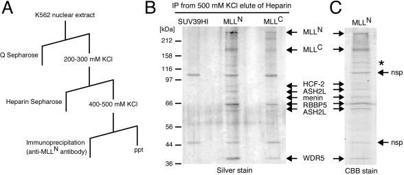
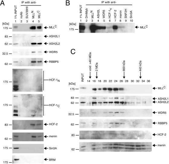
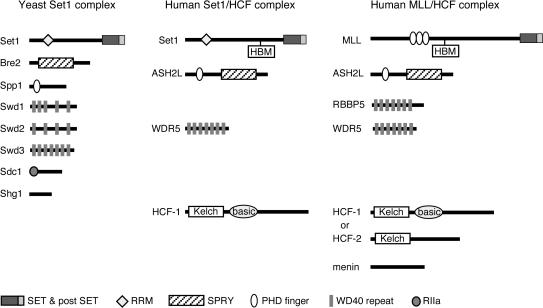
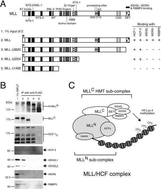
MLL (for mixed-lineage leukemia) is a proto-oncogene that is mutated in a variety of human leukemias. Its product, a homolog of Drosophila melanogaster trithorax, displays intrinsic histone methyltransferase activity and functions genetically to maintain embryonic Hox gene expression. Here we report the biochemical purification of MLL and demonstrate that it associates with a cohort of proteins shared with the yeast and human SET1 histone methyltransferase complexes, including a homolog of Ash2, another Trx-G group protein. Two other members of the novel MLL complex identified here are host cell factor 1 (HCF-1), a transcriptional coregulator, and the related HCF-2, both of which specifically interact with a conserved binding motif in the MLL(N) (p300) subunit of MLL and provide a potential mechanism for regulating its antagonistic transcriptional properties. Menin, a product of the MEN1 tumor suppressor gene, is also a component of the 1-MDa MLL complex. Abrogation of menin expression phenocopies loss of MLL and reveals a critical role for menin in the maintenance of Hox gene expression. Oncogenic mutant forms of MLL retain an ability to interact with menin but not other identified complex components. These studies link the menin tumor suppressor protein with the MLL histone methyltransferase machinery, with implications for Hox gene expression in development and leukemia pathogenesis.


Figures

FIG. 1.
FIG. 1.
Purification of MLL and identification of associated proteins. (A) The scheme for purification of MLL complex indicates the chromatography and elution conditions employed. ppt, precipitate. (B) Purification of MLL complex. The protein eluted at 500 mM KCl from heparin Sepharose (Hep500 fraction) was subjected to immunopurification (IP) using protein G beads conjugated with anti-MLLN, anti-MLLC, or anti-SUV39H1 (negative control) monoclonal antibodies as indicated at the top of the gel. Bound proteins were eluted, subjected to SDS-PAGE, and visualized by silver staining. Molecular sizes of protein markers are shown on the left. (C) A large-scale purification sample was eluted from anti-MLLN beads, fractionated in SDS-7% PAGE, and stained with Coomassie brilliant blue. Visualized bands were excised and analyzed by LC-MS/MS. Arrows indicate protein identities. *, MLL degradation product; nsp, nonspecific products that bind to beads alone.
FIG. 2.
FIG. 2.
Coprecipitation of MLL with associated factors. (A) A nuclear extract of K562 cells (lane 1 input) was subjected to immunoprecipitation (IP) using antibodies specific for MLLN (lane 4) or MLLC (lane 5). As negative controls, precipitations were also performed with an SUV39H1 antibody (lane 3) or no antibody (noAb; lane 2). The immunoprecipitates were fractionated in SDS-PAGE and then immunoblotted with the antibodies indicated to the right of the panels (anti-MLLC [mmC2.1], anti-ASH2L, anti-WDR5, anti-RBBP5 [BL766], anti-HCF-1N [N18], anti-HCF-1C [H12], anti-HCF-2, anti-menin [C19], anti-Sin3A [K-20], and anti-BRM [N-19], respectively). Molecular sizes of marker proteins are shown on the left. (B) A nuclear extract of K562 cells was subjected to immunoprecipitation analysis using antibodies specific for various components of the MLL complex as indicated at the tops of the respective lanes (anti-MLLN [rpN1], anti-ASH2L, anti-WDR5, anti-HCF-1N [N18], anti-HCF-2, and anti-menin [H-300], respectively). Precipitations were also performed with antibodies specific for SNF2H (H-300) and Sin3A (K-20) or Drosophila Myb as a negative control. The immune precipitates were separated in SDS-PAGE and then immunoblotted with a monoclonal antibody specific for MLLC (mmC2.1). (C) The protein eluate from heparin Sepharose chromatography (Hep500 fraction) obtained according to the purification scheme shown in Fig. 1 was subjected to Superose 6 gel filtration chromatography. Each fraction was concentrated by acetone precipitation, fractionated in SDS-PAGE, and immunoblotted with antibodies indicated to the right of the respective panels (anti-MLLC [mmC2.1], anti-ASH2L, anti-WDR5, anti-RBBP5 [BL766], anti-HCF-2, and anti-menin [C19], respectively). MLL-associated factors preferentially cofractionated with MLL, which peaked in fraction 24 (approximately 1 MDa). Elution of standard molecular size markers is indicated at the top.
FIG. 3.
FIG. 3.
MLL complex composition is conserved with yeast and human SET1 methyltransferase complexes. Similarities between components of the yeast SET1, human SET1/HCF-1, and human MLL/HCF complexes are shown. The protein components of each complex are shown schematically with structural domains and motifs as defined in the key at the bottom. RRM, RNA recognition motif; SET, Set domain; postSET, Set domain-associated cysteine-rich motif; PHD, PHD zinc-finger motif; SPRY, domain in SP1a and the Ryanodine receptor; RIIa, protein kinase A regulatory subunit dimerization domain motif.
FIG. 4.
FIG. 4.
Conserved methyltransferase components associate with MLLC. (A) 293 cells were transiently transfected with expression vectors encoding various MLL deletion mutants (shown schematically) containing HIS and FLAG epitope tags at their N termini. Nuclear extracts prepared from transfectants were subjected to immunoprecipitation (IP) with anti-FLAG antibody (M2). (B) Immunoprecipitates were analyzed by SDS-PAGE and were immunoblotted with antibodies indicated to the right of the respective panels (anti-His [D-8] for various MLL mutants, anti-MLLC [mmC2.1], anti-HCF-1C [H12], anti-HCF-2, anti-ASH2L, anti-WDR5, and anti-RBBP5 [BL766], respectively). Positions of molecular size markers are indicated on the left. Coprecipitation of endogenous proteins with exogenous MLL (+ or −) is indicated to the right of the schematic. (C) Composition of subcomplexes in MLL/HCF. MLL/HCF complex and chromatin with or without methylation are illustrated.
FIG. 5.
FIG. 5.
Conserved binding motif in MLLN mediates interactions with HCF-1 and HCF-2. (A and B) 293 cells were transiently transfected with expression vectors encoding wild-type MLL or a mutant lacking the HBM (shown schematically in panel A). Nuclear extracts prepared from transfectants were subjected to immunoprecipitation (IP) with anti-FLAG antibody (M2). Immune precipitates were analyzed by SDS-PAGE and were immunoblotted with antibodies indicated to the right of the respective gels in panel B (anti-His [D-8] for various MLL mutants, anti-MLLC [mmC2.1], anti-HCF-1C [H12], anti-HCF-2, anti-ASH2L, and anti-WDR5, respectively). Coprecipitation of endogenous proteins associated with MLL (+ or −) is summarized to the right of the schematic. (C) Amino acid sequence alignment of HBMs present in HSV VP16, human, mouse, and fugu MLL, human and mouse MLL2, and human SET1. The HBM consensus (D/EHXY) is highlighted in bold. (D) Dendrogram of MLL superfamily proteins summarizing their known or predicted abilities to interact with ASH2 and HCF-1 or to undergo proteolytic processing [summarized by +, −, or (+)]. + indicates that HBM is present in MLL2 but HCF-1 and -2 interactions are yet to be identified. (E) Schematic representation of HCF-1 and HCF-2 constructs assessed for their abilities to interact with MLL or Sin3A (summarized by +, −, or ND [not determined] to right of schematics). (F) Nuclear extracts were prepared from cells transduced with f-HCF-1N (lane 1), f-HCF-1Kelch (lane 2), or empty vector (lane 3). Extracts were diluted to a concentration of 100 mM KCl and were directly incubated with FLAG antibody beads (M2). Immunoprecipitates were washed and eluted from the beads with peptide and were analyzed by immunoblotting with antibodies specific for HCF-1N (top gel, N18), MLLC (middle gel, mmC2.1), or Sin3A (bottom gel, K-20). Samples were normalized to cell equivalents. NLS, nuclear localization signal.
FIG. 6.
FIG. 6.
Menin interacts with wild-type and oncogenic MLL proteins. (A and B) 293 cells were transiently transfected with expression vectors encoding various MLL deletion or fusion mutants (shown schematically in panel A) containing HIS and FLAG epitope tags at their N termini. Nuclear extracts prepared from transfectants were subjected to immunoprecipitation (IP) with anti-FLAG antibody (M2). Immune precipitates were analyzed by SDS-PAGE and were immunoblotted with antibodies indicated to the right of the respective panels (for panel B, anti-His [D-8] for various MLL mutants, anti-MLLC [mmC2.1], anti-HCF-1C [H12], and antimenin [C19], respectively). Positions of molecular size markers are indicated on the left. Coprecipitation of endogenous menin with exogenous MLL (+ or −) is indicated to the right of the schematic. (C) Nuclear extracts of REH and HB cells were subjected to immunoprecipitation using antimenin antibody (C19) or a negative control antibody (anti-DmMyb). Immune precipitates were analyzed by SDS-PAGE and were immunoblotted with anti-MLLN (mmN4.4, top gel), anti-MLLC (mmC2.1, middle gel), and anti-ENL antibodies (bottom gel) as indicated to the right of the panels. Wild-type MLL coprecipitated with menin in REH cells (lane 3). Both wild-type and fusion (MLL-ENL) MLL proteins coprecipitated with menin in HB cells (lane 6).
FIG. 7.
FIG. 7.
Menin is required for maintenance of Hox gene expression. (A and B) HeLa cells were subjected to three rounds of transfection with siRNAs specific for transcripts encoding components of MLL/HCF complex (indicated at the tops of gel lanes). RNA and protein samples were prepared 3 days after the initial transfection. Proteins were subjected to Western blot analysis with the antibodies indicated to the right of the panels (anti-MLLC [mmC2.1], anti-ASH2L, anti-WDR5, anti-HCF-1C [H12], anti-HCF-2, antimenin [C19], and antiactin [C4], respectively), which showed specific and efficient knockdown of protein expression. RNA samples were reverse transcribed and used for quantitative real-time PCR analysis for HoxA9 and GAPDH expression determined in triplicate. (B) Relative expression of HoxA9 to GAPDH is shown with error bars indicating standard deviations.

Similar articles

Cited by

References

    1. Aasland, R., T. J. Gibson, and A. F. Stewart. 1995. The PHD finger: implications for chromatin-mediated transcriptional regulation. Trends Biochem. Sci. 20:56-59. - PubMed
    1. Agarwal, S. K., S. C. Guru, C. Heppner, M. R. Erdos, R. M. Collins, S. Y. Park, S. Saggar, S. C. Chandrasekharappa, F. S. Collins, A. M. Spiegel, S. J. Marx, and A. L. Burns. 1999. Menin interacts with the AP1 transcription factor JunD and represses JunD-activated transcription. Cell 96:143-152. - PubMed
    1. Agarwal, S. K., E. A. Novotny, J. S. Crabtree, J. B. Weitzman, M. Yaniv, A. L. Burns, S. C. Chandrasekharappa, F. S. Collins, A. M. Spiegel, and S. J. Marx. 2003. Transcription factor JunD, deprived of menin, switches from growth suppressor to growth promoter. Proc. Natl. Acad. Sci. USA 100:10770-10775. - PMC - PubMed
    1. Alvarez-Venegas, R., and Z. Avramova. 2002. SET-domain proteins of the Su(var)3-9, E(z) and trithorax families. Gene 285:25-37. - PubMed
    1. Armstrong, S. A., J. E. Staunton, L. B. Silverman, R. Pieters, M. L. den Boer, M. D. Minden, S. E. Sallan, E. S. Lander, T. R. Golub, and S. J. Korsmeyer. 2002. MLL translocations specify a distinct gene expression profile that distinguishes a unique leukemia. Nat. Genet. 30:41-47. - PubMed

Publication types

MeSH terms

Substances