. 2004 Jul;24(13):5694-709.
doi: 10.1128/MCB.24.13.5694-5709.2004.

Synergistic transcription activation by Maf and Sox and their subnuclear localization are disrupted by a mutation in Maf that causes cataract

Affiliations

Synergistic transcription activation by Maf and Sox and their subnuclear localization are disrupted by a mutation in Maf that causes cataract

Nirmala Rajaram et al. Mol Cell Biol. 2004 Jul.

Abstract

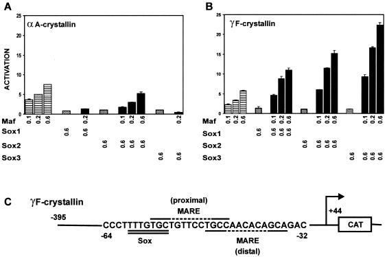
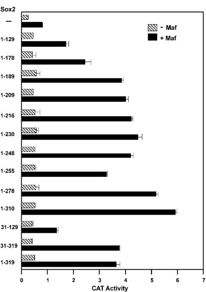
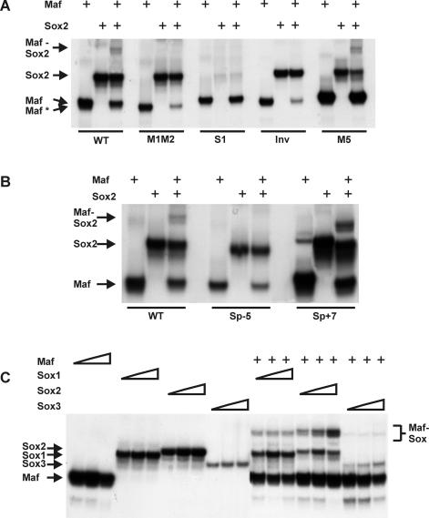
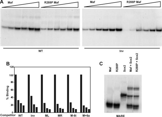
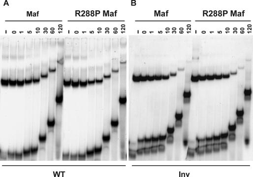
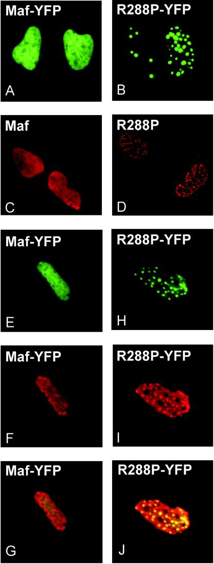
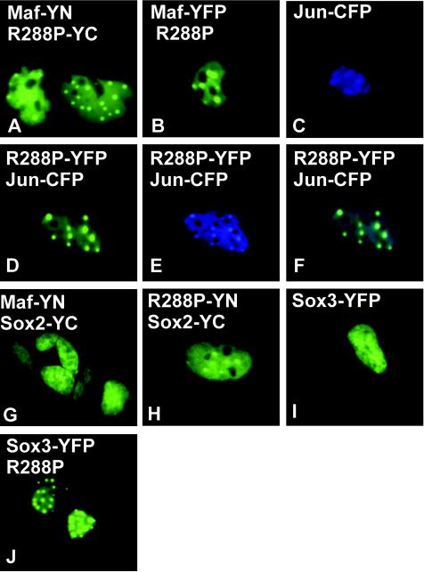
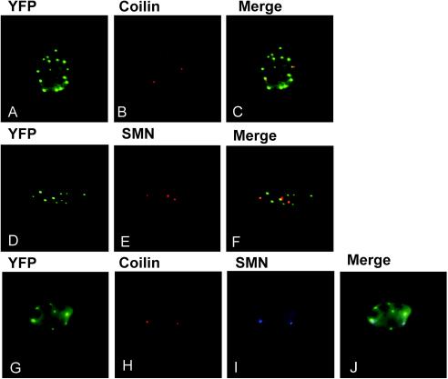
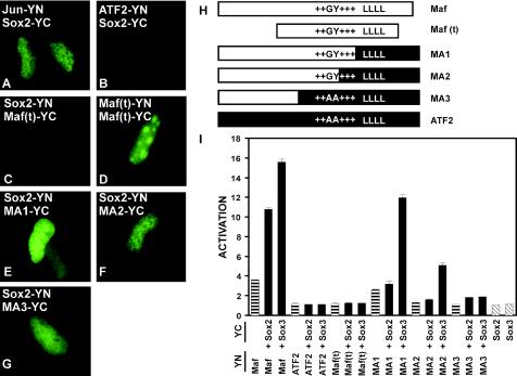
Crystallin genes are selectively expressed during lens development. Maf and Sox family proteins synergistically enhanced gammaF-crystallin promoter activity in a lens cell line. Mutational analysis of the gammaF-crystallin promoter identified a composite regulatory element containing nonconsensus Maf and Sox recognition sequences. Mutations in these recognition sequences or changes in their spacing eliminated synergistic transcription activation. The transcriptional synergy was also affected by changes in the orientation of the Maf recognition sequence that had no detectable effect on binding affinity. The interaction between Maf and Sox proteins was visualized in living cells by bimolecular fluorescence complementation analysis. The N-terminal region of Maf mediated the interaction with Sox proteins in cells. Synergistic transcription activation required the N-terminal region of Maf as well as the ancillary DNA binding domain and the unique portion of the basic region that mediate specific recognition of the gammaF-crystallin promoter element. A mutation in the ancillary DNA binding domain of Maf (R288P) that has been shown to cause cataract eliminated the transcriptional activity of Maf but had no detectable effect on DNA binding in vitro. Whereas wild-type Maf was uniformly distributed in the nucleoplasm, R288P Maf was enriched in nuclear foci. Cajal bodies and gemini of coiled bodies were closely associated with the foci occupied by R288P Maf. Wild-type Maf formed complexes with Sox proteins in the nucleoplasm, whereas R288P Maf recruited Sox proteins as well as other interaction partners to the nuclear foci. The mislocalization of normal cellular proteins to these foci provides a potential explanation for the dominant disease phenotype of the R288P mutation in Maf.


Figures

FIG. 1.
FIG. 1.
Maf and Sox proteins synergistically stimulate γF- but not αA-crystallin promoter activity. (A) Maf activation of αA-crystallin promoter activity is not stimulated by Sox proteins. The indicated amounts (in micrograms) of plasmids encoding the proteins indicated below the bars were transfected into mouse αTN4 lens epithelial cells together with a CAT reporter gene controlled by the αA crystallin promoter. The reporter gene activities were measured, and the efficiencies of transcription activation were calculated relative to cells transfected with the αA-crystallin reporter gene alone. The data in this and subsequent panels represent averages and standard deviations for at least three independent experiments, each with triplicate samples, performed on different days. (B) Maf and Sox proteins synergistically activate the γF-crystallin promoter. The efficiencies of transcription activation by the proteins indicated below the bars were measured at the γF-crystallin promoter as described for panel A. (C) Sequence of the region of the γF-crystallin promoter that contains Sox and Maf recognition elements. The Sox recognition element is double underlined, and two potential Maf recognition elements are overlined and underlined, respectively. The dashed line indicates the nonconsensus core of the Maf recognition sequences.
FIG. 2.
FIG. 2.
Regions of Sox2 required for synergistic transcription activation with Maf. The amino acid residues encoded by each Sox2 deletion derivative are indicated to the left of the bars that show the transcriptional activity of the γF-crystallin promoter in the presence of the Sox2 derivative alone (−Maf) and in combination with Maf (+Maf). The data represent averages and standard deviations from three independent experiments, each with triplicate samples.
FIG. 3.
FIG. 3.
R288P mutation in Maf that causes cataracts in humans eliminates transcription activation by Maf alone and together with Sox proteins. (A) Position of the R288P mutation within the ancillary DNA binding domain of Maf. The model shows the structure of the ancillary DNA binding domain of MafG (29) superimposed on the structure of DNA from the Skn-1-DNA complex (44). The residue corresponding to R288 is shown in ball-and-stick representation. The alignment of the ancillary DNA binding domain of Maf is likely to be different from that observed for Skn-1 (8). (B) The R288P mutation in Maf eliminates transcription activation. The efficiencies of transcription activation by the proteins indicated below the bars were measured as described for Fig. 1A. The data represent averages from two independent experiments, each with triplicate samples.
FIG. 4.
FIG. 4.
Effects of base substitutions within the γF-crystallin promoter on synergistic transcription activation by Maf and Sox proteins. The mutations indicated in each panel and shown in Table 1 were introduced into the γF-crystallin promoter, and reporter gene (CAT) activity was measured in cells expressing the proteins indicated at the bottom of the figure. The diagrams in each panel indicate the positions of the base substitutions (solid boxes). The data in each panel represent averages and standard deviations from three or more independent experiments, each with triplicate samples.
FIG. 5.
FIG. 5.
Co-occupancy of the γF-crystallin promoter by Maf and Sox2. (A and B) The oligonucleotides indicated below the lanes (sequences shown in Table 1) were incubated with the DNA binding domain of Maf (10 nM) and/or the HMG domain of Sox2 (100 nM) as indicated above the lanes, and the complexes were analyzed by polyacrylamide gel electrophoresis. Complexes formed by the DNA binding domain of Maf at different sites migrated with different mobilities (Maf and Maf*). (C) Different concentrations of the DNA binding domain of Maf (10, 20, and 50 nM) as well as the HMG domains of Sox1 (0.5, 1, 1.5 and μM), Sox2 (50, 100, and 200 nM) or Sox3 (0.5, 1, and 1.5 μM) in the presence (+) or absence (−) of Maf (20 nM) were incubated with an oligonucleotide containing the consensus MARE (sequence in Table 1). The complexes were analyzed by polyacrylamide gel electrophoresis.
FIG. 6.
FIG. 6.
Effects of the R288P mutation in Maf and of inversion of the core of the γF-crystallin element on complex formation. (A) Apparent binding affinities of the DNA binding domains of wild-type and R288P Maf at the native (WT) and inverted (Inv) γF-crystallin elements. Different concentrations (5 to 100 nM) of the proteins indicated above the lanes were incubated with the oligonucleotides indicated below the lanes, and complex formation was analyzed by polyacrylamide gel electrophoresis. (B) Relative efficiencies of competition by oligonucleotides containing symmetry-related base substitutions. The DNA binding domain of Maf was incubated with an oligonucleotide containing the γF-crystallin element. A molar excess (0-, 50-, 100-, and 250-fold) of the unlabeled oligonucleotides shown below the bars was added to the reaction, and the fraction of complexes remaining was quantified and plotted as a histogram. (C) Co-occupancy of the γF-crystallin MARE oligonucleotide by the DNA binding domain of wild-type and R288P Maf (10 nM) together with the HMG domain of Sox2 (100 nM). The proteins indicated above each lane were incubated with the γF-crystallin MARE oligonucleotide, and the complexes were analyzed by polyacrylamide gel electrophoresis.
FIG. 7.
FIG. 7.
Comparison of the rates of dissociation of the DNA binding domains of wild-type and R288P Maf from the γF-crystallin element (WT) and the element containing an inverted core (Inv). The proteins indicated above the lanes (10 nM) were incubated with the oligonucleotides indicated below the lanes. A 1,000-fold excess of unlabeled competitor oligonucleotide was added, and aliquots were loaded onto a running polyacrylamide gel at the times (in minutes) indicated above the lanes. −, no competitor added.
FIG. 8.
FIG. 8.
Effect of the R288P mutation on Maf localization. Plasmids encoding the proteins indicated in each panel were transfected into αTN4 cells. The distributions of the proteins were visualized by YFP fluorescence (A, B, E, and H) or immunolabeling with Maf-specific antibody (C, D, F, and I). Panels G and J show the overlay of the YFP fluorescence and the immunoreactivity, confirming that the immunoreactivity reflected the localization of the proteins.
FIG. 9.
FIG. 9.
R288P Maf relocalizes interaction partners in cells. Plasmids encoding the proteins indicated in each panel were transfected into αTN4 cells. The distributions of protein complexes (A, G, H, and I) or individual proteins (B to F and J) were determined by imaging YFP (A, B, D, and G to J) or CFP (C and E) fluorescence. Panel F shows an overlay of the YFP and CFP emissions.
FIG. 10.
FIG. 10.
Comparison of the distributions of R288P Maf foci with Cajal bodies and Gems. R288P Maf-YFP was expressed in HeLa cells and visualized by YFP fluorescence (A, D, and G). The cells were stained with antibodies directed against coilin (B and H) and SMN protein (E and I). The fluorescence images are superimposed in panels C, F, and J.
FIG. 11.
FIG. 11.
Regions of Maf required for interactions with Sox proteins in cells and for synergistic activation of the γF-crystallin promoter. (A to G) BiFC analysis of interactions between the proteins indicated in each panel. The proteins indicated were expressed in αTN4 cells, and the bimolecular fluorescent complexes were visualized by YFP fluorescence. The images are representative of greater than 95% of the fluorescent cells in each population. (H) Diagrams of the regions of Maf and ATF2 in each of the truncated and chimeric proteins. Maf(t) corresponds to the truncated Maf(241-344) protein, encompassing the bZIP and ancillary DNA binding domains. The unique GY dipeptide (15), the basic region (+++), and the leucine zipper (LLLL) are indicated. (I) The transcriptional activities of the protein combinations indicated below each bar at the γF-crystallin promoter were measured in αTN4 cells. The proteins were fused to the fluorescent protein fragments indicated on the left. The data represent averages from two or three independent experiments, each with duplicate samples.

Similar articles

Cited by

References

    1. Ambrosetti, D. C., H. R. Scholer, L. Dailey, and C. Basilico. 2000. Modulation of the activity of multiple transcriptional activation domains by the DNA binding domains mediates the synergistic action of Sox2 and Oct-3 on the fibroblast growth factor-4 enhancer. J. Biol. Chem. 275:23387-23397. - PubMed
    1. Borden, K. L. 2002. Pondering the promyelocytic leukemia protein (PML) puzzle: possible functions for PML nuclear bodies. Mol. Cell. Biol. 22:5259-5269. - PMC - PubMed
    1. Boyle, D. L., L. Takemoto, J. P. Brady, and E. F. Wawrousek. 2003. Morphological characterization of the AlphaA- and AlphaB-crystallin double knockout mouse lens. BMC Ophthalmol. 3:3. - PMC - PubMed
    1. Brady, J. P., D. Garland, Y. Duglas-Tabor, W. G. Robison, Jr., A. Groome, and E. F. Wawrousek. 1997. Targeted disruption of the mouse alpha A-crystallin gene induces cataract and cytoplasmic inclusion bodies containing the small heat shock protein alpha B-crystallin. Proc. Natl. Acad. Sci. USA 94:884-889. - PMC - PubMed
    1. Chen, Q., D. H. Dowhan, D. Liang, D. D. Moore, and P. A. Overbeek. 2002. CREB-binding protein/p300 co-activation of crystallin gene expression. J. Biol. Chem. 277:24081-24089. - PubMed

MeSH terms