. 2004 Jul;24(13):5835-43.
doi: 10.1128/MCB.24.13.5835-5843.2004.

Amplification of Mdmx (or Mdm4) directly contributes to tumor formation by inhibiting p53 tumor suppressor activity

Affiliations

Amplification of Mdmx (or Mdm4) directly contributes to tumor formation by inhibiting p53 tumor suppressor activity

Davide Danovi et al. Mol Cell Biol. 2004 Jul.

Erratum in

Abstract

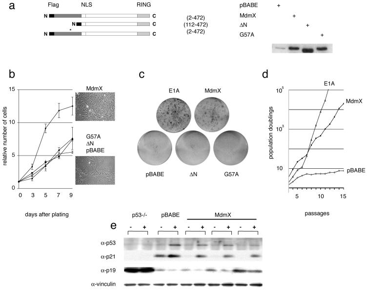
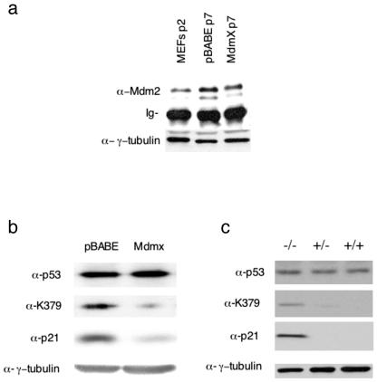
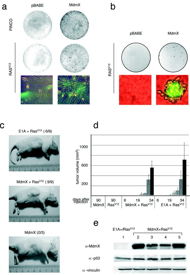
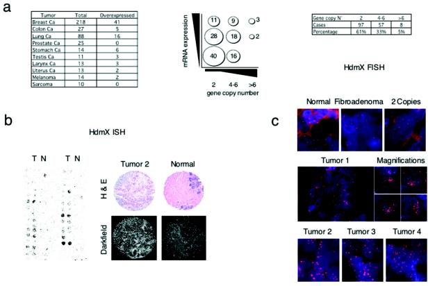
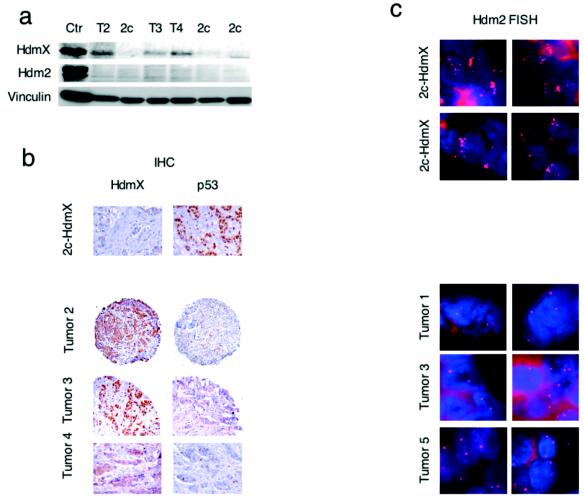
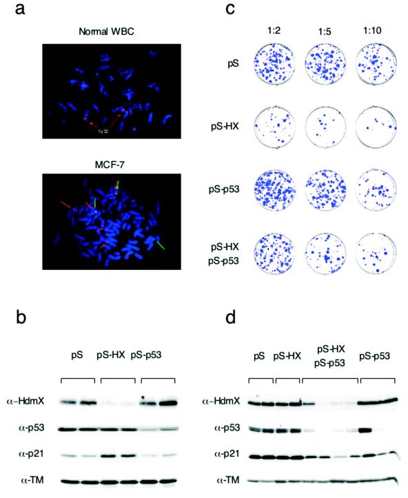
Human tumors are believed to harbor a disabled p53 tumor suppressor pathway, either through direct mutation of the p53 gene or through aberrant expression of proteins acting in the p53 pathway, such as p14(ARF) or Mdm2. A role for Mdmx (or Mdm4) as a key negative regulator of p53 function in vivo has been established. However, a direct contribution of Mdmx to tumor formation remains to be demonstrated. Here we show that retrovirus-mediated Mdmx overexpression allows primary mouse embryonic fibroblast immortalization and leads to neoplastic transformation in combination with HRas(V12). Furthermore, the human Mdmx ortholog, Hdmx, was found to be overexpressed in a significant percentage of various human tumors and amplified in 5% of primary breast tumors, all of which retained wild-type p53. Hdmx was also amplified and highly expressed in MCF-7, a breast cancer cell line harboring wild-type p53, and interfering RNA-mediated reduction of Hdmx markedly inhibited the growth potential of these cells in a p53-dependent manner. Together, these results make Hdmx a new putative drug target for cancer therapy.


Figures

FIG. 1.
FIG. 1.
Immortalization in the absence of concomitant p19ARF or p53 loss in early-passage MEFs with enforced Mdmx expression. MEFs were infected with recombinant retroviruses encoding Flag-tagged Mdmx (amino acids 2 to 472), Mdmx ΔN (N-terminal deletion of the p53-binding domain; amino acids 112 to 472), Mdmx G57A, or puromycin resistance only (pBABE). (a) The Mdmx proteins from infected MEFs were detected by Western blotting with anti-Mdmx polyclonal antibody Si-23. The asterisk indicates the position of the G57A mutation. (b) Growth curves for infected MEFs; each 100-mm dish was seeded with 105 cells. The numbers represent the means and standard deviations for two independently infected MEF cultures. Cells were photographed at a magnification of about ×30 after 7 days in cultures. (c) Colony formation assay. Infected MEFs were plated at a very low density (3 × 10³ cells/100-mm dish). (d) Proliferation of control (pBABE and E1A) or Mdmx-infected MEFs on a 3T3 schedule. The accumulated number of population doublings is shown on a log scale on the y axis. (e) Mdmx-immortalized clones were UV-C irradiated (30 J/m2), and cell extracts were prepared 24 h after treatment and subjected to Western blotting with the indicated antibodies. Passage 4 MEFs infected with pBABE alone and p53-null MEFs are shown as control cells; vinculin served as a loading control.
FIG. 2.
FIG. 2.
Mdmx/Hdmx regulates p53 acetylation. (a) Immunoprecipitation of Mdm2 from passage 7 infected MEFs with a mixture of 2A10, 4B2, and SMP14, followed by Western blot analysis with the mouse monoclonal anti-Mdm2 antibody 2A10. Mdm2 levels in extracts from early-passage, noninfected MEFs were also determined. Total lysates were analyzed with an anti-γ-tubulin antibody to show equal input in the immunoprecipitations. Ig, immunoglobulin. (b) Immunoprecipitation of p53 from passage 7 infected MEFs, followed by Western blot analysis with either a pan-p53 antibody (Ab7) or an antibody specifically recognizing acetylated lysine-379 of murine p53 [PabLys(Ac)379m]. (c) Immunoprecipitation of p53 from pooled lysates of Mdmx−/−, Mdmx+/−, and Mdmx+/+ embryos. Immunoprecipitates were first analyzed with the antibody specifically recognizing acetylated lysine-379 of murine p53 [PabLys(Ac)379m] and then analyzed, after stripping, with the pan-p53 antibody (Ab7). Total lysates were also analyzed with anti-p21 and anti-γ-tubulin antibodies.
FIG. 3.
FIG. 3.
Mdmx bypasses HRasV12-induced senescence and cooperates with HRasV12 in neoplastic transformation. (a) Coinfected MEFs overexpressing Mdmx and either EGFP (PINCO) or HRasV12 plus EGFP (RasV12) were plated for a focus formation assay. (b) MEFs from panel a were used to seed soft agar. After 2 weeks, foci were photographed at magnifications of ×5 and ×100. (c) Animals were injected with MEFs expressing RasV12, Mdmx, Mdmx/RasV12, or E1A/RasV12 at 6 to 8 weeks of age and were inspected for the presence of tumors 3 weeks later. (d) The mean tumor size was estimated at regular intervals after injection. Nine out of nine mice injected with Mdmx/RasV12-expressing MEFs and six out of nine mice injected with E1A/RasV12-expressing MEFs developed tumors. Error bars indicate standard deviations. (e) Cell extracts were prepared from an E1A/RasV12-expressing tumor mass (lane 1), from Mdmx/RasV12-infected MEFs before injection into nude mouse flanks (lane 2), from two different tumors (lanes 3 and 4), and from cells cultured ex vivo from another tumor mass (lane 5) and were subjected to Western blotting with the indicated antibodies. Vinculin served as a loading control.
FIG. 4.
FIG. 4.
Hdmx is overexpressed in human primary tumors and amplified in a subset of breast cancers. (a) Correlation between numbers of copies of the Hdmx locus as shown by FISH (x axis) and Hdmx mRNA levels (y axis). (b) The expression of Hdmx mRNA levels was determined by ISH with TMAs. The left panels show a representative portion of ISH for Hdmx with a breast cancer TMA. The bright-field panels show the morphology of tissue samples as revealed by hematoxylin and eosin (H & E) counterstaining. The dark-field panels show ISH analysis of Hdmx expression levels in breast invasive carcinoma and in normal mammary gland. (c) Subset of primary breast tumors showing amplification of Hdmx gene copy numbers as determined by FISH analysis. (Top panel, from left to right) Normal breast tissue sample, fibroadenoma, and carcinoma containing normal copy numbers for the Hdmx gene. (Middle and bottom panels) Representative breast carcinoma tissues showing significant amplification of the Hdmx locus (>6 to 8 copies).
FIG. 5.
FIG. 5.
Tumors showing Hdmx amplification overexpress Hdmx protein and retain wild-type p53 and two copies of Hdm2. (a) High levels of Hdmx protein were detected in tumors showing amplification of the Hdmx locus with 6B1A (Hdmx) or 4B2 (Hdm2) antibodies. Ctr, control; T2, T3, and T4, tumors 2, 3, and 4, respectively; 2c, two copies. (b) Detection of Hdmx and p53 by IHC analysis of selected breast tumor samples with 6B1A (Hdmx) or PAb1801 (p53) antibodies. (c) Hdm2 FISH (two fields per tumor) analysis of breast cancer samples. (Bottom panels) Samples carrying multiple copies of Hdmx. (Top panels) Hdm2 amplification in two samples carrying normal Hdmx copy numbers.
FIG. 6.
FIG. 6.
The RNA interference-mediated reduction of Hdmx protein levels reduces the colony-forming efficiency of a breast cancer cell line in a p53-dependent manner. (a) FISH analysis of normal white blood cells (WBC) and MCF-7 cells. (Top panel) The probe (BAC clone RP11-430C7) recognizes the Hdmx locus on chromosome 1q32. (Bottom panel) MCF-7 cells harbor multiple copies of the Hdmx locus (red arrows) and three copies of a neighboring, more telomeric region (BAC clone 14B15) (green arrows). (b) MCF-7 cells were transiently transfected by electroporation as described previously (4) with 4 μg of the indicated plasmids and harvested 40 h later. Lysates were analyzed by Western blotting. Blots were probed with antitropomyosin (α-TM) as a loading control. (c) MCF-7 cells were transfected with pBABE-Puro and either pS, pS-Hx, or pS-p53 alone or pS-Hx together with pS-p53; the final DNA amount was adjusted to a total of 6 μg with pS. (d) Puromycin-resistant colonies were isolated and established as cell lines. The expression of Hdmx, p53, p21, and tropomyosin (loading control) was analyzed by Western blotting.

Similar articles

Cited by

References

    1. Barlev, N. A., L. Liu, N. H. Chehab, K. Mansfield, K. G. Harris, T. D. Halazonetis, and S. L. Berger. 2001. Acetylation of p53 activates transcription through recruitment of coactivators/histone acetyltransferases. Mol. Cell 8:1243-1254. - PubMed
    1. Bottger, V., A., Bottger, C., Garcia-Echeverria, Y. F., Ramos, A. J. van der Eb, A. G. Jochemsen, and D. P. Lane. 1999. Comparative study of the p53-mdm2 and p53-MDMX interfaces. Oncogene 18:189-199. - PubMed
    1. Boyd, S. D., K. Y. Tsai, and T. Jacks. 2000. An intact HDM2 RING-finger domain is required for nuclear exclusion of p53. Nat. Cell Biol. 2:563-568. - PubMed
    1. Brummelkamp, T. R., R. Bernards, and R. Agami. 2002. A system for stable expression of short interfering RNAs in mammalian cells. Science 296:550-553. - PubMed
    1. Colombo, E., J. C. Marine, D. Danovi, B. Falini, and P. G. Pelicci. 2002. Nucleophosmin regulates the stability and transcriptional activity of p53. Nat. Cell Biol. 4:529-533. - PubMed

Publication types

MeSH terms