. 2004 Jul;24(13):5844-9.
doi: 10.1128/MCB.24.13.5844-5849.2004.

The ETS transcription factor GABPalpha is essential for early embryogenesis

Affiliations

The ETS transcription factor GABPalpha is essential for early embryogenesis

Sika Ristevski et al. Mol Cell Biol. 2004 Jul.

Abstract

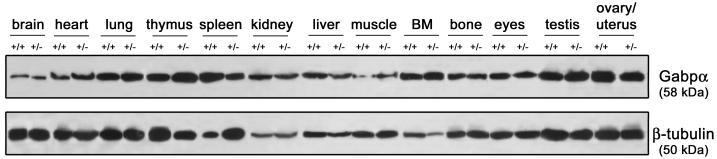
The ETS transcription factor complex GABP consists of the GABPalpha protein, containing an ETS DNA binding domain, and an unrelated GABPbeta protein, containing a transactivation domain and nuclear localization signal. GABP has been shown in vitro to regulate the expression of nuclear genes involved in mitochondrial respiration and neuromuscular signaling. We investigated the in vivo function of GABP by generating a null mutation in the murine Gabpalpha gene. Embryos homozygous for the null Gabpalpha allele die prior to implantation, consistent with the broad expression of Gabpalpha throughout embryogenesis and in embryonic stem cells. Gabpalpha(+/-) mice demonstrated no detectable phenotype and unaltered protein levels in the panel of tissues examined. This indicates that Gabpalpha protein levels are tightly regulated to protect cells from the effects of loss of Gabp complex function. These results show that Gabpalpha function is essential and is not compensated for by other ETS transcription factors in the mouse, and they are consistent with a specific requirement for Gabp expression for the maintenance of target genes involved in essential mitochondrial cellular functions during early cleavage events of the embryo.


Figures

FIG. 1.
FIG. 1.
Genomic structure and targeted disruption of mouse Gabpα. (A) Schematic of mouse Gabpα genomic structure. Asterisks indicate the first coding exons of Gabpα and the closely linked gene ATP synthase coupling factor 6. (B) Intron-exon boundary sequences of Gabpα and comparison of human (12) and mouse intron sizes. Coding sequences are capitalized, introns are lowercased, and the first coding exon is indicated by an asterisk. Splice sites (gt-ag) are boldfaced. (C) Wild-type (wt) and targeted mouse Gabpα alleles. An IRES-LacZ-neomycin cassette was inserted into exon 2, immediately downstream of the start codon, and the position of the targeting construct (from the XbaI to the HindIII site) is indicated. The positions of the intron-3 probe (external to the targeting construct) used for Southern blot analysis (E3-4) and of PCR primers are indicated. Restriction endonuclease sites are as follows: B, BamHI; Bl, BalI; H, HindIII; N, NotI; X, XbaI. (D) Southern blot analysis of ES cell clones by BamHI digestion and hybridization with the E3-4 probe detecting 7-kb wild-type and 13-kb targeted alleles. (E) PCR screen of wild-type (+/+) and heterozygous (+/−) genomic DNA performed using a 5′ primer within intron 1 (F3) or the neomycin cassette (F8) and a common 3′ primer (B3) immediately downstream of exon 2 to generate 192-bp wild-type and 700-bp targeted PCR products.
FIG. 2.
FIG. 2.
Gabpα expression during embryogenesis. (A) Western blot analysis of Gabpα and β-tubulin protein levels in wild-type (+/+) and Gabpα heterozygous (+/−) ES cells. (B through D) Whole-mount mRNA in situ hybridization analysis of Gabpα was performed at E8.0 (B), E9.0 (C), and E10 (D). Arrows indicate areas of relatively abundant expression. A and P indicate the anterior and posterior sides of the embryo in each panel. Tissues are as follows: NF, neural fold; N, neural pore; YS, yolk sac; Mes, mesencephalon; Met, metencephalon; MA, mandibular arch; Mye, myelencephalon; Pro, prosencephalon; ONP, oronasopharyngeal region; Myo, myotome; MT, mesonephric tubule; Cer, cerebellum; Tel, telencephalon; HA, hyoid arch; HLB, hindlimb bud; FLB, forelimb bud.
FIG. 3.
FIG. 3.
Protein expression in adult Gabpα-heterozygous mice. Shown are results of Western blot analysis of Gabpα protein in lysates from Gabpα wild-type (+/+) and heterozygous (+/−) mouse tissues, pooled from four animals of each sex for each genotype at the age of 6 to 8 weeks. The β-tubulin protein was used as a loading control. Muscle was sampled from the quadriceps. BM, bone marrow.

Similar articles

Cited by

References

    1. Aurrekoetxea-Hernández, K., and E. Buetti. 2000. Synergistic action of GA-binding protein and glucocorticoid receptor in transcription from the mouse mammary tumor virus promoter. J. Virol. 74:4988-4998. - PMC - PubMed
    1. Avots, A., A. Hoffmeyer, E. Flory, A. Cimanis, U. R. Rapp, and E. Serfling. 1997. GABP factors bind to a distal interleukin 2 (IL-2) enhancer and contribute to c-Raf-mediated increase in IL-2 induction. Mol. Cell. Biol. 17:4381-4389. - PMC - PubMed
    1. Bannert, N., A. Avots, M. Baier, E. Serfling, and R. Kurth. 1999. GA-binding protein factors, in concert with the coactivator CREB binding protein/p300, control the induction of the interleukin 16 promoter in T lymphocytes. Proc. Natl. Acad. Sci. USA 96:1541-1546. - PMC - PubMed
    1. Bister, K., M. Nunn, C. Moscovici, B. Perbal, M. A. Baluda, and P. H. Duesberg. 1982. Acute leukemia viruses E26 and avian myeloblastosis virus have related transformation-specific RNA sequences but different genetic structures, gene products, and oncogenic properties. Proc. Natl. Acad. Sci. USA 79:3677-3681. - PMC - PubMed
    1. Boccia, L. M., D. Lillicrap, K. Newcombe, and C. R. Mueller. 1996. Binding of the Ets factor GA-binding protein to an upstream site in the factor IX promoter is a critical event in transactivation. Mol. Cell. Biol. 16:1929-1935. - PMC - PubMed

LinkOut - more resources