. 2004 Jul;24(13):5887-99.
doi: 10.1128/MCB.24.13.5887-5899.2004.

Targeted inactivation of testicular nuclear orphan receptor 4 delays and disrupts late meiotic prophase and subsequent meiotic divisions of spermatogenesis

Affiliations

Targeted inactivation of testicular nuclear orphan receptor 4 delays and disrupts late meiotic prophase and subsequent meiotic divisions of spermatogenesis

Xiaomin Mu et al. Mol Cell Biol. 2004 Jul.

Abstract

Testicular orphan nuclear receptor 4 (TR4) is specifically and stage-dependently expressed in late-stage pachytene spermatocytes and round spermatids. In the developing mouse testis, the highest expression of TR4 can be detected at postnatal days 16 to 21 when the first wave of spermatogenesis progresses to late meiotic prophase. Using a knockout strategy to delete TR4 in mice, we found that sperm production in TR4(-/-) mice is reduced. The comparison of testes from developing TR4(+/+) and TR4(-/-) mice shows that spermatogenesis in TR4(-/-) mice is delayed. Analysis of the first wave of spermatogenesis shows that the delay can be due to delay and disruption of spermatogenesis at the end of late meiotic prophase and subsequent meiotic divisions. Seminiferous tubule staging shows that stages X to XII, where late meiotic prophase and meiotic divisions take place, are delayed and disrupted in TR4(-/-) mice. Histological examination of testis sections from TR4(-/-) mice shows degenerated primary spermatocytes and some necrotic tubules. Testis-specific gene analyses show that the expression of sperm 1 and cyclin A1, which are genes expressed at the end of meiotic prophase, was delayed and decreased in TR4(-/-) mouse testes. Taken together, results from TR4(+/+) and TR4(-/-) mice indicate that TR4 is essential for normal spermatogenesis in mice.


Figures
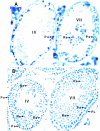
FIG. 1.
FIG. 1.
TR4 is cell-specifically expressed in pachytene spermatocytes and stage-dependently expressed in round spermatids. (A) In situ hybridization of normal adult testis sections with antisense TR4 digoxigenin-labeled probe. Seminiferous tubules in stages IV and VII, which show positive signals in pachytene spermatocytes and in both pachytene spermatocytes and spermatids, respectively, are shown. (B) PAS and hematoxylin staining of two seminiferous tubules in panel A, in a consecutively cut section. Sections from three TR4+/+ and TR4−/− mice were examined. A representative slide is shown in each panel. Ps, pachytene spermatocytes; Rs, round spermatids; PL, prelepotene spermatocytes. Magnification, ×400.
FIG. 2.
FIG. 2.
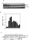
Time course of TR4 expression during testis development. (A) Total RNA was isolated from testes of mice at different postnatal days as indicated. RT-PCR was performed, with β-actin as an internal control. (B) Total RNA was isolated from testes of mice at different postnatal days as indicated, and real-time quantitative RT-PCR was performed. Results are means ± standard deviations from two or three RNA samples from two or three different mice. All sets of results show the same trend, and one set of data is shown. (C) Timetable of first wave of spermatogenesis, including spermatogonia (Sg), the prelepotene (PL), lepotene (L), zygotene (Zs), pachytene, and diplotene (Ds) stages of germ cell differentiation.
FIG. 3.
FIG. 3.
Confirmation of knockout of TR4 gene in TR4−/− mice. (A) PCR analysis of mouse genomic DNA. The wild-type and target alleles give 455- and 760-bp PCR products, respectively. (B) RT-PCR analysis of TR4 in TR4+/+, TR4+/−, and TR4−/− mouse testes. Total RNAs from TR4+/+, TR4+/−, and TR4−/− mice were extracted, and RT-PCR was performed. (C) Southern blot analyses of mouse testis DNA from TR4+/+, TR4+/−, and TR4−/− mice. DNA was digested with EcoRI and hybridized with the probe indicated. The expected fragments after EcoRI digestion are 8 kb for the wild-type allele and 4.9 kb for the mutant allele.
FIG. 4.
FIG. 4.
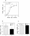
Testis weight and sperm production in TR4+/+ and TR4−/− mice. (A) Comparison of testis weights from TR4+/+ and TR4−/− mice. Testes from TR4+/+ mice and TR4−/− mice were removed, and the weights of testes were determined. At the different age points, two or three mice were used. (B and C) Comparison of sperm counts (B) and motility (C) from caudae epididymides between TR4+/+ and TR4−/− mice. The sperm from caudae epididymides from more than five TR4+/+ and TR4−/− mice, all at 2 to 3 months of age, were counted by hemacytometer under phase-contrast microscopy for total sperm (B) and sperm motility (C). Results are means ± standard deviations of at least five mice.
FIG. 5.
FIG. 5.
Delayed spermatogenesis in TR4−/− mice. (A) PAS-hematoxylin staining of testes from 6-week-old TR4+/+ mouse. Note that the stage VII seminiferous tubules were most frequently observed. (B) PAS-hematoxylin staining of testes from 6-week-old TR4−/− mouse. Note that testes lack stage VII seminiferous tubules and that the tubules at stages X to XII were most frequently observed. (C) Morphology of epididymides of TR4+/+ mice at 6 weeks of age. Note that epididymides were full of sperm (arrows). (D) Morphology of epididymides from 6-week-old TR4−/− mice. Note that very few sperm can be seen in epididymides. Each panel shows a representative slide from examination of three TR4−/− or three TR4+/+ mice. Magnification, ×400.
FIG. 6.
FIG. 6.
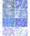
Delayed and disrupted late meiotic prophase and subsequent meiotic divisions in first wave of spermatogenesis in TR4−/− mice. Morphology of seminiferous tubules from TR4+/+ (A, C, and E) and TR4−/− (B, D, F, G, and H) mice at postnatal days 14 (A and B), 22 (C and D), 28 (E and F), and 31 (G and H) is shown. At day 14, germ cell differentiation progresses to mostly zygotene (Zs) stage in both TR4+/+ and TR4−/− mice. At day 22, in TR4+/+ mice, meiosis has been completed and many round spermatids (Rs) are produced, with a few of them being differentiated into elongated spermatids (Es). In TR4−/− mice cells were still arrested in pachytene (Ps) or diplotene stages, no round spermatids were produced, and tubules contained multinucleated giant cells (SY) and primary spermatocytes with increased cytosol (Psb). Vacuoles (V) in the cytosol of pachytene spermatocytes can be frequently observed. At day 28, adluminal cells are primarily round and elongated spermatids in TR4+/+ mice, while adluminal cells are still primarily pachytene spermatocytes or diplotene spermatocytes in TR4−/− mice. At day 31, meiosis has been completed and round spermatids appear in some tubules of TR4−/− mice, but quite a few multinucleated giant cells (i.e., SY) can be observed. Sections from at least three TR4+/+ and TR4−/− mice at indicated ages were examined, and a representative section is shown. Magnifications: ×1,000 (A, B, C, D, and H); ×400 (E to G). (I and J) DNA flow cytometry analysis of testicular cell suspension obtained from TR4+/+ (I) and TR4−/− (J) mice at postnatal day 35. HC, elongated spermatids; 1C, round spermatids; 2C, spermatogonia and nongerm cells; S-ph, spermatogonia synthesizing DNA; 4C, pachytene spermatocytes and G2 spermatogonia. The arrows indicate the differences between TR4+/+ and TR4−/− mice.
FIG. 6.
FIG. 6.
Delayed and disrupted late meiotic prophase and subsequent meiotic divisions in first wave of spermatogenesis in TR4−/− mice. Morphology of seminiferous tubules from TR4+/+ (A, C, and E) and TR4−/− (B, D, F, G, and H) mice at postnatal days 14 (A and B), 22 (C and D), 28 (E and F), and 31 (G and H) is shown. At day 14, germ cell differentiation progresses to mostly zygotene (Zs) stage in both TR4+/+ and TR4−/− mice. At day 22, in TR4+/+ mice, meiosis has been completed and many round spermatids (Rs) are produced, with a few of them being differentiated into elongated spermatids (Es). In TR4−/− mice cells were still arrested in pachytene (Ps) or diplotene stages, no round spermatids were produced, and tubules contained multinucleated giant cells (SY) and primary spermatocytes with increased cytosol (Psb). Vacuoles (V) in the cytosol of pachytene spermatocytes can be frequently observed. At day 28, adluminal cells are primarily round and elongated spermatids in TR4+/+ mice, while adluminal cells are still primarily pachytene spermatocytes or diplotene spermatocytes in TR4−/− mice. At day 31, meiosis has been completed and round spermatids appear in some tubules of TR4−/− mice, but quite a few multinucleated giant cells (i.e., SY) can be observed. Sections from at least three TR4+/+ and TR4−/− mice at indicated ages were examined, and a representative section is shown. Magnifications: ×1,000 (A, B, C, D, and H); ×400 (E to G). (I and J) DNA flow cytometry analysis of testicular cell suspension obtained from TR4+/+ (I) and TR4−/− (J) mice at postnatal day 35. HC, elongated spermatids; 1C, round spermatids; 2C, spermatogonia and nongerm cells; S-ph, spermatogonia synthesizing DNA; 4C, pachytene spermatocytes and G2 spermatogonia. The arrows indicate the differences between TR4+/+ and TR4−/− mice.
FIG. 7.
FIG. 7.
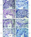
Disrupted and prolonged tubule stages XI to XII of testes from adult TR4−/− mice. (A) Tubule at stage III from TR4+/+ mice. (B) Tubule at stage III from TR4−/− mice. Note the proacrosome granules in round spermatids stained by PAS in both panel A and panel B. (C) Tubule at stage VII from TR4+/+ mice. (D) Tubule at stage VII from TR4−/− mice. Note that stage VII tubules from both TR4+/+ and TR4−/− mice can produce step 16 testis mature spermatids (16S). No histological difference can be found between panels A and B or panels C and D. (E) Tubule at stage XII from TR4+/+ mice. (F to H) Tubules at stages XI to XII from TR4−/− mice. Note prolonged metaphase cells, SY, and primary spermatocytes devoid of chromosome structure (Psc). (A to H) Sections from at least three TR4+/+ and TR4−/− mice aged 2.5 to 3 months were examined, and a representative section is shown. Magnification, ×1,000. (I) Numbers of stage X to XII tubules and total tubules from each of six testis sections from TR4+/+ and TR4−/− mice stained with PAS and hematoxylin were counted, and the ratios of stage X to XII tubules to total tubules were calculated. Results are means ± standard deviations of at least three repeated experiments.
FIG. 7.
FIG. 7.
Disrupted and prolonged tubule stages XI to XII of testes from adult TR4−/− mice. (A) Tubule at stage III from TR4+/+ mice. (B) Tubule at stage III from TR4−/− mice. Note the proacrosome granules in round spermatids stained by PAS in both panel A and panel B. (C) Tubule at stage VII from TR4+/+ mice. (D) Tubule at stage VII from TR4−/− mice. Note that stage VII tubules from both TR4+/+ and TR4−/− mice can produce step 16 testis mature spermatids (16S). No histological difference can be found between panels A and B or panels C and D. (E) Tubule at stage XII from TR4+/+ mice. (F to H) Tubules at stages XI to XII from TR4−/− mice. Note prolonged metaphase cells, SY, and primary spermatocytes devoid of chromosome structure (Psc). (A to H) Sections from at least three TR4+/+ and TR4−/− mice aged 2.5 to 3 months were examined, and a representative section is shown. Magnification, ×1,000. (I) Numbers of stage X to XII tubules and total tubules from each of six testis sections from TR4+/+ and TR4−/− mice stained with PAS and hematoxylin were counted, and the ratios of stage X to XII tubules to total tubules were calculated. Results are means ± standard deviations of at least three repeated experiments.
FIG. 8.
FIG. 8.
Degeneration and apoptosis of primary spermatocytes and necrosis of some tubules. (A) Degeneration of primary spermatocytes. V, vacuoles. (B) Necrosis of seminiferous tubules from TR4−/− mice. *, necrotic tubules. (C and D) Detection of apoptosis (arrows) from TR4−/− mice (C) and TR4+/+ mice (D). Slides from three TR4−/− or TR4+/+ mice were examined, and a representative slide is pictured. Magnification, ×400.
FIG. 9.
FIG. 9.
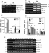
Analysis of testis-specific gene expression in TR4−/− mice. RT-PCR and real-time quantitative RT-PCR of testis-specific genes were performed. (A) Expression patterns of premeiosis-expressed genes proacrosin, Hsp 70-2, and histone H1t in TR4+/+ and TR4−/− mice at indicated ages. W, weeks; M, months. (B) Expression patterns of postmeiosis-expressed genes TP1, TP2, Prm 1, and Prm 2 in TR4+/+ and TR4−/− mice at indicated ages. (C) Quantitative analysis of TP1, TP2, prtm1, and prtm2 in TR4+/+ and TR4−/− mice at indicated ages. (D) Quantitative analysis of the end of meiotic prophase-expressed genes sperm 1 and cyclin A1 in TR4+/+, TR4+/−, and TR4−/− mice at indicated ages. (E) Comparison of sperm 1 and cyclin A1 expression patterns between TR4+/+ and TR4−/− mice at various developmental and adult stages as indicated by RT-PCR. w, weeks; d, days; C, control. (F) Quantitative analysis of sperm 1 expression pattern in TR4+/+ and TR4−/− mice at various developmental and adult stages by real-time RT-PCR. Total RNA was isolated from testes of mice at different ages as indicated in the same manner as for panel E, and real-time quantitative RT-PCR was performed. (G) Quantitative analysis of cyclin A1 expression pattern in TR4+/+ and TR4−/− mice at various developmental and adult stages by real-time RT-PCR. Total RNA was isolated from testes of mice at different ages as indicated in the same manner as for panel E, and real-time quantitative RT-PCR was performed. (H) Quantitative analysis of Cyp24a1 expression pattern in adult TR4+/+ and TR4−/− mice by real-time RT-PCR. Total RNA was isolated from testes of mice at ages indicated, and real-time quantitative RT-PCR was performed. (A to H) All RT-PCR experiments were repeated three times with RNA samples from two or three different mice. Mouse ages are indicated as postnatal days. β-Actin mRNA was used as internal controls. All real-time RT-PCRs were triplicated and repeated twice with two or three RNA samples from different mice, and all results are normalized with β-actin. All sets of results show the same trend, and one set of data is shown.
FIG. 9.
FIG. 9.
Analysis of testis-specific gene expression in TR4−/− mice. RT-PCR and real-time quantitative RT-PCR of testis-specific genes were performed. (A) Expression patterns of premeiosis-expressed genes proacrosin, Hsp 70-2, and histone H1t in TR4+/+ and TR4−/− mice at indicated ages. W, weeks; M, months. (B) Expression patterns of postmeiosis-expressed genes TP1, TP2, Prm 1, and Prm 2 in TR4+/+ and TR4−/− mice at indicated ages. (C) Quantitative analysis of TP1, TP2, prtm1, and prtm2 in TR4+/+ and TR4−/− mice at indicated ages. (D) Quantitative analysis of the end of meiotic prophase-expressed genes sperm 1 and cyclin A1 in TR4+/+, TR4+/−, and TR4−/− mice at indicated ages. (E) Comparison of sperm 1 and cyclin A1 expression patterns between TR4+/+ and TR4−/− mice at various developmental and adult stages as indicated by RT-PCR. w, weeks; d, days; C, control. (F) Quantitative analysis of sperm 1 expression pattern in TR4+/+ and TR4−/− mice at various developmental and adult stages by real-time RT-PCR. Total RNA was isolated from testes of mice at different ages as indicated in the same manner as for panel E, and real-time quantitative RT-PCR was performed. (G) Quantitative analysis of cyclin A1 expression pattern in TR4+/+ and TR4−/− mice at various developmental and adult stages by real-time RT-PCR. Total RNA was isolated from testes of mice at different ages as indicated in the same manner as for panel E, and real-time quantitative RT-PCR was performed. (H) Quantitative analysis of Cyp24a1 expression pattern in adult TR4+/+ and TR4−/− mice by real-time RT-PCR. Total RNA was isolated from testes of mice at ages indicated, and real-time quantitative RT-PCR was performed. (A to H) All RT-PCR experiments were repeated three times with RNA samples from two or three different mice. Mouse ages are indicated as postnatal days. β-Actin mRNA was used as internal controls. All real-time RT-PCRs were triplicated and repeated twice with two or three RNA samples from different mice, and all results are normalized with β-actin. All sets of results show the same trend, and one set of data is shown.
FIG. 10.
FIG. 10.
Diagram of germ cell progression, corresponding to spermatogenesis phase and tubule stages. The black boxes at the top represent the stage showing expression of TR4; end of meiotic prophase-expressed genes sperm 1 and cyclin A1; premeiosis-expressed genes proacrosin, Hsp 70-2, and histone H1t; postmeiosis-expressed genes TP1, TP2, Prm 1, and Prm 2. Note that in TR4−/− mice, late meiotic prophase and subsequent meiotic divisions in late pachytene diplotene and metaphase spermatocytes of tubule stages X to XII were delayed, prolonged, and disrupted. A, type A spermatogonia; In, intermediate spermatogonia; B, type B spermatogonia; PL, preleptotene spermatocytes; L, leptotene spermatocytes; Zs, zygotene spermatocytes; EPs, early pachytene spermatocytes; LPs, late pachytene spermatocytes; Ds, diplotene spermatocytes; MI, first meiosis; MII, second meiosis; Rs, round spermatids; Es, elongated spermatids; S16, step 16 spermatids; E, early; L, late.

Similar articles

Cited by

References

    1. Anderson, B., R. V. Pearse II, P. N. Schlegel, Z. Cichon, M. B. Schonemann, C. W. Bardin, and M. G. Rosenfeld. 1993. Sperm 1: a POU-domain gene transiently expressed immediately before meiosis I in the male germ cells. Proc. Natl. Acad. Sci. USA 90:11084-11088. - PMC - PubMed
    1. Bellve, A. R., J. C. Cavicchia, C. F. Millette, D. A. O'Brien, Y. M. Bhatnagar, and M. Dym. 1977. Spermatogenesis cells of the prepuberal mouse. J. Cell Biol. 74:68-85. - PMC - PubMed
    1. Chang, C., S. F. Da Silva, R. Ideta, Y. F. Lee, S. Y. Yeh, and J. P. H. Burbach. 1994. Human and rat TR4 orphan receptor specify a subclass of the steroid receptor superfamily. Proc. Natl. Acad. Sci. USA 91:6040-6044. - PMC - PubMed
    1. Dix, D. J., J. W. Allen, B. W. Collin, P. Poorman-Allen, C. Mori, D. R. Blizard, P. R. Brown, E. H. Goulding, B. D. Strong, and E. M. Eddy. 1997. HSP70-2 is required for desynapsis of synaponemal complexes during meiotic prophase in juvenile and adult mouse spermatocytes. Development 124:4595-4605. - PubMed
    1. Drabent, B., C. Bode, B. Bramlage, and D. Doenecke. 1996. Expression of the mouse testicular histone gene H1t during spermatogenesis. Histochem. Cell Biol. 106:247-251. - PubMed

Publication types