. 2004 Jul;24(13):5900-13.
doi: 10.1128/MCB.24.13.5900-5913.2004.

BRCA1 inhibits membrane estrogen and growth factor receptor signaling to cell proliferation in breast cancer

Affiliations

BRCA1 inhibits membrane estrogen and growth factor receptor signaling to cell proliferation in breast cancer

Mahnaz Razandi et al. Mol Cell Biol. 2004 Jul.

Abstract

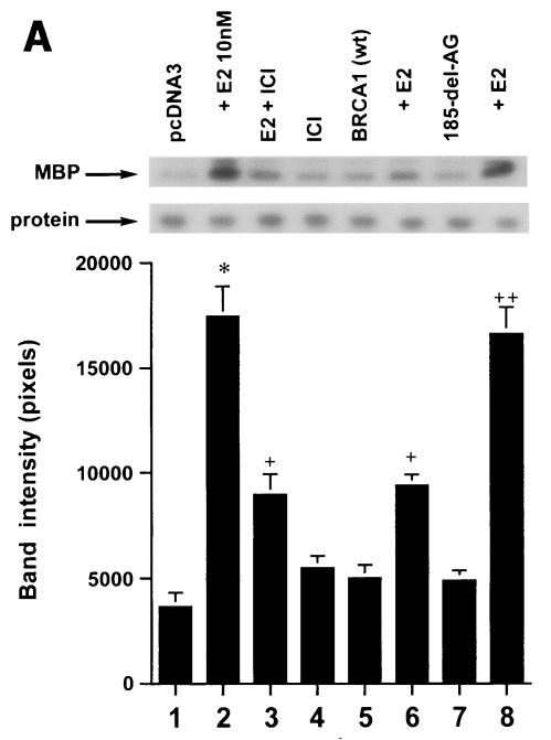
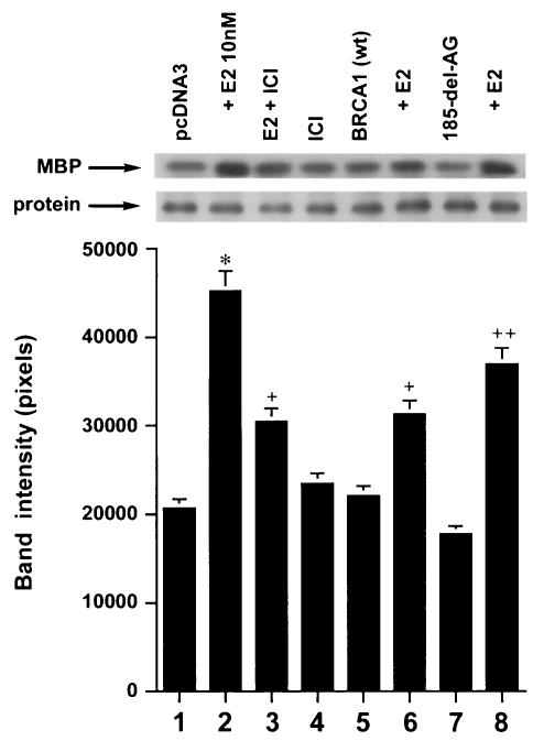
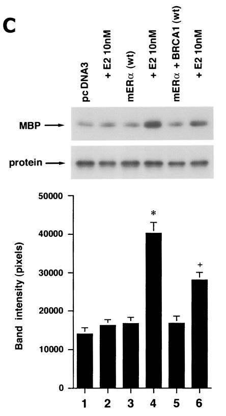
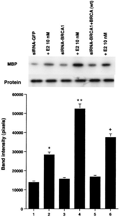
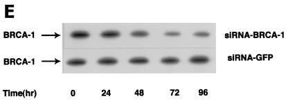
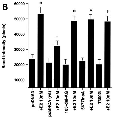
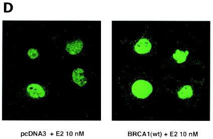
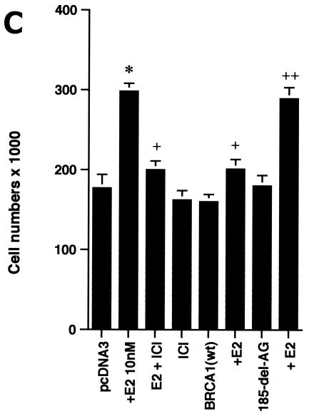
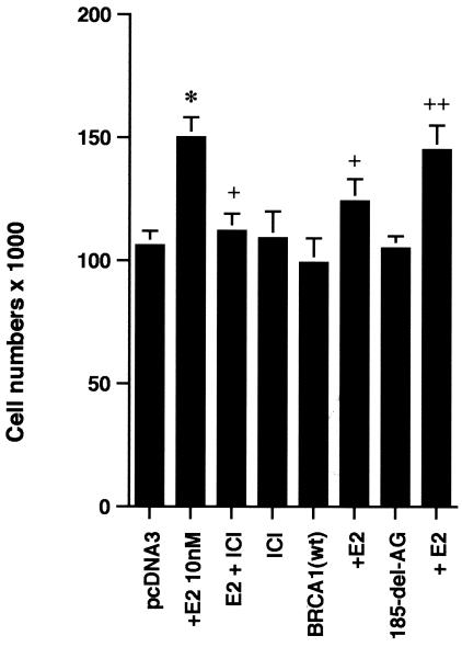
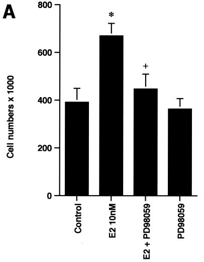
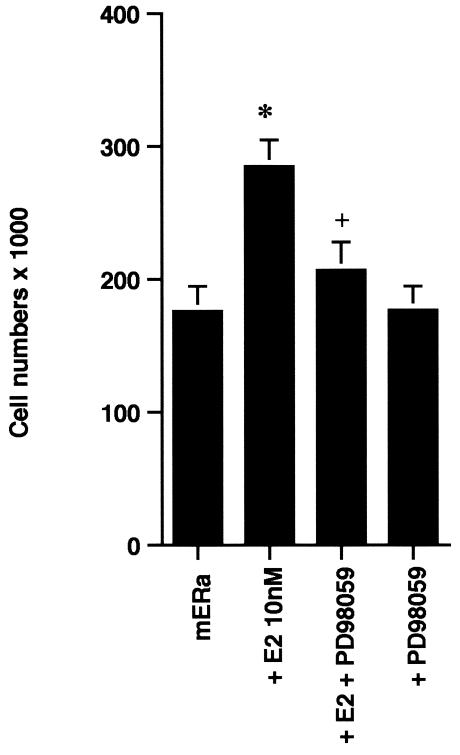
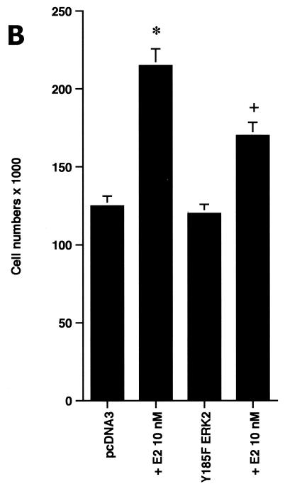
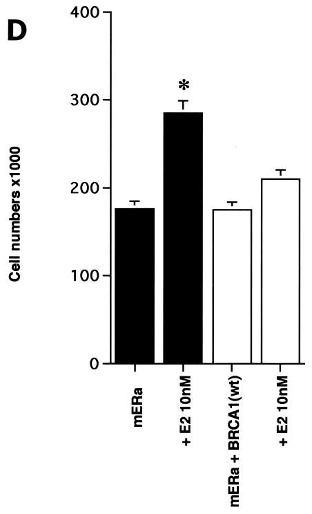
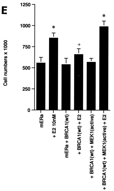
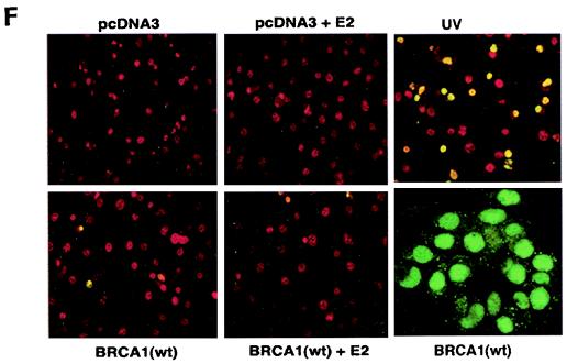
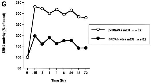
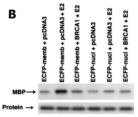
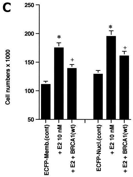
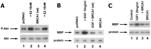
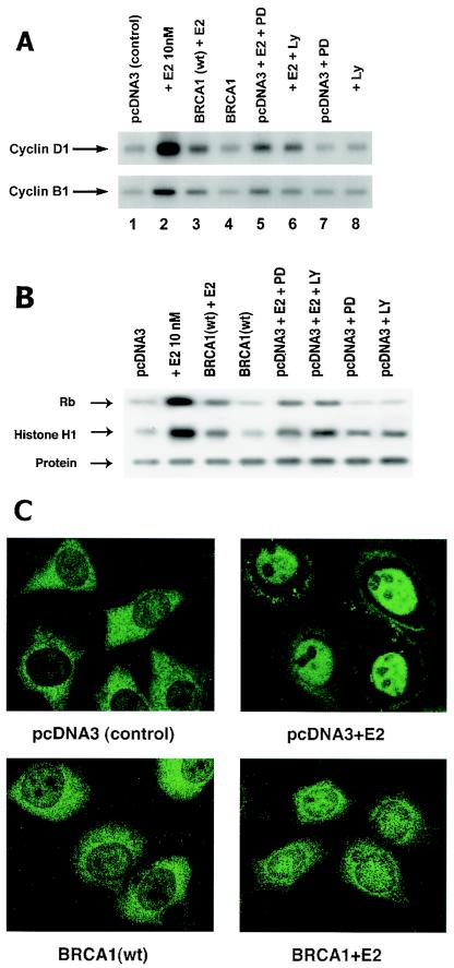
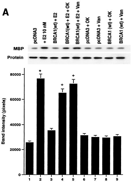
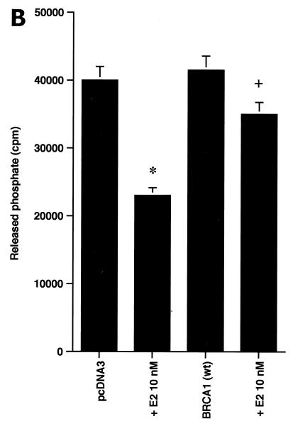
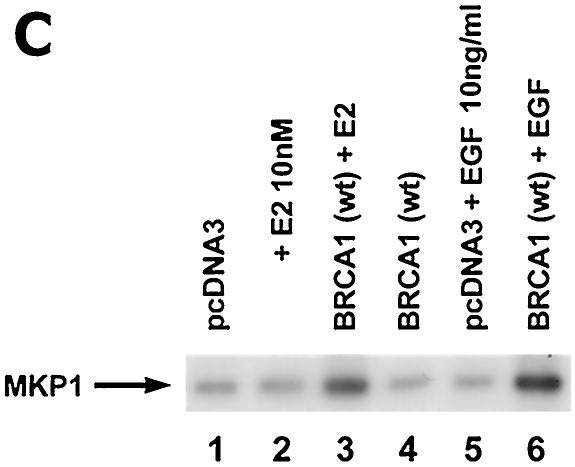
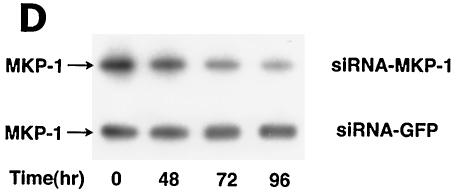
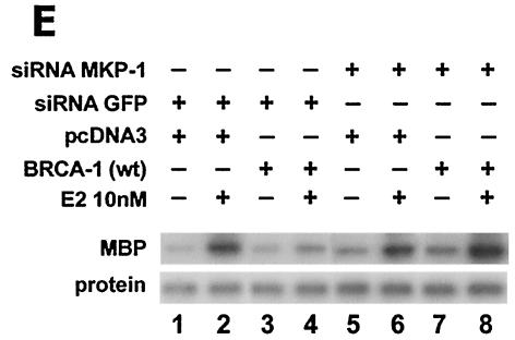
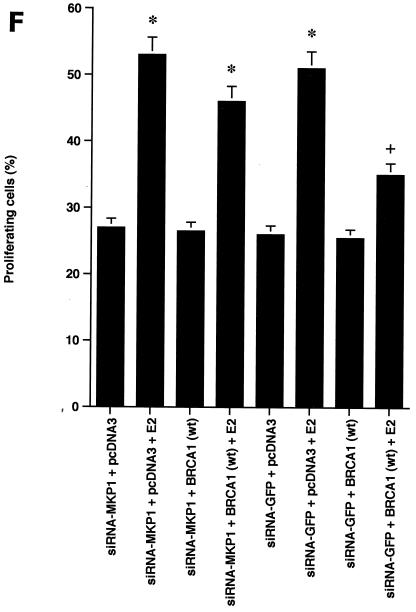
BRCA1 mutations and estrogen use are risk factors for the development of breast cancer. Recent work has identified estrogen receptors localized at the plasma membrane that signal to cell biology. We examined the impact of BRCA1 on membrane estrogen and growth factor receptor signaling to breast cancer cell proliferation. MCF-7 and ZR-75-1 cells showed a rapid and sustained activation of extracellular signal-related kinase (ERK) in response to estradiol (E2) that was substantially prevented by wild-type (wt) but not mutant BRCA1. The proliferation of MCF-7 cells induced by E2 was significantly inhibited by PD98059, a specific ERK inhibitor, or by dominant negative ERK2 expression and by expression of wt BRCA1 (but not mutant BRCA1). E2 induced the synthesis of cyclins D1 and B1, the activity of cyclin-dependent kinases Cdk4 and CDK1, and G(1)/S and G(2)/M cell cycle progression. The intact tumor suppressor inhibited all of these. wt BRCA1 also inhibited epidermal growth factor and insulin-like growth factor I-induced ERK and cell proliferation. The inhibition of ERK and cell proliferation by BRCA1 was prevented by phosphatase inhibitors and by interfering RNA knockdown of the ERK phosphatase, mitogen-activated kinase phosphatase 1. Our findings support a novel tumor suppressor function of BRCA1 that is relevant to breast cancer and identify a potential interactive risk factor for women with BRCA1 mutations.


Figures

FIG. 1.
FIG. 1.
wt BRCA1 but not mutant BRCA1 inhibits E2-induced ERK activation. (A) MCF-7 (left) and ZR-75-1 (right) cells, both ER positive, were transfected to express wt BRCA1 or 185delAG mutant BRCA1 or were transfected with pcDNA3 (control), recovered in serum, synchronized overnight without serum, and then incubated with 10 nM E2 for 9 min. ERK activity against myelin basic protein was then determined. ERK immunoblots are shown below activity to normalize activity for total ERK protein. The bar graph shows the combined results of three experiments. Values are means ± standard errors of the means, determined by analysis of variance plus Scheffe's test (P values of <0.05 are considered significant). Significance of results: *, P of <0.05 for pcDNA3 versus same plus E2; +, P of <0.05 for pcDNA3 plus E2 versus BRCA1 (wt) plus E2 or versus E2 plus ICI182780; ++, P of <0.05 for the 185delAG BRCA1 mutant versus same plus E2. (B) MCF-7 cells were transfected to express pcDNA3 (control), wt BRCA1, or the 185delAG, 5677insA, or T300G BRCA1 mutant, and E2-induced ERK activity was determined. Significance of results: *, P of <0.05 for pcDNA3 or mutant alone versus same plus E2; +, P of <0.05 for pcDNA3 plus E2 versus BRCA1 (wt) plus E2. (C) (Left) HCC-1937 cells were transfected with pcDNA3 or ERα, with or without a wt BRCA1 expression plasmid, and E2-induced ERK activity was determined. Significance of results: *, P of <0.05 for mouse ERα (mERa)-expressing cells versus same incubated with E2; +, P of <0.05 for ER-expressing cells plus E2 versus ER plus wt BRCA1-expressing cells plus E2. The data are from three experiments. (Right) Expression levels of wt BRCA1 and mutant BRCA1 proteins in MCF-7 cells are comparable. MCF-7 cells were transfected with pcDNA3 (control) or expression plasmids for wt BRCA1 or two mutant BRCA1 proteins, 5677insA BRCA1 (InsA) and T300G BRCA1. The cells were recovered, and 24 h later, Western blotting of the cell lysate was carried out with an N-terminal-directed antibody to BRCA1. (D) Endogenous or expressed BRCA1 localizes to the nucleus of MCF-7 cells. MCF-7 cells on coverslips were transfected with pcDNA3 or wt BRCA1, incubated with 10 nM E2 for 36 h, and then fixed for 10 min, as described in Materials and Methods. BRCA1 expression was determined by immunofluorescent confocal microscopy using a first antibody directed against the C terminus and a second antibody conjugated to fluorescein isothiocyanate. The study was repeated twice. (E) Endogenous BRCA1 restrains E2/ER signaling to ERK. (Top) Time course of BRCA1 protein knockdown by siRNA. MCF-7 cells were transfected to express an interfering RNA for BRCA1 or GFP (control), and BRCA1 protein knockdown was determined by Western blotting. (Bottom) Expression of siRNA for BRCA1 augments E2-induced ERK. MCF-7 cells transfected to express siRNA for BRCA1 or GFP were incubated 72 h later with 5 nM E2, and ERK activity was determined. wt BRCA1 was also cotransfected under one condition. A bar graph reflecting the combined results of three experiments is shown. Significance of results: *, P of <0.05 for siRNA-GFP versus same plus E2; **, P of <0.05 for siRNABRCA1 versus same plus E2; +, P of <0.05 for siRNA-BRCA1 plus E2 versus same plus wt BRCA1.
FIG. 1.
FIG. 1.
wt BRCA1 but not mutant BRCA1 inhibits E2-induced ERK activation. (A) MCF-7 (left) and ZR-75-1 (right) cells, both ER positive, were transfected to express wt BRCA1 or 185delAG mutant BRCA1 or were transfected with pcDNA3 (control), recovered in serum, synchronized overnight without serum, and then incubated with 10 nM E2 for 9 min. ERK activity against myelin basic protein was then determined. ERK immunoblots are shown below activity to normalize activity for total ERK protein. The bar graph shows the combined results of three experiments. Values are means ± standard errors of the means, determined by analysis of variance plus Scheffe's test (P values of <0.05 are considered significant). Significance of results: *, P of <0.05 for pcDNA3 versus same plus E2; +, P of <0.05 for pcDNA3 plus E2 versus BRCA1 (wt) plus E2 or versus E2 plus ICI182780; ++, P of <0.05 for the 185delAG BRCA1 mutant versus same plus E2. (B) MCF-7 cells were transfected to express pcDNA3 (control), wt BRCA1, or the 185delAG, 5677insA, or T300G BRCA1 mutant, and E2-induced ERK activity was determined. Significance of results: *, P of <0.05 for pcDNA3 or mutant alone versus same plus E2; +, P of <0.05 for pcDNA3 plus E2 versus BRCA1 (wt) plus E2. (C) (Left) HCC-1937 cells were transfected with pcDNA3 or ERα, with or without a wt BRCA1 expression plasmid, and E2-induced ERK activity was determined. Significance of results: *, P of <0.05 for mouse ERα (mERa)-expressing cells versus same incubated with E2; +, P of <0.05 for ER-expressing cells plus E2 versus ER plus wt BRCA1-expressing cells plus E2. The data are from three experiments. (Right) Expression levels of wt BRCA1 and mutant BRCA1 proteins in MCF-7 cells are comparable. MCF-7 cells were transfected with pcDNA3 (control) or expression plasmids for wt BRCA1 or two mutant BRCA1 proteins, 5677insA BRCA1 (InsA) and T300G BRCA1. The cells were recovered, and 24 h later, Western blotting of the cell lysate was carried out with an N-terminal-directed antibody to BRCA1. (D) Endogenous or expressed BRCA1 localizes to the nucleus of MCF-7 cells. MCF-7 cells on coverslips were transfected with pcDNA3 or wt BRCA1, incubated with 10 nM E2 for 36 h, and then fixed for 10 min, as described in Materials and Methods. BRCA1 expression was determined by immunofluorescent confocal microscopy using a first antibody directed against the C terminus and a second antibody conjugated to fluorescein isothiocyanate. The study was repeated twice. (E) Endogenous BRCA1 restrains E2/ER signaling to ERK. (Top) Time course of BRCA1 protein knockdown by siRNA. MCF-7 cells were transfected to express an interfering RNA for BRCA1 or GFP (control), and BRCA1 protein knockdown was determined by Western blotting. (Bottom) Expression of siRNA for BRCA1 augments E2-induced ERK. MCF-7 cells transfected to express siRNA for BRCA1 or GFP were incubated 72 h later with 5 nM E2, and ERK activity was determined. wt BRCA1 was also cotransfected under one condition. A bar graph reflecting the combined results of three experiments is shown. Significance of results: *, P of <0.05 for siRNA-GFP versus same plus E2; **, P of <0.05 for siRNABRCA1 versus same plus E2; +, P of <0.05 for siRNA-BRCA1 plus E2 versus same plus wt BRCA1.
FIG. 2.
FIG. 2.
ERK activation by E2 is important for cell proliferation. (A) MCF-7 (left) or HCC-1937 (right) cells, the latter transfected to express ERα, were incubated with 0.2% serum (control) with or without 10 nM E2 and with or without the MEK-1 inhibitor PD98059. After 3 days, the cells were trypsinized and counted. The bar graph represents the combined results from triplicate wells for each of two experiments. Significance of results: *, P of <0.05 for control versus E2; +, P of <0.05 for E2 versus E2 plus PD98059. (B) Dominant negative ERK2(Y185F) prevents E2-induced cell proliferation. Cells were transfected with dominant negative ERK2(Y185F) or pcDNA3 (control), recovered, and then incubated with 10 nM E2 for 3 days. The results are from three experiments (n = 3). Significance of results: *, P of <0.05 for control versus E2, +, P of <0.05 for E2 versus ERK2(Y185F) plus E2. (C) Cell proliferation induced by E2 is prevented by wt BRCA1. MCF-7 (left) or ZR-75-1 (right) cells were transfected with pcDNA3, wt BRCA1, or 185delAG mutant BRCA1, recovered, and incubated with 10 nM E2 for 3 days. To some wells, 1 μM ICI182780 (ER antagonist) was added (n = 3). Significance of results: *, P of <0.05 for control versus E2; +, P of <0.05 for E2 versus same plus ICI182780 or E2 plus wt BRCA1. ++, P of <0.05 for BRCA1 (wt) plus E2 versus 185delAG mutant BRCA1 plus E2. (D) wt BRCA1 expression inhibits E2-induced proliferation in HCC-1937 cells. These cells were transfected with mERa plus pcDNA3 or wt BRCA1 and incubated with 10 nM E2 for 3 days. (E) Active MEK-1 reverses BRCA1 inhibition of E2-induced proliferation. HCC-1937 cells were transfected with mERa plus pcDNA3, or mERa plus wt BRCA1, with and without active MEK-1 (n = 3). Significance of results: *, P of <0.05 for control versus E2, or ERα plus BRCA1 (wt) plus MEK-1 versus same in the presence of E2 (last two bars); +, P of <0.05 for E2 versus same plus BRCA1 in the presence of ERα for both. (F) wt BRCA1 does not induce apoptosis of E2-treated MCF-7 cells. MCF-7 cells were transfected to express pcDNA3 or wt BRCA1 and then recovered and incubated with and without 10 nM E2 for 72 h. Other cells were exposed to UV irradiation (50 J over 2 min). BRCA1 immunostaining revealed a 60% transfection efficiency. Apoptosis was determined by TUNEL staining. (G) Temporal kinetics of E2-induced ERK activity. HCC-1937 cells were transfected with mERa without (open circles) or with (closed circles) wt BRCA1. E2 was added daily in fresh medium, and ERK activity, normalized for ERK protein, was determined over 72 h. (H) ERK activation by E2 is unaffected by transcription inhibition. MCF-7 cells were transfected with a wt BRCA1 expression plasmid, recovered, and then incubated with the transcription inhibitor actinomycin D, at 4 μM, for 6 h prior to the addition of 10 nM E2. The cells were subsequently processed for ERK activity at 9 min after E2 addition. Total ERK2 protein is shown in this representative study of two experiments. (I) ERα protein is not modulated by wt BRCA1 expression. MCF-7 cells were transfected to express pcDNA3 (control) or wt BRCA1, recovered, and then incubated with 10 nM E2 for 24 or 48 h. Membrane and nuclear fractions were isolated by sucrose gradient centrifugation, and Western blotting was carried out using the H-222 antibody (directed against the ligand binding domain).
FIG. 2.
FIG. 2.
ERK activation by E2 is important for cell proliferation. (A) MCF-7 (left) or HCC-1937 (right) cells, the latter transfected to express ERα, were incubated with 0.2% serum (control) with or without 10 nM E2 and with or without the MEK-1 inhibitor PD98059. After 3 days, the cells were trypsinized and counted. The bar graph represents the combined results from triplicate wells for each of two experiments. Significance of results: *, P of <0.05 for control versus E2; +, P of <0.05 for E2 versus E2 plus PD98059. (B) Dominant negative ERK2(Y185F) prevents E2-induced cell proliferation. Cells were transfected with dominant negative ERK2(Y185F) or pcDNA3 (control), recovered, and then incubated with 10 nM E2 for 3 days. The results are from three experiments (n = 3). Significance of results: *, P of <0.05 for control versus E2, +, P of <0.05 for E2 versus ERK2(Y185F) plus E2. (C) Cell proliferation induced by E2 is prevented by wt BRCA1. MCF-7 (left) or ZR-75-1 (right) cells were transfected with pcDNA3, wt BRCA1, or 185delAG mutant BRCA1, recovered, and incubated with 10 nM E2 for 3 days. To some wells, 1 μM ICI182780 (ER antagonist) was added (n = 3). Significance of results: *, P of <0.05 for control versus E2; +, P of <0.05 for E2 versus same plus ICI182780 or E2 plus wt BRCA1. ++, P of <0.05 for BRCA1 (wt) plus E2 versus 185delAG mutant BRCA1 plus E2. (D) wt BRCA1 expression inhibits E2-induced proliferation in HCC-1937 cells. These cells were transfected with mERa plus pcDNA3 or wt BRCA1 and incubated with 10 nM E2 for 3 days. (E) Active MEK-1 reverses BRCA1 inhibition of E2-induced proliferation. HCC-1937 cells were transfected with mERa plus pcDNA3, or mERa plus wt BRCA1, with and without active MEK-1 (n = 3). Significance of results: *, P of <0.05 for control versus E2, or ERα plus BRCA1 (wt) plus MEK-1 versus same in the presence of E2 (last two bars); +, P of <0.05 for E2 versus same plus BRCA1 in the presence of ERα for both. (F) wt BRCA1 does not induce apoptosis of E2-treated MCF-7 cells. MCF-7 cells were transfected to express pcDNA3 or wt BRCA1 and then recovered and incubated with and without 10 nM E2 for 72 h. Other cells were exposed to UV irradiation (50 J over 2 min). BRCA1 immunostaining revealed a 60% transfection efficiency. Apoptosis was determined by TUNEL staining. (G) Temporal kinetics of E2-induced ERK activity. HCC-1937 cells were transfected with mERa without (open circles) or with (closed circles) wt BRCA1. E2 was added daily in fresh medium, and ERK activity, normalized for ERK protein, was determined over 72 h. (H) ERK activation by E2 is unaffected by transcription inhibition. MCF-7 cells were transfected with a wt BRCA1 expression plasmid, recovered, and then incubated with the transcription inhibitor actinomycin D, at 4 μM, for 6 h prior to the addition of 10 nM E2. The cells were subsequently processed for ERK activity at 9 min after E2 addition. Total ERK2 protein is shown in this representative study of two experiments. (I) ERα protein is not modulated by wt BRCA1 expression. MCF-7 cells were transfected to express pcDNA3 (control) or wt BRCA1, recovered, and then incubated with 10 nM E2 for 24 or 48 h. Membrane and nuclear fractions were isolated by sucrose gradient centrifugation, and Western blotting was carried out using the H-222 antibody (directed against the ligand binding domain).
FIG. 3.
FIG. 3.
(A) Distribution in HCC-1937 cells of the transfected E domain of ERα, targeted to the membrane (E-Mem-ECFP) or nucleus (E-Nuc-ECFP), or nontargeted (E-ECFP), as revealed by immunofluorescent confocal microscopy. Note that the membrane-targeted E domain appears only at the cell surface, while the nucleus-targeted E domain is localized only to the nucleus. (B) The membrane-targeted but not the nucleus-targeted E domain of ERα supports E2-induced ERK. HCC-1937 cells were transfected with one of two targeted E-domain vectors, pcDNA3 or wt BRCA1, recovered overnight, and then treated with 10 nM E2 for 9 min. ERK activity was then determined. The results of a representative study, repeated twice, are shown. (C) Membrane- and nucleus-targeted E domain contributes to E2-induced cell proliferation. Transfected HCC-1937 cells were recovered and then synchronized in the absence of serum or E2 for 16 h. The cells were then exposed to 0.2% serum plus 10 nM E2 for 3 days. Some cells were transfected to also express wt BRCA1. The bar graph represents the combined results of three experiments. Significance of results: *, P of <0.05 for control versus E2; +, P of <0.005 for E2 versus same plus BRCA1.
FIG. 3.
FIG. 3.
(A) Distribution in HCC-1937 cells of the transfected E domain of ERα, targeted to the membrane (E-Mem-ECFP) or nucleus (E-Nuc-ECFP), or nontargeted (E-ECFP), as revealed by immunofluorescent confocal microscopy. Note that the membrane-targeted E domain appears only at the cell surface, while the nucleus-targeted E domain is localized only to the nucleus. (B) The membrane-targeted but not the nucleus-targeted E domain of ERα supports E2-induced ERK. HCC-1937 cells were transfected with one of two targeted E-domain vectors, pcDNA3 or wt BRCA1, recovered overnight, and then treated with 10 nM E2 for 9 min. ERK activity was then determined. The results of a representative study, repeated twice, are shown. (C) Membrane- and nucleus-targeted E domain contributes to E2-induced cell proliferation. Transfected HCC-1937 cells were recovered and then synchronized in the absence of serum or E2 for 16 h. The cells were then exposed to 0.2% serum plus 10 nM E2 for 3 days. Some cells were transfected to also express wt BRCA1. The bar graph represents the combined results of three experiments. Significance of results: *, P of <0.05 for control versus E2; +, P of <0.005 for E2 versus same plus BRCA1.
FIG. 4.
FIG. 4.
BRCA1 does not block E2-induced AKT activity. (A) MCF-7 cells were transfected to express pcDNA3 or wt BRCA1 and recovered, and E2-induced AKT activation and phosphorylation on serine 473 were determined by immunoblotting after 15 min of incubation. Protein loading of total AKT is shown below the activity immunoblot; the study was repeated twice. (B) EGF-induced ERK is inhibited by wt BRCA1 expression. MCF-7 cells were transfected to express pcDNA3 (control) or wt BRCA1 and then treated with 10 ng of EGF/ml for 9 min, and ERK activity was determined. (C) IGF-I-induced ERK in MCF-7 cells is inhibited by BRCA1. MCF-7 cells were transfected to express pcDNA3 or wt BRCA1. After recovery, the cells were incubated with IGF-I at 20 ng/ml for 9 min, and ERK was determined. The results from a representative study, repeated twice, are shown.
FIG. 5.
FIG. 5.
BRCA1 inhibits key cell cycle actions induced by E2. (A) MCF-7 cells were transfected with wt BRCA1 or pcDNA3 (control) and then incubated with or without 10 nM E2 for 16 h (cyclin D1) or 36 h (cyclin B1). For some cells, 1 μM PD98059, a MEK inhibitor, or 10 μM LY294002, a PI3K inhibitor, was added. Cyclins were detected by Western blotting. (B) MCF-7 cells were incubated with E2 after recovery from transfection. Cdk4 activity was determined after 16 h of incubation by immunoprecipitating the kinase from cell lysates and adding a portion to an in vitro activity assay utilizing retinoblastoma protein as the substrate. CDK1 activity was determined after 36 h of incubation by an assay with histone H1 as the substrate. CDK1 total protein blots are shown below each sample. The results of three experiments are represented. (C) Transfected MCF-7 cells were incubated with or without E2 for 36 h. Cyclin B1 cell distribution was determined by immunofluorescent microscopy during G2/M.
FIG. 6.
FIG. 6.
BRCA1 activates phosphatase activity to downregulate ERK activity. (A) Phosphatase inhibitors reverse BRCA1 inhibition of E2-stimulated ERK. HCC-1937 cells were transfected to express ER plus pcDNA3 or wt BRCA1, recovered and synchronized for 48 h, and then exposed (or not exposed [pcDNA3, lane 1]) to 10 nM E2 for 9 min. Under some conditions, the cells were also incubated with 1 μM of sodium vanadate (Van) or 0.1 μM okadaic acid (OK) 20 min prior to E2 addition. Phosphatase inhibitors or BRCA1 alone had little effect when ERK activity was normalized for protein. The bar graph reflects the combined results of three experiments. Significance of results: *, P of <0.05 for pcDNA3 versus same plus E2; +, P of <0.05 for BRCA1 (wt) plus E2 versus same plus okadaic acid or same plus Van. (B) BRCA1 prevents E2-induced downregulation of ERK phosphatase activity. 32P-labeled ERK2 was prepared as described in Materials and Methods and used as a substrate for an in vitro phosphatase assay. MCF-7 cells transfected with pcDNA3 (control) or wt BRCA1 were incubated with or without 10 nM E2 for 9 min, and the cells were then lysed. Equal protein aliquots of cell lysate were then added to 32P-labeled ERK2 protein aliquots, and phosphatase activity was determined. The data are from triplicate determinations for each condition and are representative of two separate studies. (C) BRCA1 upregulates MKP-1 protein. MCF-7 cells were transfected with pcDNA3 (control) or wt BRCA1, recovered, synchronized in the absence of steroid or serum, and then incubated in medium with or without 10 nM E2 for 24 h. Western blot analyses from cell lysates were carried out for immunoprecipitated MKP-1. The representative study was repeated twice. (D) siRNA for MKP-1 downregulates MKP-1 protein. MCF-7 cells were transfected with annealed, double-stranded RNA for MKP-1 or GFP by using Oligofectamine, as described in Materials and Methods. Western blot analyses for MKP-1 were accomplished in cells lysed at 48, 72, and 96 h posttransfection. (E) siRNA for MKP-1 reverses BRCA1 inhibition of E2-induced ERK. MCF-7 cells were sequentially transfected with 0.3 μg of double-stranded RNA oligomers (to GFP or MKP-1) on day 1, recovered, and then transfected 24 h later with pcDNA3 or wt BRCA1. After recovery and synchronization over 48 h in 0.2% serum, E2 was added for 9 min. ERK activity was determined 72 h after siRNA transfection. The experiment was repeated twice. (F) siRNA for MKP-1 prevents wt BRCA1 inhibition of E2-induced proliferation. MCF-7 cells were transfected with pcDNA3 or wt BRCA1, recovered, and then transfected with siRNA for GFP or MKP-1. The cells were recovered and then incubated with 10 nM E2 for 23 h, followed by BrdU pulsing for 1 h. The cells were fixed, and BrdU labeling was detected as described in Materials and Methods. The data are the means ± standard errors of the means for results with 1,000 cells counted per condition in each of three separate experiments and then combined.
FIG. 6.
FIG. 6.
BRCA1 activates phosphatase activity to downregulate ERK activity. (A) Phosphatase inhibitors reverse BRCA1 inhibition of E2-stimulated ERK. HCC-1937 cells were transfected to express ER plus pcDNA3 or wt BRCA1, recovered and synchronized for 48 h, and then exposed (or not exposed [pcDNA3, lane 1]) to 10 nM E2 for 9 min. Under some conditions, the cells were also incubated with 1 μM of sodium vanadate (Van) or 0.1 μM okadaic acid (OK) 20 min prior to E2 addition. Phosphatase inhibitors or BRCA1 alone had little effect when ERK activity was normalized for protein. The bar graph reflects the combined results of three experiments. Significance of results: *, P of <0.05 for pcDNA3 versus same plus E2; +, P of <0.05 for BRCA1 (wt) plus E2 versus same plus okadaic acid or same plus Van. (B) BRCA1 prevents E2-induced downregulation of ERK phosphatase activity. 32P-labeled ERK2 was prepared as described in Materials and Methods and used as a substrate for an in vitro phosphatase assay. MCF-7 cells transfected with pcDNA3 (control) or wt BRCA1 were incubated with or without 10 nM E2 for 9 min, and the cells were then lysed. Equal protein aliquots of cell lysate were then added to 32P-labeled ERK2 protein aliquots, and phosphatase activity was determined. The data are from triplicate determinations for each condition and are representative of two separate studies. (C) BRCA1 upregulates MKP-1 protein. MCF-7 cells were transfected with pcDNA3 (control) or wt BRCA1, recovered, synchronized in the absence of steroid or serum, and then incubated in medium with or without 10 nM E2 for 24 h. Western blot analyses from cell lysates were carried out for immunoprecipitated MKP-1. The representative study was repeated twice. (D) siRNA for MKP-1 downregulates MKP-1 protein. MCF-7 cells were transfected with annealed, double-stranded RNA for MKP-1 or GFP by using Oligofectamine, as described in Materials and Methods. Western blot analyses for MKP-1 were accomplished in cells lysed at 48, 72, and 96 h posttransfection. (E) siRNA for MKP-1 reverses BRCA1 inhibition of E2-induced ERK. MCF-7 cells were sequentially transfected with 0.3 μg of double-stranded RNA oligomers (to GFP or MKP-1) on day 1, recovered, and then transfected 24 h later with pcDNA3 or wt BRCA1. After recovery and synchronization over 48 h in 0.2% serum, E2 was added for 9 min. ERK activity was determined 72 h after siRNA transfection. The experiment was repeated twice. (F) siRNA for MKP-1 prevents wt BRCA1 inhibition of E2-induced proliferation. MCF-7 cells were transfected with pcDNA3 or wt BRCA1, recovered, and then transfected with siRNA for GFP or MKP-1. The cells were recovered and then incubated with 10 nM E2 for 23 h, followed by BrdU pulsing for 1 h. The cells were fixed, and BrdU labeling was detected as described in Materials and Methods. The data are the means ± standard errors of the means for results with 1,000 cells counted per condition in each of three separate experiments and then combined.

Similar articles

Cited by

References

    1. Baer, R., and T. Ludwig. 2002. The BRCA1/BARD1 heterodimer, a tumor suppressor complex with ubiquitin E3 ligase activity. Curr. Opin. Genet. Dev. 12:86-91. - PubMed
    1. Casey, G. 1995. The BRCA1 and BRCA2 breast cancer genes. Curr. Opin. Oncol. 9:88-93. - PubMed
    1. Chen, Y., A. A. Farmer, C.-F. Chen, D. C. Jones, P.-L. Chen, and W.-H. Lee. 1996. BRCA1 is a 220-kDa nuclear phosphoprotein that is expressed and phosphorylated in a cell cycle-dependent manner. Cancer Res. 56:3168-3172. - PubMed
    1. Chu, Y., P. A. Solski, R. Khosravi-Far, C. J. Der, and K. Kelly. 1996. The mitogen-activated protein kinase phosphatases PAC1, MKP-1, and MKP-2 have unique substrate specificities and reduced activity in vivo toward the ERK2 sevenmaker mutation. J. Biol. Chem. 271:6497-6501. - PubMed
    1. Couse, J. F., S. W. Curtis, T. F. Washburn, J. Lindzey, T. S. Golding, D. B. Lubahn, O. Smithies, and K. S. Korach. 1995. Analysis of transcription and estrogen insensitivity in the female mouse after targeted disruption of the estrogen receptor gene. Mol. Endocrinol. 9:1441-1454. - PubMed

Publication types

MeSH terms