. 2004 Jul;24(13):6011-20.
doi: 10.1128/MCB.24.13.6011-6020.2004.

Phosphorylation by protein kinase CK2 changes the DNA binding properties of the human chromatin protein DEK

Affiliations

Phosphorylation by protein kinase CK2 changes the DNA binding properties of the human chromatin protein DEK

Ferdinand Kappes et al. Mol Cell Biol. 2004 Jul.

Abstract

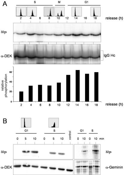
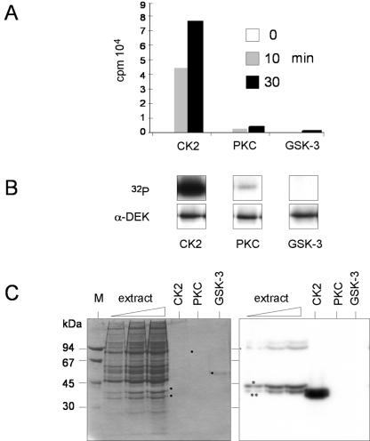
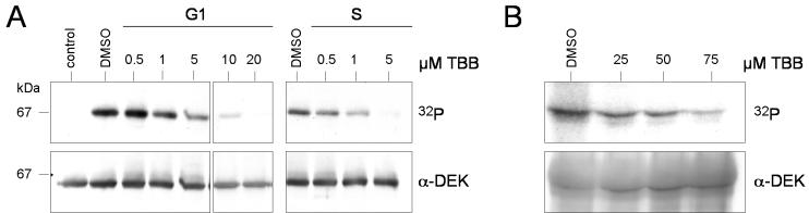
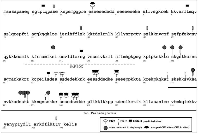
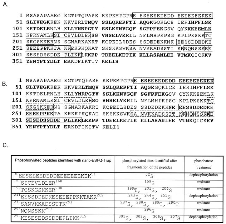
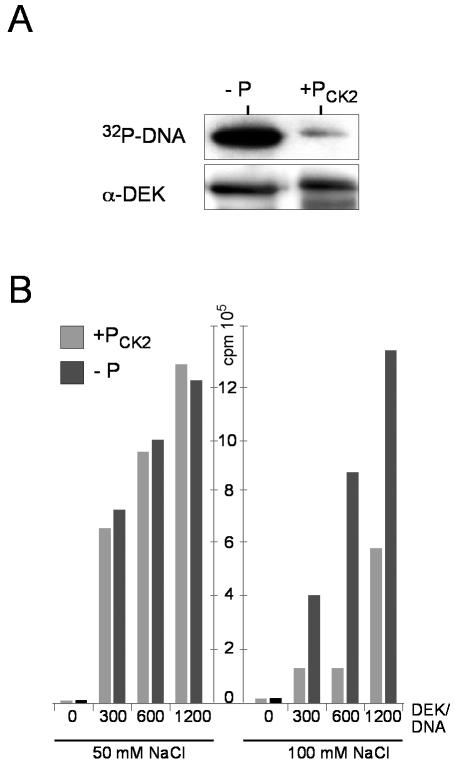
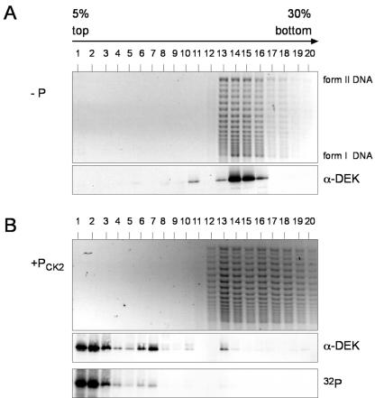
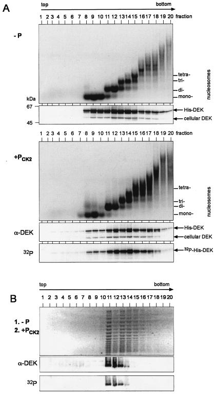
We have examined the posttranslational modification of the human chromatin protein DEK and found that DEK is phosphorylated by the protein kinase CK2 in vitro and in vivo. Phosphorylation sites were mapped by quadrupole ion trap mass spectrometry and found to be clustered in the C-terminal region of the DEK protein. Phosphorylation fluctuates during the cell cycle with a moderate peak during G(1) phase. Filter binding assays, as well as Southwestern analysis, demonstrate that phosphorylation weakens the binding of DEK to DNA. In vivo, however, phosphorylated DEK remains on chromatin. We present evidence that phosphorylated DEK is tethered to chromatin throughout the cell cycle by the un- or underphosphorylated form of DEK.


Figures

FIG. 1.
FIG. 1.
Phosphorylation of DEK in vivo and in vitro. (A) Phosphorylation in vivo. HeLa cells were synchronized by a double-thymidine block and radiolabeled with 32Pi for 2 h at the indicated times after release. Cells were fractionated, and the 450 mM NaCl eluate was treated with DEK antibodies. After electrophoresis, the immunocomplexes were analyzed by autoradiography (32P) and Western blotting (α-DEK). For FACS analysis, cells were treated in parallel and harvested at the indicated time points (top). For densitometric analysis, bands were scanned and analyzed by NIH Imager and the ratio of the autoradiograph to the Western blot was plotted against the time of release (bottom). IgG (immunoglobulin G) Hc indicates the position of the antibody heavy chain. (B) Phosphorylation in vitro. Recombinant, dephosphorylated, His-tagged DEK protein (600 ng) was incubated with G1 phase (15.5 h after release) and S phase (6 h after release) extracts in the presence of [γ-32P]ATP (control lane, without extract). FACS analysis was taken from cells treated in the same way (top). Reactions were stopped at the indicated times, and DEK was purified from the samples with Ni-NTA-agarose. The eluates were precipitated and analyzed by autoradiography (32P) and Western blotting (α-DEK). As a control, human geminin was incubated in the same extracts, immunoprecipitated, and processed as described above.
FIG. 2.
FIG. 2.
DEK is phosphorylated by CK2. (A) Filter binding assay. After dephosphorylation with λ-phosphatase, recombinant His-DEK (5 pmol) was incubated in the presence of [γ-32P]ATP and purified protein kinases (10 U). Reactions were stopped at the indicated time points, transferred to nitrocellulose filters, washed, and then counted. Incorporated radioactivity for the individual kinases is given in counts per minute (cpm, 104). (B) Products from the filter binding assay were analyzed by SDS-PAGE, autoradiography (32P), and Western blotting (α-DEK). (C) In-gel kinase assay. Increasing amounts of HeLa S-20 extract, together with purified CK2, PKC, and GSK-3, were subjected to electrophoresis in a 10% polyacrylamide gel that was polymerized in the presence of 40 μg of His-DEK per ml. The proteins were renatured, and the gel was incubated with [γ-32P]ATP. Shown are the Coomassie-stained gel (left) and the autoradiograph (right). Dots indicate the positions of purified kinases. The asterisk and double asterisk indicate the positions of the CK2 α and α′ subunits, respectively.
FIG. 3.
FIG. 3.
Inhibition of DEK phosphorylation by TBB. (A) In vitro phosphorylation. Recombinant His-DEK (600 ng) was dephosphorylated with λ-phosphatase and incubated with S-20 extracts from HeLa cells (150 μg; G1 and S phases), [γ-32P]ATP, and increasing amounts of TBB. Dimethyl sulfoxide (DMSO), at the highest concentration, was used as a control (control lane, without extract). Reactions were stopped after 10 min, and DEK was purified with Ni-NTA-agarose. The eluates were subjected to SDS-PAGE and analyzed by autoradiography (32P) and Western blotting (α-DEK). The molecular masses of markers are indicated in kilodaltons. (B) Inhibition of DEK phosphorylation in vivo. Asynchronously growing HeLa cells were incubated with the indicated amounts of TBB for 1 h in phosphate-free medium. Cells were washed and incubated with TBB and 200 μCi of 32Pi for another 2 h. DEK was immunoprecipitated from the 450 mM NaCl eluate. The autoradiograph (32P) and the immunoblot (α-DEK) are shown.
FIG. 4.
FIG. 4.
Phosphorylation sites mapped by in vitro phosphorylation with purified CK2. The DEK protein sequence is shown. The broken line indicates the position of the SAF-box DNA binding motif, and the solid line indicates the position of the second DNA binding domain.
FIG. 5.
FIG. 5.
Coverage and identified peptides. (A) Sequence coverage of DEK protein phosphorylated with CK2 identified after nano-ESI-Q-TRAP measurements. Phosphorylated peptides are in the black box. Unphosphorylated peptides are in boldface and cover 33% of the whole protein. Taking phosphorylation and oxidation of methionine into account, the coverage increased to 99%. (B) Sequence coverage of DEK protein dephosphorylated with λ-phosphatase identified after nano-ESI-Q-TRAP measurements. The dephosphorylated peptides that were formerly phosphorylated are in the broken-line box. Taking into consideration the newly dephosphorylated peptides, the coverage of the protein increased to 45%. The phosphorylated peptides resistant to dephosphorylation are shown in the black box. Taking phosphorylation and oxidation of methionine into account, the coverage increased to 70%. (C) Phosphorylated peptides identified by nano-ESI-Q-TRAP.
FIG. 6.
FIG. 6.
Influence of DEK phosphorylation on DNA binding. (A) Southwestern analysis. DEK was transferred to a nitrocellulose membrane and incubated with radioactively labeled, HinfI-digested SV40 DNA. The autoradiograph (32P-DNA) and the Western blot (α-DEK) are shown (−P, dephosphorylated DEK; +Pck2, DEK phosphorylated by CK2 in vitro). (B) Filter binding assay. 32P-labeled, HinfI-digested SV40 DNA was incubated in the absence (0) or presence of increasing amounts of either dephosphorylated (−P) or phosphorylated (+Pck2) DEK. The assay was performed with either 50 or 100 mM NaCl. DEK/DNA molar ratios are indicated.
FIG. 7.
FIG. 7.
Interaction of dephosphorylated and phosphorylated DEK with salt-treated SV40 minichromosomes. (A) Salt-treated SV40 minichromosomes were incubated with dephosphorylated DEK (−P) (at a ratio of 60 mol of DEK/mol of DNA) in the presence of topoisomerase I and separated on 5 to 30% sucrose gradients for 3 h. Individual fractions were analyzed by agarose gel electrophoresis (top) and Western blotting (α-DEK). I, supercoiled DNA; II, relaxed, closed circular, and nicked DNA. (B) Dephosphorylated His-DEK was incubated with salt-treated SV40 minichromosomes as in panel A, followed by in situ phosphorylation with CK2 in the presence of [γ-32P]ATP, and processed as described above (+Pck2). 32P indicates the autoradiograph.
FIG. 8.
FIG. 8.
Association of dephosphorylated and phosphorylated DEK with native oligonucleosomes. (A) HeLa cell nuclei were digested for 10 min at 14°C with micrococcal nuclease. Released oligonucleosomes were incubated with dephosphorylated (−P) and CK2-phosphorylated (+PCK2) DEK and separated on 5 to 40% sucrose gradients for 14 h. Purified DNA from the individual fractions was analyzed by agarose gel electrophoresis and ethidium bromide staining (top). Positions of DNA fragments corresponding to mono-, di-, tri-, and tetranucleosomes are indicated. Proteins were separated by SDS-PAGE and analyzed by Western blotting with DEK-specific antibodies (α-DEK). 32P-labeled DEK was detected by autoradiography (32P). Positions of endogenous DEK, cellular DEK, and exogenously added DEK (His-DEK) are indicated. Marker molecular masses are indicated in kilodaltons. (B) Salt-treated SV40 minichromosomes were incubated with dephosphorylated DEK (at a ratio of 30 mol of DEK/mol of DNA) in the presence of topoisomerase I for 1 h at 37°C (1. −P). In vitro CK2-phosphorylated DEK was added (at a ratio of 30 mol of DEK/mol of DNA), the mixture was incubated for another hour at 37°C (2. +PCK2), and samples were processed as described for Fig. 7. Shown are the agarose gel (top), the Western blot (α-DEK), and the autoradiograph (32P).

Similar articles

Cited by

References

    1. Adams, B. S., H. C. Cha, J. Cleary, T. Haiying, H. Wang, K. Sitwala, and D. M. Markovitz. 2003. DEK binding to class II MHC Y-box sequences is gene- and allele-specific. Arthritis Res. Ther. 5:R226-R233. - PMC - PubMed
    1. Agresti, A., and M. E. Bianchi. 2003. HMGB proteins and gene expression. Curr. Opin. Genet. Dev. 13:170-178. - PubMed
    1. Ahmed, K., D. A. Gerber, and C. Cochet. 2002. Joining the cell survival squad: an emerging role for protein kinase CK2. Trends Cell Biol. 12:226-230. - PubMed
    1. Alexiadis, V., T. Waldmann, J. Andersen, M. Mann, R. Knippers, and C. Gruss. 2000. The protein encoded by the proto-oncogene DEK changes the topology of chromatin and reduces the replication efficiency of DNA replication in a chromatin-specific manner. Genes Dev. 14:1308-1312. - PMC - PubMed
    1. Allende, J. E., and C. C. Allende. 1995. Protein kinases. 4. Protein kinase CK2: an enzyme with multiple substrates and a puzzling regulation. FASEB J. 9:313-323. - PubMed

Publication types

LinkOut - more resources