. 2004 Jul;24(13):6049-57.
doi: 10.1128/MCB.24.13.6049-6057.2004.

Development of a functional skin matrix requires deposition of collagen V heterotrimers

Affiliations

Development of a functional skin matrix requires deposition of collagen V heterotrimers

Hélène Chanut-Delalande et al. Mol Cell Biol. 2004 Jul.

Abstract

Collagen V is a minor component of the heterotypic I/III/V collagen fibrils and the defective product in most cases of classical Ehlers Danlos syndrome (EDS). The present study was undertaken to elucidate the impact of collagen V mutations on skin development, the most severely affected EDS tissues, using mice harboring a targeted deletion of the alpha2(V) collagen gene (Col5a2). Contrary to the original report, our studies indicate that the Col5a2 deletion (a.k.a. the pN allele) represents a functionally null mutation that affects matrix assembly through a complex sequence of events. First the mutation impairs assembly and/or secretion of the alpha1(V)(2)alpha2(V) heterotrimer with the result that the alpha1(V) homotrimer is the predominant species deposited into the matrix. Second, the alpha1(V) homotrimer is excluded from incorporation into the heterotypic collagen fibrils and this in turn severely impairs matrix organization. Third, the mutant matrix stimulates a compensatory loop by the alpha1(V) collagen gene that leads to additional deposition of alpha1(V) homotrimers. These data therefore underscore the importance of the collagen V heterotrimer in dermal fibrillogenesis. Furthermore, reduced thickness of the basement membranes underlying the epidermis and increased apoptosis of the stromal fibroblasts in pN/pN skin strongly indicate additional roles of collagen V in the development of a functional skin matrix.


Figures
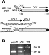
FIG. 1.
FIG. 1.
(A) Schematic illustration of the incidence of exon 6 deletion on the pro-α2(V) chain structure. The exon 6 deletion caused the juxtaposition of the COL1 and COL2 domains. Only an arginine residue from the NC2 domain coded by exon 5 (in boldface) persisted between the two collagenous (COL) domains. NC, noncollagenous domain. (B) Ethidium bromide visualization of RT-PCR products amplified with exon 5 and 7 primers, using total RNA samples purified from wild-type (WT) and heterozygous (pN/+) and homozygous (pN/pN) mouse tails. The size of the smaller product is consistent with the loss of the 78 bp corresponding to exon 6.
FIG. 2.
FIG. 2.
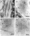
Ultrastructure of the skin (A and B) and of the extracellular matrix produced by primary dermal fibroblast cultures (C and D). Transmission electron micrographs of cross-sections of dermis show bundles of banded fibrils in the wild-type (A, arrows), whereas only rare fibrils are observed in the mutant (B, arrow). Proteoglycan dense aggregates and collagen VI beaded filaments are observed in the entire extracellular space (B, arrowheads). Cross-sections of the fibril network (arrows) produced by wild-type (C) and pN/pN (D) primary dermal fibroblasts are shown.
FIG. 3.
FIG. 3.
Collagen composition of the extracellular matrix produced by wild-type (WT) and mutant (pN/pN) dermal fibroblast cultures. Cultured fibroblasts were metabolically labeled with [14C]proline. Cell layers (A and B) and cell media (C) were pepsinized and separated by SDS-PAGE (6% polyacrylamide) under reduced (A and C) or unreduced (B) conditions. β forms (brackets) correspond to nonreducible dimers of the different collagen chains. Molecular mass standards are indicated on the left.
FIG. 4.
FIG. 4.
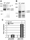
Prevalence of the collagen V homotrimer in mutant skin. (A and B) Electrophoretic patterns of purified collagen V from skin (A) and from cell layers of dermal fibroblast cultures (B) in wild-type (WT) and mutant (pN/pN) mice. Collagens were extracted by pepsin digestion, and collagen V was purified by salt fractionation prior to SDS-PAGE (6% polyacrylamide) separation and Coomassie blue staining. β forms (bracket) correspond to nonreducible α dimers; the lower β form is missing in the pN/pN sample (A, arrow). Molecular mass standards are indicated on the left. (C) Expression level of proα2(V) and proα1(V) transcripts in wild-type (WT) and mutant (pN/pN) dermal fibroblasts determined by real-time PCR. Expression levels were derived from three independent experiments. Fold expression variations were normalized to the relative expression obtained for wild-type samples.
FIG. 5.
FIG. 5.
Localization of collagen I and V in fibrils produced by mutant dermal fibroblast cultures. Electron micrographs show immunogold localization of collagen I (A) and collagen V (B and C). Using collagen I antibodies, gold particles are periodically arranged along the fibrils (A, arrows). Collagen V (B and C) is localized as sparse gold particles along the fibrils and as thin filaments that bound fibrils (arrows).
FIG. 6.
FIG. 6.
Morphology of wild-type (A) and mutant (B) cultured dermal fibroblasts. Wild-type fibroblasts (A) reach confluence in a few days, whereas mutant fibroblasts (B) remain dispersed and numerous pieces of debris are present in the medium. (C and D) TUNEL stain of dermal fibroblasts. No staining is observed in wild-type dermal fibroblasts (C), whereas numerous dermal fibroblasts (D, arrows) are TUNEL positive. Magnifications: A and B, ×100; C and D, ×250. (E to H) Transmission electron micrographs of wild-type (E) and pN/pN (F to H) dermal fibroblasts. Compare to the wild-type control (E), cells exhibit morphological signs of apopotosis: peripheral chromatin condensation (F), fragmented nuclear morphology (G), and abundance of vacuoles and fragmentation of cytoplasm (H).
FIG. 7.
FIG. 7.
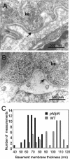
Cross-sections of the basement membrane (arrows) underlying epidermis from wild-type (A) and pN/pN (B) mouse skin. ke, keratinocytes. (C) Histograms show basement membrane thickness distributions of wild-type (WT; gray bars) and pN/pN (black bars) mouse skin.
FIG. 8.
FIG. 8.
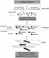
Theoretical model of the consequences of the pN mutation for matrix assembly in skin.

Similar articles

Cited by

References

    1. Andrikopoulos, K., X. Liu, D. R. Keene, R. Jaenisch, and F. Ramirez. 1995. Targeted mutation in the col5a2 gene reveals a regulatory role for type V collagen during matrix assembly. Nat. Genet. 9:31-36. - PubMed
    1. Arteaga-Solis, E., B. Gayraud, S. Y. Lee, L. Shum, L. Sakai, and F. Ramirez. 2001. Regulation of limb patterning by extracellular microfibrils. J. Cell Biol. 154:275-281. - PMC - PubMed
    1. Birk, D. E. 2001. Type V collagen: heterotypic type I/V collagen interactions in the regulation of fibril assembly. Micron 32:223-237. - PubMed
    1. Birk, D. E., J. M. Fitch, J. P. Babiarz, K. J. Doane, and T. F. Linsenmayer. 1990. Collagen fibrillogenesis in vitro: interaction of types I and V collagen regulates fibril diameter. J. Cell Sci. 95:649-657. - PubMed
    1. Birk, D. E., J. M. Fitch, and T. F. Linsenmayer. 1986. Organization of collagen types I and V in the embryonic chicken cornea. Investig. Ophthalmol. Vis. Sci. 27:1470-1477. - PubMed

Publication types

Substances