. 2004 Jun;113(12):1692-700.
doi: 10.1172/JCI20384.

Disruption of Fgf10/Fgfr2b-coordinated epithelial-mesenchymal interactions causes cleft palate

Affiliations

Disruption of Fgf10/Fgfr2b-coordinated epithelial-mesenchymal interactions causes cleft palate

Ritva Rice et al. J Clin Invest. 2004 Jun.

Abstract

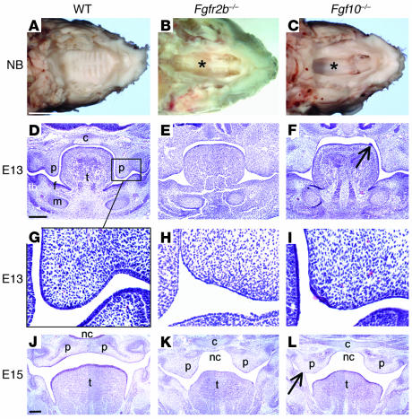
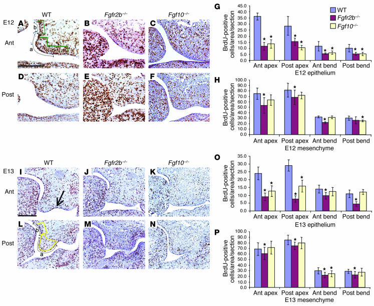
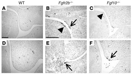
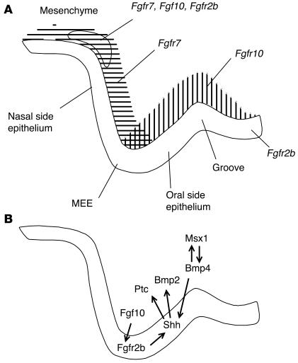
Classical research has suggested that early palate formation develops via epithelial-mesenchymal interactions, and in this study we reveal which signals control this process. Using Fgf10-/-, FGF receptor 2b-/- (Fgfr2b-/-), and Sonic hedgehog (Shh) mutant mice, which all exhibit cleft palate, we show that Shh is a downstream target of Fgf10/Fgfr2b signaling. Our results demonstrate that mesenchymal Fgf10 regulates the epithelial expression of Shh, which in turn signals back to the mesenchyme. This was confirmed by demonstrating that cell proliferation is decreased not only in the palatal epithelium but also in the mesenchyme of Fgfr2b-/- mice. These results reveal a new role for Fgf signaling in mammalian palate development. We show that coordinated epithelial-mesenchymal interactions are essential during the initial stages of palate development and require an Fgf-Shh signaling network.


Figures

Figure 1
Figure 1
Palatal abnormalities in Fgfr2b_/_ and Fgf10_/_ mice. (A_C) View of the palate with mandible removed from NB mice. (A) WT palate was completely closed with clear symmetrical rugae. (B and C) Fgfr2b_/_ and Fgf10_/_ mutants exhibited a similar wide cleft of the secondary palate, with a view into the nasal cavity (asterisks). (D_L) E13 and E15 hematoxylin stained coronal sections through the oral cavity. (G_I) Higher-magnification views of D_F. By E13 the epithelium of WT mice had begun to thicken and stratify into a squamous pattern. That of both mutants was thin and lacked organization. Occasional patches of thicker epithelium did form, notably in Fgf10_/_ mice either at the MEE (I) or as outgrowths from the dorsum of the tongue (F, arrow). However, cells in these patches underwent apoptosis (see Figure 4). (K and L) Palatal shelves were positioned above the tongue but neither met nor fused. (L) Epithelial fusion between the palatal shelf and mandible (arrow). C, cranial base; f, floor of the mouth; m, Meckel’s cartilage; nc, nasal cavity; p, palatal shelf; t, tongue; tb, molar tooth bud. Scale bars: A, 2 mm (A_C, same magnification); D, 200 ∝m (D_F, same magnification); and J, 200 ∝m (J_L, same magnification).
Figure 2
Figure 2
Detection of Fgfr2b, Fgf10, and Fgf7 mRNA during palatogenesis by in situ hybridization. (A) At E12, Fgfr2b was expressed throughout the oral epithelium. (B) Fgf10 was detected in mesenchyme immediately adjacent to the epithelium in the early palatal outgrowths, the tongue, and the floor of the mouth. (D, E, G, and H) This pattern continued at E13 and E14, with Fgfr2b being strongly expressed in the epithelium of the palate and floor of the mouth. Fgf10 was mainly expressed in mesenchyme underlying the MEE and the oral epithelial surface of the palate. (D) Fgfr2b (weakly), (E) Fgf10, and (F) Fgf7 transcripts were also detected in the mesenchyme in the bend region between the palatal shelf and the cranial base (arrows) (arrowhead indicates the anterior-posterior groove between the palatal process and the body of the maxilla). (C and F) At E12 and E13 Fgf7 was mainly expressed in the palatal mesenchyme immediately adjacent to the future nasal epithelium. (I) After shelf elevation, Fgf7 was also expressed in the mesenchyme adjacent to the oral epithelium. f, floor of the mouth. All images are the same magnification; scale bar: 200 ∝m.
Figure 3
Figure 3
Cell proliferation in the developing palate in WT, Fgfr2b_/_, and Fgf10_/_ mice. (A_H) BrdU analysis of E12 palate. (A_F) Histological sections of BrdU staining. Compared to that in WT animals, proliferation in Fgfr2b_/_ and Fgf10_/_ mutants was reduced in all areas analyzed. This was significant in all epithelial readings, and in general the reduction was greater in Fgfr2b_/_ compared with Fgf10_/_ mutants. (I_P) BrdU analysis of E13 palate. (I_N) Histological sections of BrdU staining. Proliferation rates were significantly reduced in all epithelial areas analyzed except in Fgf10_/_ mutants in the bend region at the junction of the nasal aspect of the palatal shelf and the cranial base. Mesenchymal proliferation rates were also reduced in both mutants, though not to the degree seen in the epithelium. (I) BrdU incorporation was particularly noticeable in the groove between the maxilla and the palatal process (arrow). All sections are the same magnification; scale bar, 200 ∝m. The y axes in G, H, O, and P indicate the mean values of BrdU incorporation of each area assayed. Error bars represent standard deviation; asterisk denotes a significant finding (P < 0.005) compared with the WT value. In A and L, dotted lines indicate length of epithelium measured, and green and yellow areas indicate the mesenchymal areas assayed in the apex (a) and bend (b) regions. Ant, anterior; Post, posterior.
Figure 4
Figure 4
Detection of TUNEL-positive cells in the posterior oral cavity of WT, Fgfr2b_/_, and Fgf10_/_ mice at E13. (A and D) TUNEL-positive cells were not detected in WT oral epithelium. (B, C, E, and F) TUNEL-positive cells were detected in the dorsum of the tongue (B and C, arrowheads), in the palatal bend (B, arrow), in the floor of the mouth (E, arrow), and in the MEE (F, arrow) in both Fgfr2b_/_ and Fgf10_/_ mice. TUNEL-positive cells were also occasionally detected in the palatal mesenchyme (B). D and F are high magnifications of the palatal shelf apex in A and C, respectively. A and C are the same magnification, as are B and E; scale bars: 100 ∝m.
Figure 5
Figure 5
Shh is a target of Fgf10/Fgfr2b signaling. (A) FGF10-impregnated bead stimulates palatal epithelial proliferation (arrow). (B) BSA-impregnated bead had no effect on proliferation. (C) Shh bead stimulated proliferation in isolated palatal mesenchymal explants. (D) FGF10 induced Shh in vitro, in the oral side of WT palatal epithelium (arrow), but not in Fgfr2b_/_ mutant mice (E). (F) FGF10 also induced Ptc1 in the mesenchyme immediately adjacent to the bead in palatal explants (arrows). Ptc1 is a known target of Shh and an indicator of the level of hedgehog signaling. (G_L) In situ hybridization to detect Shh mRNA. Shh was expressed in the MEE and the oral side of the palatal epithelium, as well as in the dorsum of the tongue. (H, I, K, and L) In Fgfr2b_/_ and Fgf10_/_ mutants, Shh expression was reduced and concentrated to discrete patches of palatal epithelium. Shh remained expressed throughout the tongue dorsum epithelium (arrow). (M and N) Frontal histological sections through the posterior oral region of NB WT (M) and K14-Cre;Shhc/n (N) mice, stained with Ladewig’s trichrome. (M) In the WT mouse, the palatal shelves had elevated and fused in the midline forming a barrier between the oral cavity and the nasopharynx (asterisk). (N) The palatal shelves of K14-Cre;Shhc/n mutants failed to develop beyond rudimentary processes. (O) In situ hybridization to detect Smo mRNA at E13. Transcripts were detected in the mesenchyme of the palate (arrow). G_I, L and O, same magnification; scale bars: 200 ∝m. J and K: original magnification, ∞12.5.
Figure 6
Figure 6
Molecular control of early palate development. (A) Schematic diagram showing the mRNA expression of Fgfr2b, Fgf10, and Fgf7 in the E13 mouse palate. Fgfr2b was expressed in the oral epithelium and at a low level in the mesenchyme in the bend area between the cranial base and palatal shelf. Fgf10 and Fgf7 were also expressed at this site. In addition, Fgf10 was mainly expressed in the mesenchyme on the oral side of the developing palate and Fgf7 on the nasal aspect. In the mesenchyme adjacent to the MEE, Fgf10 and Fgf7 expression domains overlapped. “Groove” indicates the anterior-posterior groove between the palatal process and the body of the maxilla. (B) Schematic diagram illustrating the proposed Fgf10/Fgfr2b epithelial-mesenchymal interactions and possible downstream signaling in the developing palate.

Comment in

Similar articles

Cited by

References

    1. Gorlin, R.J., Cohen, M.M., Jr., and Hennekam, R.C.M. 2001. Syndromes of the head and neck. 4th edition. Oxford University Press. New York, New York, USA. 850–853.
    1. Jones MC. Etiology of facial clefts: prospective evaluation of 428 patients. Cleft Palate J. 1988;25:16–20. - PubMed
    1. Murray J. Gene/environment causes of cleft lip and/or palate. Clin. Genet. 2002;61:248–256. - PubMed
    1. Wilkie AO, Morriss-Kay GM. Genetics of craniofacial development and malformation. Nat. Rev. Genet. 2001;2:458–468. - PubMed
    1. Rice DP, et al. Integration of FGF and TWIST in calvarial bone and suture development. Development. 2000;127:1845–1855. - PubMed

Publication types

MeSH terms