. 2004 Jun;113(12):1701-10.
doi: 10.1172/JCI20935.

Specific induction of neuronal cells from bone marrow stromal cells and application for autologous transplantation

Affiliations

Specific induction of neuronal cells from bone marrow stromal cells and application for autologous transplantation

Mari Dezawa et al. J Clin Invest. 2004 Jun.

Abstract

Bone marrow stromal cells (MSCs) have the capability under specific conditions of differentiating into various cell types such as osteocytes, chondrocytes, and adipocytes. Here we demonstrate a highly efficient and specific induction of cells with neuronal characteristics, without glial differentiation, from both rat and human MSCs using gene transfection with Notch intracellular domain (NICD) and subsequent treatment with bFGF, forskolin, and ciliary neurotrophic factor. MSCs expressed markers related to neural stem cells after transfection with NICD, and subsequent trophic factor administration induced neuronal cells. Some of them showed voltage-gated fast sodium and delayed rectifier potassium currents and action potentials compatible with characteristics of functional neurons. Further treatment of the induced neuronal cells with glial cell line-derived neurotrophic factor (GDNF) increased the proportion of tyrosine hydroxylase-positive and dopamine-producing cells. Transplantation of these GDNF-treated cells showed improvement in apomorphine-induced rotational behavior and adjusting step and paw-reaching tests following intrastriatal implantation in a 6-hydroxy dopamine rat model of Parkinson disease. This study shows that a population of neuronal cells can be specifically generated from MSCs and that induced cells may allow for a neuroreconstructive approach.


Figures

Figure 1
Figure 1
Characterization of isolated MSCs. (A) FACS analysis of rat MSCs. Numbers in panels represent mean fluorescent intensity of the cells expressing each marker. (B and C) Phase-contrast microscopy of rat (B) and human (C) MSCs. (DK) Immunocytochemistry of CD29 (D), CD90 (E), and CD34 (F) of human MSCs and PDGF receptor β (PDGFR) (G), and smooth muscle actin (sm-actin) (H), CD31 (I), CD45 (J), and neurofilamen-M (K) of rat MSCs. Bars = 50 ∝m. (LP) Adipogenic (L and M), chondrogenic (N), and osteogenic (O) induction from human (LN) and rat (O) MSCs. Micrographs in (M) show oil red staining of lipid droplet in adipocytes in (N) immunocytochemistry of collagen type II of chondrogenic induction and in (O) alkaline phosphatase in osteocytes. (L) Phase-contrast image of adipocytes. Bars = 50 ∝m. (P) Alkaline phosphatase (ALP) activity of rat and human MSCs before and after osteogenic induction. **P < 0.01.
Figure 2
Figure 2
Phenotype of N-MSCs. (A) Immuno-cytochemistry of GLAST, 3-PGDH, and nestin in rat MSCs and N-MSCs. Bar = 50 ∝m. (B) Promoter activity of 3-PGDH in rat MSCs and N-MSCs (NICD transfection followed by 11 days of G418 selection). Both full-length and truncated (M1965) forms of 3-PGDH showed nine- to tenfold increases in promoter activity in N-MSCs. **P < 0.01.
Figure 3
Figure 3
Analysis of TF-MSCs (5 days after trophic factor induction). (AC) Phase contrast of TF-MSCs from rats (A and B) and humans (C). Bars, A = 200 ∝m, B and C = 50 ∝m. (DF and HJ) Immunocytochemical analysis of neuronal and glial markers in rat (F, HJ) and human (D and E) TF-MSCs. MAP-2ab (D), neurofilament-M (E), and β-tubulin isotype 3 (F) were detected. None of the cells were reactive to GFAP (H), GalC (I), and O4 (J). (G) The Brd-U labeling of rat TF-MSCs. MAP-2ab–positive cells (green) did not incorporate Brd-U (red), whereas negative cells were occasionally incorporated with Brd-U. Bars in DJ = 100 ∝m. (K) Western blot analysis of MAP-2ab and GFAP rat samples. Brain, positive control; TF-MSC. β-tubulin (tub) as a loading control. (LQ) Patch clamp. K+ current increased with trophic factor induction up to approximately 1,600 pA and 4,000 pA in rat (L) and human (M) TF-MSCs, respectively. (N) Phase contrast of human TF-MSCs recorded in (M). (O) Voltage-gated inward current recorded in rat BDNF + NGF–treated TF-MSCs. A series of Na current to show the process of block by TTX. Capacity current was blanked. (P) Action potentials from rat BDNF + NGF–treated TF-MSCs; subthreshold, threshold, and suprathreshold current injections were made. (Q) Immunocytochemistry of sodium channel (green). Bar = 30 –m. (R) Relative promoter activities of NeuroD and GFAP in rat MSCs, N-MSCs, and TF-MSCs.
Figure 4
Figure 4
Transcription factors during induction. (A) RT-PCR of rat MSCs, N-MSCs, 24 hours and 5 days after trophic factor induction in TF-MSCs, and positive control. (B) Western blot of Notch extracellular domain (NECD) and NICD in rat MSCs, N-MSCs, TF-MSCs (5 days after trophic factor induction), and positive control from brain. β-tubulin (tub) as a loading control. (C and D) Generation of MAP-2ab+ cells (green in D) by JAK/STAT inhibitor WHI-P131 administration and subsequent trophic factor induction in rat MSCs. (E and F) Rat MSCs cotransfected with active form of STAT3 (anti-FLAG; green) and NICD, followed by trophic factor induction. Only a small number of cells were positive for MAP-2ab (red). (G and H) Hes1/5 transfection to rat MSCs. Bars in CH = 80 ∝m.
Figure 5
Figure 5
Induction of TH-positive cells. (A) The percentage of each transmitter in rat TF-MSCs and TH in rat G-MSCs. NPY, neuropeptide Y; CGRP, calcitonin gene-related peptide; VACht, vesicular acetylcholine transporter; SP, substance P; Glu, glutamate; Ser, serotonin transporter; Vip, vasoactive intestinal peptide; GABA, γ-aminobutyric acid. (BD) TH expression (green) in rat (B) and human G-MSCs (C and D). Bars in B and C = 100 ∝m; D = 30 ∝m. (E) RT-PCR of each factor in rat cells. Upregulation is recognized in G-MSCs compared with TF-MSCs. (F) Western blot of TH. Adrenal medulla (Ad M), as a positive control. β-tubulin (tub) as a loading control. (G) Analysis of apomorphine-induced rotations after transplantation of rat cells. Sham operation group (diamonds), MSC group (triangles), TF-MSC group (circles), and G-MSC group (squares) (*0.01 < P < 0.05; **P < 0.01, statistical difference to sham group). (H) Apomorphine-induced rotation in sham-operated rats (triangles) and after the transplantation of human MSCs (hMSCs; squares) and human G-MSCs (hG-MSCs; circles). (I and J) Adjusting step test (I) and paw-reaching test (J) after transplantation of rat G-MSCs. Adjusting step test: the ratios of the number of steps of the lesioned side to the contralateral intact side paw. Paw-reaching test: the ratios of the number of pellets eaten by the lesioned side paw to the contralateral intact side paw. Control group received no grafting after 6-OHDA administration.
Figure 6
Figure 6
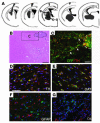
Histological analysis of striatum 10 weeks after transplantation in G-MSC group. (A) GFP-labeled rat transplanted cells were found in the gray regions in the schematic diagrams. (B) Box B in panel A is subjected to HE staining. Implantation zone is indicated by a dotted line. Bar = 100 ∝m. (C) Immunostaining of anti-GFP (green) and –TH (red) of box C in panel B from an adjacent section of panel B. TH-positive processes are extending to the outside of the implantation zone (arrows). Bar = 30 ∝m. (DG) Immunostaining for TH (D), DAT (E), GFAP (F), and O4 (G) (all red color-coded) taken from the area of boxes DG in panel A. Bar = 50 ∝m.

Similar articles

Cited by

References

    1. Kawasaki H, et al. Induction of midbrain dopaminergic neurons from ES cells by stromal cell-derived inducing activity. Neuron. 2000;28:31–40. - PubMed
    1. Pittenger MF, et al. Multilineage potential of adult human mesenchymal stem cells. Science. 1999;284:143–147. - PubMed
    1. Woodbury D, Schwarz EJ, Prockop DJ, Black IB. Adult rat and human bone marrow stromal cells differentiate into neurons. J. Neurosci. Res. 2000;61:364–370. - PubMed
    1. Sanchez-Ramos JR. Neural cells derived from adult bone marrow and umbilical cord blood. J. Neurosci. Res. 2002;69:880–893. - PubMed
    1. Jiang Y, et al. Pluripotency of mesenchymal stem cells derived from adult marrow. Nature. 2002;418:41–49. - PubMed

Publication types

MeSH terms