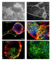
Derivation and characterization of monkey embryonic stem cells
- PMID: 15200688
- PMCID: PMC455691
- DOI: 10.1186/1477-7827-2-41
Derivation and characterization of monkey embryonic stem cells
Abstract
Embryonic stem (ES) cell based therapy carries great potential in the treatment of neurodegenerative diseases. However, before clinical application is realized, the safety, efficacy and feasibility of this therapeutic approach must be established in animal models. The rhesus macaque is physiologically and phylogenetically similar to the human, and therefore, is a clinically relevant animal model for biomedical research, especially that focused on neurodegenerative conditions. Undifferentiated monkey ES cells can be maintained in a pluripotent state for many passages, as characterized by a collective repertoire of markers representing embryonic cell surface molecules, enzymes and transcriptional factors. They can also be differentiated into lineage-specific phenotypes of all three embryonic germ layers by epigenetic protocols. For cell-based therapy, however, the quality of ES cells and their progeny must be ensured during the process of ES cell propagation and differentiation. While only a limited number of primate ES cell lines have been studied, it is likely that substantial inter-line variability exists. This implies that diverse ES cell lines may differ in developmental stages, lineage commitment, karyotypic normalcy, gene expression, or differentiation potential. These variables, inherited genetically and/or induced epigenetically, carry obvious complications to therapeutic applications. Our laboratory has characterized and isolated rhesus monkey ES cell lines from in vitro produced blastocysts. All tested cell lines carry the potential to form pluripotent embryoid bodies and nestin-positive progenitor cells. These ES cell progeny can be differentiated into phenotypes representing the endodermal, mesodermal and ectodermal lineages. This review article describes the derivation of monkey ES cell lines, characterization of the undifferentiated phenotype, and their differentiation into lineage-specific, particularly neural, phenotypes. The promises and limitations of primate ES cell-based therapy are also discussed.
Figures


Similar articles
-
Embryonic stem cell lines of nonhuman primates.ScientificWorldJournal. 2002 Jun 26;2:1762-73. doi: 10.1100/tsw.2002.829. ScientificWorldJournal. 2002. PMID: 12806169 Free PMC article. Review.
-
Differentiation of monkey embryonic stem cells into neural lineages.Biol Reprod. 2003 May;68(5):1727-35. doi: 10.1095/biolreprod.102.012195. Epub 2002 Dec 11. Biol Reprod. 2003. PMID: 12606331
-
Derivation and characterization of chondrocytes from embryonic stem cells in vitro.Methods Mol Biol. 2006;330:171-90. doi: 10.1385/1-59745-036-7:171. Methods Mol Biol. 2006. PMID: 16846025
-
Isolation and characterization of novel rhesus monkey embryonic stem cell lines.Stem Cells. 2006 Oct;24(10):2177-86. doi: 10.1634/stemcells.2006-0125. Epub 2006 Jun 1. Stem Cells. 2006. PMID: 16741224
-
Progress with nonhuman primate embryonic stem cells.Biol Reprod. 2004 Dec;71(6):1766-71. doi: 10.1095/biolreprod.104.029413. Epub 2004 May 5. Biol Reprod. 2004. PMID: 15128597 Review.
Cited by
-
Fluorescence in situ hybridization using an old world monkey Y chromosome specific probe combined with immunofluorescence staining on rhesus monkey tissues.J Histochem Cytochem. 2007 Nov;55(11):1115-21. doi: 10.1369/jhc.7A7216.2007. Epub 2007 Jun 26. J Histochem Cytochem. 2007. PMID: 17595337 Free PMC article.
-
Differentiation of nonhuman primate embryonic stem cells along neural lineages.Differentiation. 2009 Mar;77(3):229-38. doi: 10.1016/j.diff.2008.10.014. Epub 2008 Dec 2. Differentiation. 2009. PMID: 19272521 Free PMC article.
-
Mitochondria in stem cells.Mitochondrion. 2007 Sep;7(5):289-96. doi: 10.1016/j.mito.2007.05.002. Epub 2007 May 23. Mitochondrion. 2007. PMID: 17588828 Free PMC article. Review.
-
Semiquantitative histopathology and 3D magnetic resonance microscopy as collaborative platforms for tissue identification and comparison within teratomas derived from pedigreed primate embryonic stem cells.Stem Cell Res. 2010 Nov;5(3):201-11. doi: 10.1016/j.scr.2010.07.005. Epub 2010 Aug 6. Stem Cell Res. 2010. PMID: 20864427 Free PMC article.
-
Derivation of induced pluripotent stem cells from the baboon: a nonhuman primate model for preclinical testing of stem cell therapies.Cell Reprogram. 2013 Dec;15(6):495-502. doi: 10.1089/cell.2012.0093. Epub 2013 Nov 4. Cell Reprogram. 2013. PMID: 24182315 Free PMC article.
References
-
- Evans MJ, Kaufman MH. Establishment in culture of pluripotential cells from mouse embryos. Nature. 1981;292:154–156. - PubMed
-
- Thomson JA, Kalishman J, Golos TG, During M, Harris CP, Hearn JP. Pluripotent cell lines from common marmoset (Callithrix jacchus) blastocysts. Biol Reprod. 1996;55:254–259. - PubMed
Publication types
MeSH terms
Grants and funding
LinkOut - more resources
Full Text Sources
Other Literature Sources
Medical
Research Materials

