. 2004 Jun 16;24(24):5525-36.
doi: 10.1523/JNEUROSCI.0309-04.2004.

Synaptic contributions to focal and widespread spatiotemporal dynamics in the isolated rat subiculum in vitro

Affiliations

Synaptic contributions to focal and widespread spatiotemporal dynamics in the isolated rat subiculum in vitro

L Menendez de la Prida et al. J Neurosci. .

Abstract

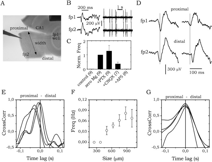
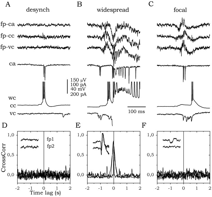
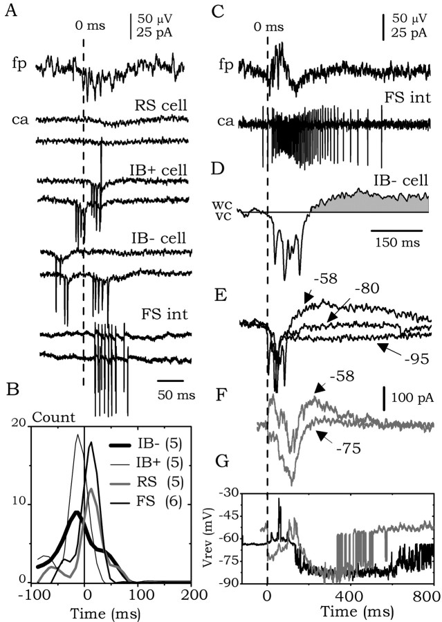
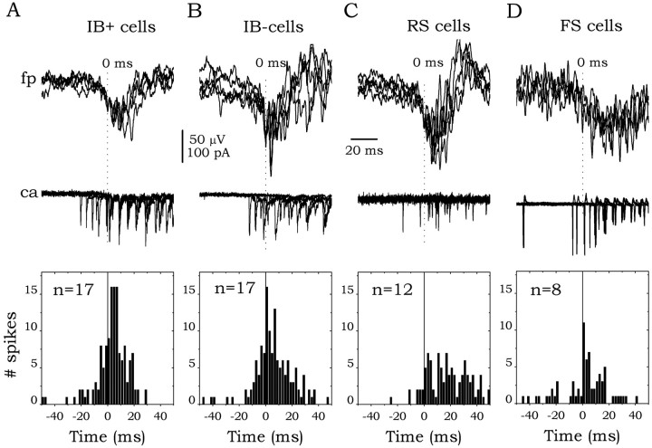
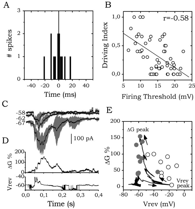
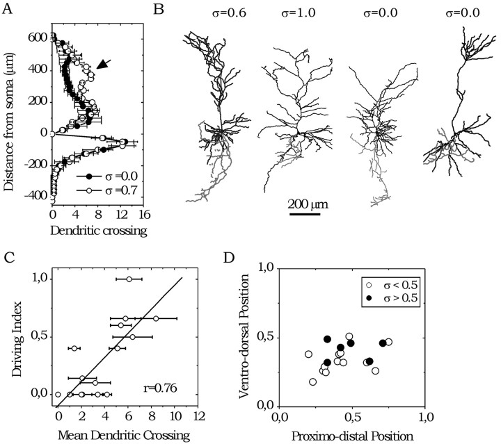
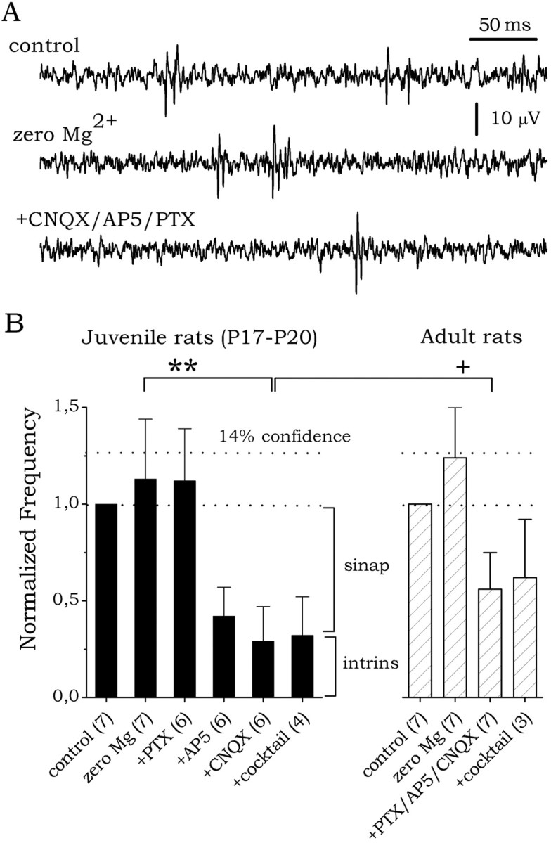
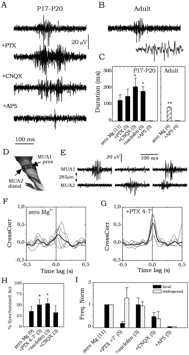
The subiculum, which has a strategic position in controlling hippocampal activity, is receiving significant attention in epilepsy research. However, the functional organization of subicular circuits remains unknown. Here, we combined different recording and analytical methods to study focal and widespread population activity in the isolated subiculum in zero Mg2+ media. Patch and field recordings were combined to examine the contribution of different cell types to population activity. The properties of cells leading field activity were examined. Predictive factors for a cell to behave as leader included exhibiting the bursting phenotype, displaying a low firing threshold, and having more distal apical dendrites. A subset of bursting cells constituted the first glutamatergic type that led a recruitment process that subsequently activated additional excitatory as well as inhibitory cells. This defined a sequence of synaptic excitation and inhibition that was studied by measuring the associated conductance changes and the evolution of the composite reversal potential. It is shown that inhibition was time-locked to excitation, which shunted excitatory inputs and suppressed firing during focal activity. This was recorded extracellularly as a multi-unit ensemble of active cells, the spatial boundaries of which were controlled by inhibition in contrast to widespread epileptiform activity. Focal activity was not dependent on the preparation or the developmental state because it was also recorded under 5 mm [K+]o and in adult tissue. Our data indicate that the subicular networks can be spontaneously organized as leader-follower local circuits in which excitation is mainly driven by a subset of bursting cells and inhibition controls spatiotemporal firing.


Figures

Figure 1.
Figure 1.
Experimental preparation. A, Isolated subicular minislices of different sizes (widths) were prepared from juvenile and adult rats. The proximal (near CA1) and distal (near pre-subiculum) borders are shown. Simultaneous field (fp1 and fp2) and whole-cell (wc) recordings were performed. B, Epileptiform field events were simultaneously recorded at the proximal (fp1) and distal (fp2) borders. Epileptiform events are depicted at high (left) and low (right) temporal resolution. C, Pharmacological dependency of epileptiform activity. The number of slices is indicated. D, Consecutive epileptiform events exhibited a large variability, with no clear leading region. Two consecutive events from a representative minislice are shown. E, Cross-correlation analysis of dual field recordings from a minislice ∼1000 μm wide (data from 4 consecutive events). Interelectrode distance, ∼300 μm. F, Frequency of epileptiform events versus minislice size (width). Data are from n = 29 minislices. G, Cross-correlation analysis of dual recordings (4 consecutive events) from the minislice shown in E, after re-sectioning (∼650 μm wide). Same interelectrode distance before and after cutting, ∼300 μm.
Figure 2.
Figure 2.
Simultaneous field (fp) and patch recordings. Cell responses were examined using cell-attached (ca) and whole-cell (wc) configurations in the current-clamp (cc) and voltage-clamp (vc) modes. Field recordings corresponding to ca, cc, and vctraces are depicted. A, Simultaneous field and patch recordings during desynchronized field activity. This cell was classified as weak bursting (IB-). B, Simultaneous field and patch recordings during widespread field activity. C, Simultaneous field and patch recordings during focal field activity. D, Cross-correlation analysis of simultaneous field recordings. Results are shown from three consecutive episodes of desynchronized field activity. E, Widespread activity. F, Focal activity. Insets show examples of dual field recordings (fp1 and fp2).
Figure 3.
Figure 3.
Cellular and synaptic behavior underlying focal activity. A, Cell responses in cell-attached (ca) configuration during focal activity were examined using field-triggered data segments (fp trace). Two consecutive episodes are shown for each cell (RS, IB+, IB-, and FS cells). B, Histograms of cell firing from each cell type. C, Representative response from another FS interneuron (ca trace) triggered by the onset of focal field activity (fp trace). Note the different time scale compared with A. D, Field-triggered synaptic currents from an IB-cell. E, Field-triggered synaptic currents from the cell shown in D at three different holding potentials (-58, -80, and -95 mV). F, Field-triggered synaptic currents from a different IB-cell at two holding potentials. G, Field-triggered composite reversal potential from cells shown in E (black trace) and F (gray trace). E-G share the same time scale (shown in G).
Figure 4.
Figure 4.
Cell behavior underlying widespread activity. Field-triggered (fp) cell responses (5 consecutive events are depicted) in a cell-attached configuration (ca) from representative IB+ (A), IB-(B), RS (C), and FS (D) cells. Firing histograms from all cells in each group are shown at the bottom.
Figure 5.
Figure 5.
Electrophysiological properties and the driving index. A, Example of firing histograms from one cell (IB+ cell) in cell-attached recordings during field-triggered activity (data from 13 consecutive episodes). The driving index measures the probability of a given cell to fire before the field (see Materials and Methods). B, Statistically significant correlation (p < 0.05; r =-0.58) between the firing threshold and the driving index (n = 46 glutamatergic cells). C, Field-triggered averages (black traces) of synaptic currents during focal activity at three holding potentials. SDs are shown in gray. D, Conductance changes (up) and composite reversal potential (bottom) computed from data in C. E, Vrev-ΔG phase plots (see Materials and Methods and Borg-Graham et al. 1998). ΔG(t) is plotted as a function of the Vrev(t) during the first milliseconds before reaching the conductance peak. Arrows mark this temporal evolution. Gray circles denote the VrevG values at conductance peak. Open circles denote the VrevG values at the Vrev peak. Note the shunting effect of peak conductance increases (gray dots).
Figure 6.
Figure 6.
Morphological properties and the driving index. A, Scholl diagram of apical and basal dendritic arbors of cells exhibiting low (σ = 0.0 ± 0.0; n = 5) and high (σ = 0.7 ± 0.2; n = 4) driving index. Statistical difference was evident at distal apical dendritic crossings (arrow). B, Representative examples of cells in each category. C, Correlation between the mean apical dendritic crossings at 250-450 μm from the soma and the driving index in all cells examined (n = 15). D, Spatial distribution of cells in each category. Positions are normalized.
Figure 7.
Figure 7.
Simultaneous field and MUA recordings during desynchronized (desynch; A), widespread (B), and focal (C) field activity. Insets a-c show additional details of firing activity.
Figure 8.
Figure 8.
Pharmacological control of MUA firing during desynchronized activity. A, Representative MUA traces under control, zero Mg2+, and zero Mg2+ plus CNQX/AP5/PTX. B, Summary of results from pharmacological control of MUA firing in juvenile (left) and adult (right) rats. The number of slices is indicated. Statistical differences are shown: **p < 0.001; +p < 0.05.
Figure 9.
Figure 9.
Pharmacological dissection of synaptic factors mediating focal activity. A, Field-triggered MUA ensembles during focal activity from juvenile tissue in zero Mg2+ media and after bath application of PTX, CNQX, and AP-5. B, Field-triggered MUA ensembles during focal activity from adult tissue in zero Mg2+ media. The inset shows additional details of firing activity. C, Summary of pharmacological results. The number of slices is indicated. D, Nissl-stained subicular minislice. A mark denoted the position of tungsten electrodes. E, Simultaneous MUA recordings during focal activity. F, Cross-correlation analysis of simultaneously recorded MUA firing rate. Results from five consecutive episodes are shown in gray; the mean is shown in black. G, Same as in F after 4-7 min of PTX application. Note the significant increase of cross-correlation. H, Summary of the cross-correlation analysis under different conditions: zero Mg2+, PTX (4-7 min), saclofen, and CNQX. The number of slices is indicated. I, Pharmacological dependence of the frequency of focal and widespread field events.

Similar articles

Cited by

References

    1. Anderson JS, Carandini M, Ferster D (2000) Orientation tuning of input conductance, excitation, and inhibition in cat primary visual cortex. J Neurophysiol 84: 909-926. - PubMed
    1. Behr J, Heinemann U (1996) Low Mg2+ induced epileptiform activity in the subiculum before and after disconnection from rat hippocamapl and entorhinal cortex slices. Neurosci Lett 205: 25-28. - PubMed
    1. Behr J, Gloveli T, Heinemann U (1998) The perforant path projection from the medial entorhinal cortex layer III to the subiculum in the rat combined hippocampal-entorhinal cortex slice. Eur J Neurosci 10: 1011-1018. - PubMed
    1. Berger TW, Swanson GW, Milner TA, Lynch GS, Thompson RF (1980) Reciprocal anatomical connections between hippocampus and subiculum in the rabbit evidence for subicular innervation of regio superior. Brain Res 183: 265-276. - PubMed
    1. Borg-Graham LJ (2001) The computation of directional selectivity in the retina occurs presynaptic to the ganglion cell. Nat Neurosci 4: 176-183. - PubMed

Publication types

LinkOut - more resources