. 2004 Jun 16;24(24):5537-48.
doi: 10.1523/JNEUROSCI.5658-03.2004.

Uropathic observations in mice expressing a constitutively active point mutation in the 5-HT3A receptor subunit

Affiliations

Uropathic observations in mice expressing a constitutively active point mutation in the 5-HT3A receptor subunit

Anindya Bhattacharya et al. J Neurosci. .

Abstract

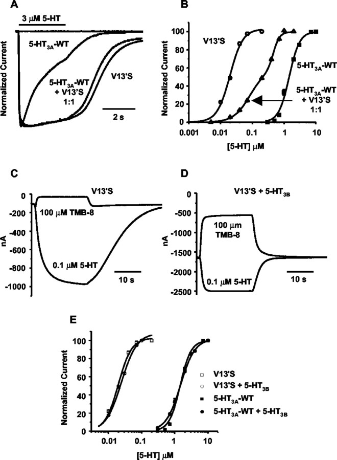
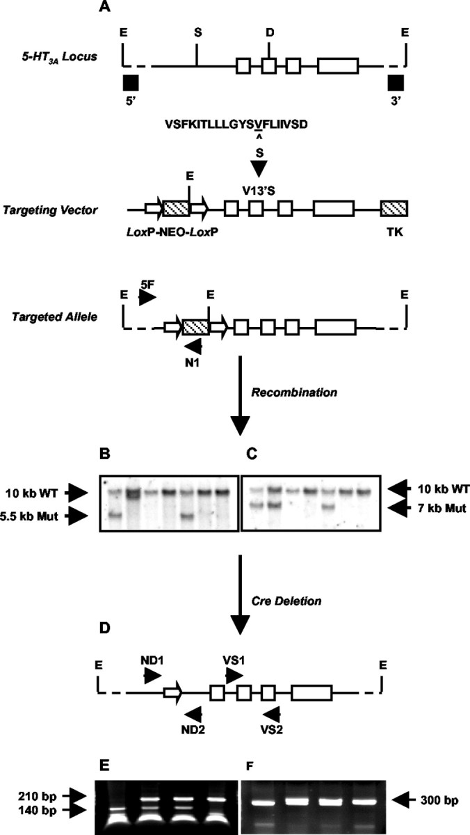
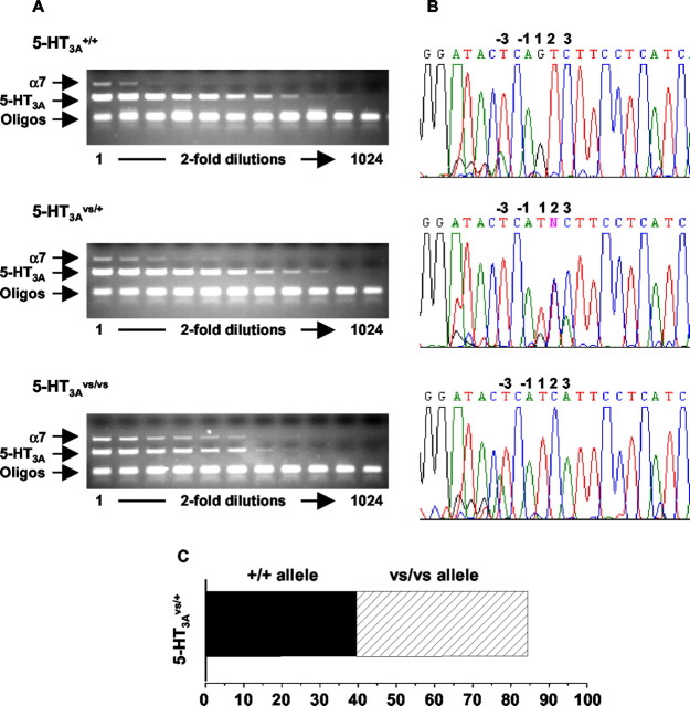
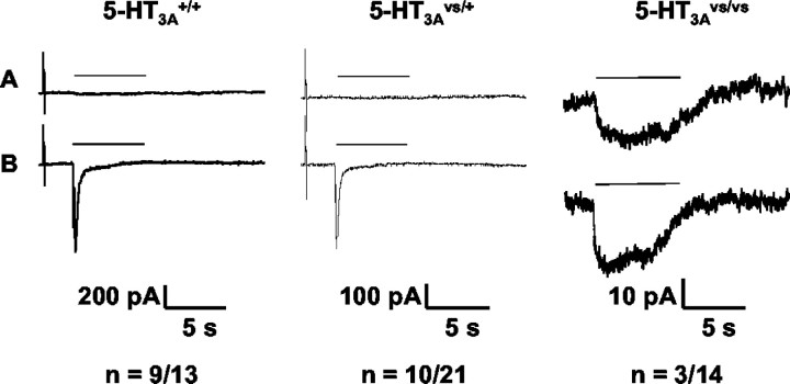
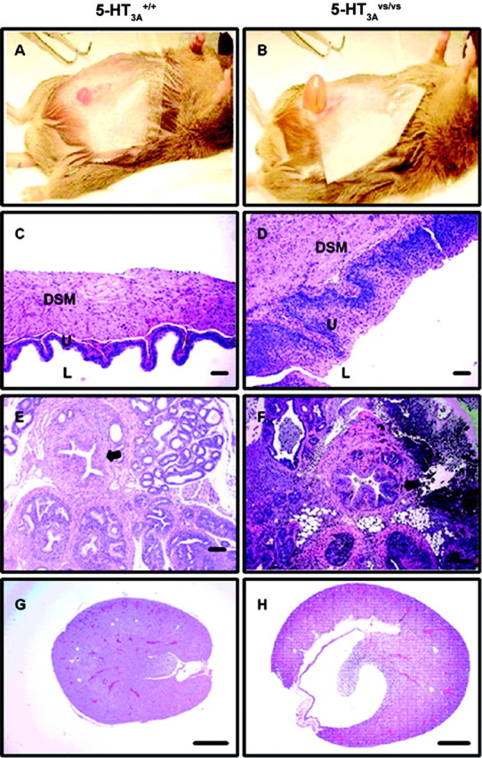
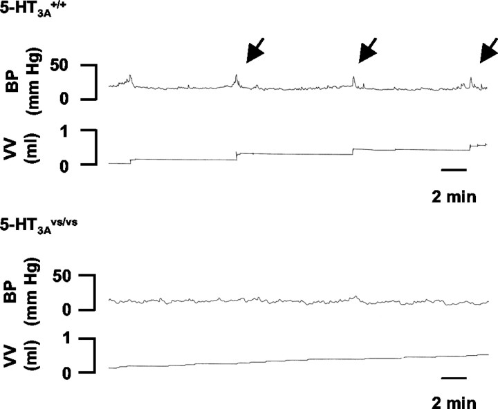
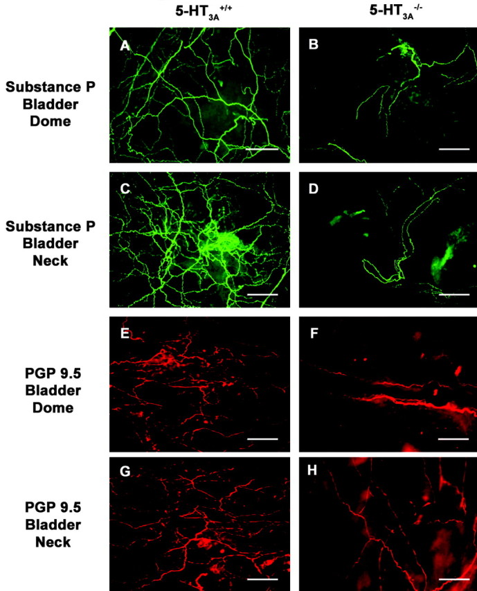
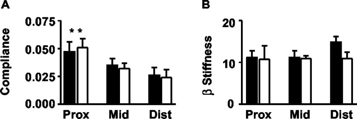
Mutant mice with a hypersensitive serotonin (5-HT)3A receptor were generated through targeted exon replacement. A valine to serine mutation (V13'S) in the channel-lining M2 domain of the 5-HT3A receptor subunit rendered the 5-HT3 receptor 70-fold more sensitive to serotonin and produced constitutive activity when combined with the 5-HT3B subunit. Mice homozygous for the mutant allele (5-HT3Avs/vs) had decreased levels of 5-HT3A mRNA. Measurements on sympathetic ganglion cells in these mice showed that whole-cell serotonin responses were reduced, and that the remaining 5-HT3 receptors were hypersensitive. Male 5-HT3Avs/vs mice died at 2-3 months of age, and heterozygous (5-HT3Avs/+) males and homozygous mutant females died at 4-6 months of age from an obstructive uropathy. Both male and female 5-HT3A mutant mice had urinary bladder mucosal and smooth muscle hyperplasia and hypertrophy, whereas male mutant mice had additional prostatic smooth muscle and urethral hyperplasia. 5-HT3A mutant mice had marked voiding dysfunction characterized by a loss of micturition contractions with overflow incontinence. Detrusor strips from 5-HT3Avs/vs mice failed to contract to neurogenic stimulation, despite overall normal responses to a cholinergic agonist, suggestive of altered neuronal signaling in mutant mouse bladders. Consistent with this hypothesis, decreased nerve fiber immunoreactivity was observed in the urinary bladders of 5-HT3Avs/vs compared with 5-HT3A wild-type (5-HT3A+/+) mice. These data suggest that persistent activation of the hypersensitive and constitutively active 5-HT3A receptor in vivo may lead to excitotoxic neuronal cell death and functional changes in the urinary bladder, resulting in bladder hyperdistension, urinary retention, and overflow incontinence.


Figures

Figure 1.
Figure 1.
Spontaneous and 5-HT-evoked currents from various combinations of 5-HT3A, V13′S, and 5-HT3B receptor subunits expressed in Xenopus oocytes. A, Representative traces of normalized currents evoked by 3 μm 5-HT at -60 mV. Traces are shown for 5-HT3A-WT, V13′S, and a 1:1 mixture of 5-HT3-WT and V13′S cRNAs. The horizontal bar above the traces represents the duration of ligand perfusion through the recording chamber. The flat trace at the baseline level is typical of responses from both the WT and mutant in the presence of 100 nm tropisetron, a 5-HT3 receptor antagonist. B, Normalized dose-response relationships of oocytes injected with 5-HT3A-WT (solid squares), V13′S (open circles), and a 1:1 mixture of 5-HT3-WT and V13′S (open triangles) cRNAs. C, D, Typical voltage-clamp currents from oocytes injected with V13′S cRNA (C) or with V13′S plus 5-HT3B cRNA 1:1 (D). Holding potential, -60 mV. In each panel, the top trace shows the response to 100 μm TMB-8, and the bottom trace shows the response to 0.1 μm 5-HT. Note the larger leak current at the beginning and end, as well as faster onset and washout to 0.1 μm serotonin in (D). The TMB-8-sensitive leak current denotes spontaneous activation of the receptor. E, Normalized dose-response relationships of homomultimeric (5-HT3A-WT or V13′S only; squares) and heteromultimeric (circles) receptors. Receptors included either the 5-HT3A-WT subunit (solid symbols) or V13′S subunit (open symbols).
Figure 2.
Figure 2.
Strategy for generating 5-HT3A knock-in mice through targeted exon replacement. A, A 6 kb genomic fragment of the mouse 5-HT3A receptor gene (solid bar) was used to introduce a point mutation (V13′S) into exon (open boxes) 7 coding for the M2 domain of the 5-HT3A receptor. The sequence of the mouse 5-HT3A receptor M2 region (positions 1′ to 20′) is shown. The Val residue at 13′, corresponding to amino acid position 290 in the entire sequence, is underlined. In the 5-HT3A knock-in mouse this is mutated to Ser (S). Neo and TK selection markers (hatched boxes) were inserted into the SwaI site (S) in the large intron preceding exon 6 and at the 3′ end of the construct, respectively. The Neo sequence was flanked by two LoxP sites (open arrowheads). The linearized targeting vector was used for transfection of embryonic stem cells, and the resulting targeted allele was screened as described in Materials and Methods. B, C, Representative Southern blots of EcoRI (E)-digested genomic DNA were probed with either a 5′-flanking probe (B) or a 3′-flanking probe (C) to identify the wild-type and mutant (Mut) alleles. D, 5-HT3A-targeted mice were crossed to Cre-expressing transgenic mice as described in Materials and Methods, resulting in a Neo deleted mutant allele with one 34 bp LoxP site remaining in intron 5. E, F, Representative PCR screening of genomic DNA from Neo-deleted mice. E, Screening across the mutation site using primers VS1 and VS2 followed by DdeI (D) digestion. F, Screening across the single LoxP site after removal of the Neo cassette, using primers ND1 and ND2.
Figure 3.
Figure 3.
RT-PCR analysis of 5-HT3A mRNA levels in mutant and wild-type mice. A, Diminished 5-HT3A mRNA levels in the 5-HT3Avs/vs mice. Total RNA was isolated from the SCG of 5-HT3A+/+, 5-HT3Avs/+, and 5-HT3Avs/vs mice as described in Materials and Methods. Serial dilutions were prepared as indicated, and 1 μl of RNA was used as template for RT-PCR analysis using primers specific for the 5-HT3A receptor (amplicon of ∼300 bp) and theα7 nACh receptor (amplicon of ∼700 bp) as an internal control. Assuming that α7 nACh receptor mRNA did not change, 5-HT3A mRNA was compared with α7 nAChR mRNA at the lowest dilution where signals could be detected. B, 5-HT3Avs/+ mice express comparable levels of the 5-HT3A mutant and wild-type mRNA. RT-PCR sequencing (B) of the products shown in A were used to estimate the relative abundance of wild-type and mutant mRNA in 5-HT3Avs/+ mice as described in Materials and Methods. The relative abundance of wild-type (solid bars) and mutant (hashed bars) sequences in the RT-PCR products from 5-HT3Avs/+ mice was determined by comparing the representation of the pure alleles, and the averaged percentage of the products is shown in the bar graph in C. Oligo, Oligonucleotide.
Figure 4.
Figure 4.
Serotonin-induced whole-cell currents from SCG neurons of 5-HT3A+/+, 5-HT3Avs/+, and 5-HT3Avs/vs mice. Whole-cell patch-clamp recordings were obtained from primary cultures of SCG neurons. Representative voltage-clamped current responses from 5-HT3A+/+ (left panel), 5-HT3Avs/+ (middle panel), and 5-HT3Avs/vs (right panel) mice to low (A) and high (B) concentrations of serotonin are shown. The low concentration of serotonin for 5-HT3A+/+ and 5-HT3Avs/+ SCG neurons was 0.3 μm, and that for 5-HT3Avs/vs neurons was 0.1 μm, whereas the high concentration was 10 μm in all three. The number of 5-HT3A receptors positive over successfully patched neurons for each genotype is shown below each panel.
Figure 5.
Figure 5.
A-H, Histopathology of the lower urinary tract from 5-HT3A+/+ (A, C, E, G) and 5-HT3Avs/vs (B, D, F, H) mice. A, B, Representative photograph of the hyperdistended urinary bladders seen in 5-HT3Avs/vs (B) compared with 5-HT3A+/+ (A) mice. C, D, H and E stained sections of the urinary bladder wall highlighting the lumen (L), urothelial mucosal layer (U), and the detrusor smooth muscle layer (DSM). E, F, H and E stained sections of the prostatic urethra and surrounding prostatic tissue. Note the increased thickness of the urinary bladder wall (D vs C) and prostatic urethra (arrow in F vs E) in 5-HT3Avs/vs compared with 5-HT3A+/+ mice, with associated mucosal hyperplasia and smooth muscle hypertrophy. In addition, extensive glandular and periglandular inflammatory cell infiltrate can be seen in the prostatic sections of male 5-HT3Avs/vsmice(F).G,H, Longitudinal sections of kidney from 5-HT3A+/+ (G) and5-HT3Avs/vs (H) mice showing the markedly distended renal pelvis in 5-HT3A mutant mice as a consequence of chronic lower urinary tract obstruction. Scale bars: C, D, 50 μm; E-H, 100 μm.
Figure 6.
Figure 6.
Open-filling cystometry in 5-HT3A+/+ and 5-HT3Avs/vs mice. Representative cystometrograms from conscious 8-week-old male 5-HT3A+/+ (top panel) and 5-HT3Avs/vs (bottom panel) mice. Traces illustrate bladder pressure (BP) recorded in response to a constant infusion of saline and accumulated void volumes (VV) recorded with each micturition. Micturition voiding contractions (arrows) were observed in 5-HT3A+/+ mice. In contrast, 5-HT3Avs/vs mice had a complete loss of micturition contractions, consistent with a phenotype of dribbling overflow incontinence.
Figure 7.
Figure 7.
Neurogenic-mediated contractions of detrusor smooth muscle from 5-HT3A+/+, 5-HT3Avs/+, and 5-HT3Avs/vs mice of various ages. A, Detrusor strips from male and female 6-week-old 5-HT3A+/+ mice (closed squares and closed circles) responded to electrical field stimulation, whereas bladder strips from male and female 6-week-old 5-HT3Avs/vs mice (open squares and open circles) showed minimal responses to neurogenic-mediated contraction. B, C, At 8 (B) and 12 (C) weeks of age, neurogenic-mediated contractions were completely absent in detrusor strips from male 5-HT3Avs/+ and 5-HT3Avs/vs mice (closed circles and open squares), whereas male 5-HT3A+/+ mice responded to electrical field stimulation (closed squares). Contractile responses were plotted as a percentage of KCl (67 mm)-induced maximal force. Points represent mean contraction ± SEM for n = 3-4 animals per group.
Figure 8.
Figure 8.
Effect of carbachol on detrusor smooth muscle contraction in male 5-HT3A+/+, 5-HT3Avs/+, and 5-HT3Avs/vs mice. A, B, Carbachol induced concentration-dependent contractions of bladder smooth muscle in vitro in both 8-week-old (A) and 12-week-old (B) 5-HT3A+/+ (closed squares), 5-HT3Avs/+ (closed circles), and 5-HT3Avs/vs (open squares) mice. C, D, In conscious open filling cystometry, carbachol induced concentration-dependent increases in bladder pressure (BP) in 8-week-old (C) and 12-week-old (D) 5-HT3A+/+, 5-HT3Avs/+, and 5-HT3Avs/vs mice (symbols as above). In both experiments, no differences were seen in carbachol-induced contractions between wild-type and mutant mice, except at the data points indicated (*). Contraction and bladder pressure changes were plotted as a percentage of KCl (67 mm)-induced maximal force (A, B) and increases from baseline bladder pressure (C, D), respectively. Points represent mean ± SEM for n = 3-6 animals per group. *p < 0.05, statistical significance versus 5-HT3A+/+ mice.
Figure 9.
Figure 9.
Decreased innervation density in the bladder urothelium of 5-HT3Avs/vs mice. A-H, Representative images of whole-mount bladder immunostaining from 8- to 10-week-old 5-HT3A+/+ (A, C, E, G) and 5-HT3Avs/vs (B, D, F, H) mice (n=3) for Substance P (A-D, green) and PGP 9.5 (E-H, red). Note the decreased density of the fine nerve fibers coursing through the urothelium in both the dome (A, B, E, F) and the neck (C, D, G, H) regions of the urinary bladder. Scale bar: A-H, 50 μm.
Figure 10.
Figure 10.
Urethral compliance and β-stiffness measurements in female 5-HT3A+/+ and 5-HT3Avs/vs mice. A, B, Data shown represent low pressure (0-6 mmHg)-induced changes in the compliance (A) and β stiffness (B) of the proximal (prox), middle (mid), and distal (dist) urethra in 5-HT3A+/+ (closed bars) and 5-HT3Avs/vs (open bars) mice. Data represent the mean ± SEM for n = 4-5 animals per group. **p < 0.01, statistically significant difference in the compliance of the proximal urethra compared with the middle and distal portions in both 5-HT3A+/+ and 5-HT3Avs/vs mice.

Similar articles

Cited by

References

    1. Alhaider AA, Lei SZ, Wilcox GL (1991) Spinal 5-HT3 receptor mediated antinociception: possible release of GABA. J Neurosci 11: 1881-1888. - PMC - PubMed
    1. Barras M, Van der Graaf PH, Angel I (1996) Characterization of the 5-HT receptor potentiating neurotransmission in rabbit bladder. Eur J Pharmacol 318: 425-428. - PubMed
    1. Bassuk JA, Grady R, Mitchell M (2000) The molecular era of bladder research: transgenic mice as experimental tools in the study of outlet obstruction. J Urol 164: 170-179. - PubMed
    1. Broide RS, Salas R, Ji D, Paylor R, Patrick JW, Dani JA, De Biasi M (2002) Increased sensitivity to nicotine-induced seizures in mice expressing the L250T α7 nicotinic acetylcholine receptor mutation. Mol Pharmacol 61: 695-705. - PubMed
    1. Charlton RG, Morley AR, Chambers P, Gillespie JI (1999) Focal changes in nerve, muscle and connective tissue in normal and unstable human bladder. BJU International 84: 953-960. - PubMed

Publication types

MeSH terms

Substances