. 2004 Jun 16;24(24):5549-59.
doi: 10.1523/JNEUROSCI.2719-03.2004.

Morphine-induced changes in delta opioid receptor trafficking are linked to somatosensory processing in the rat spinal cord

Affiliations

Morphine-induced changes in delta opioid receptor trafficking are linked to somatosensory processing in the rat spinal cord

Anne Morinville et al. J Neurosci. .

Abstract

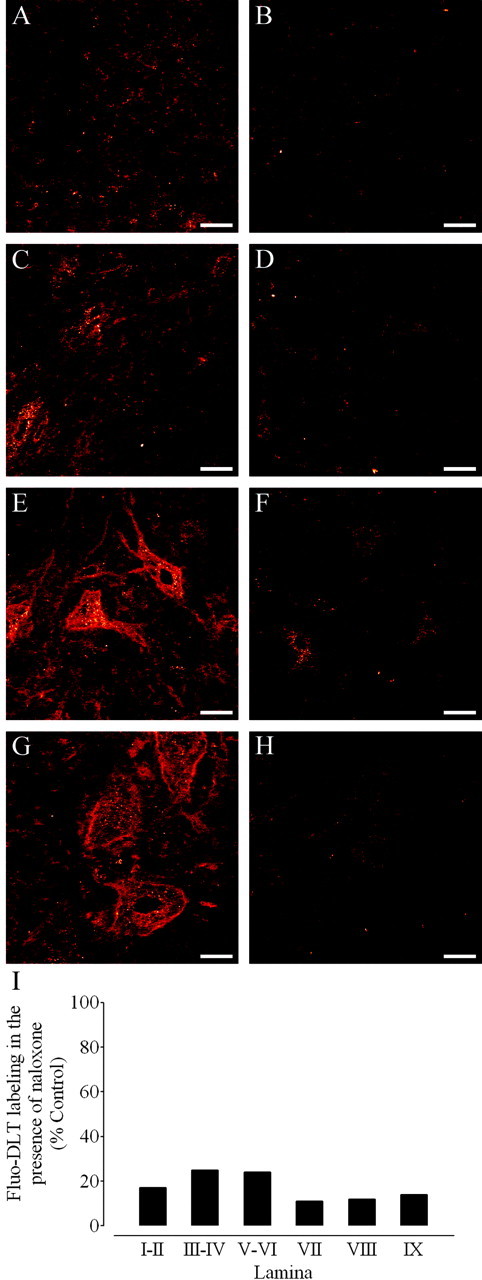
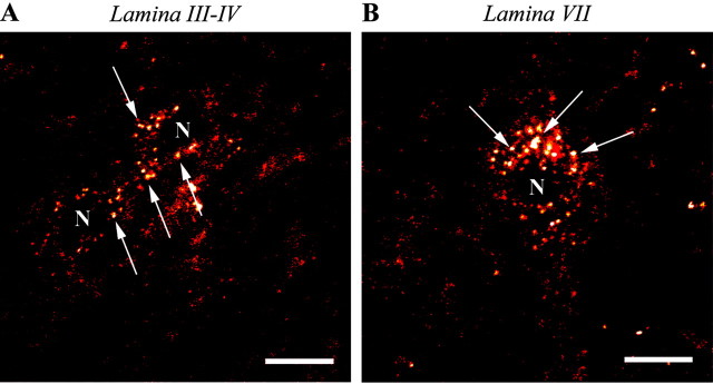
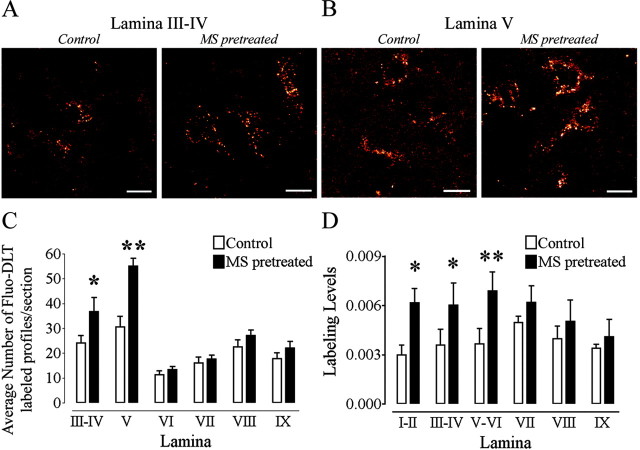
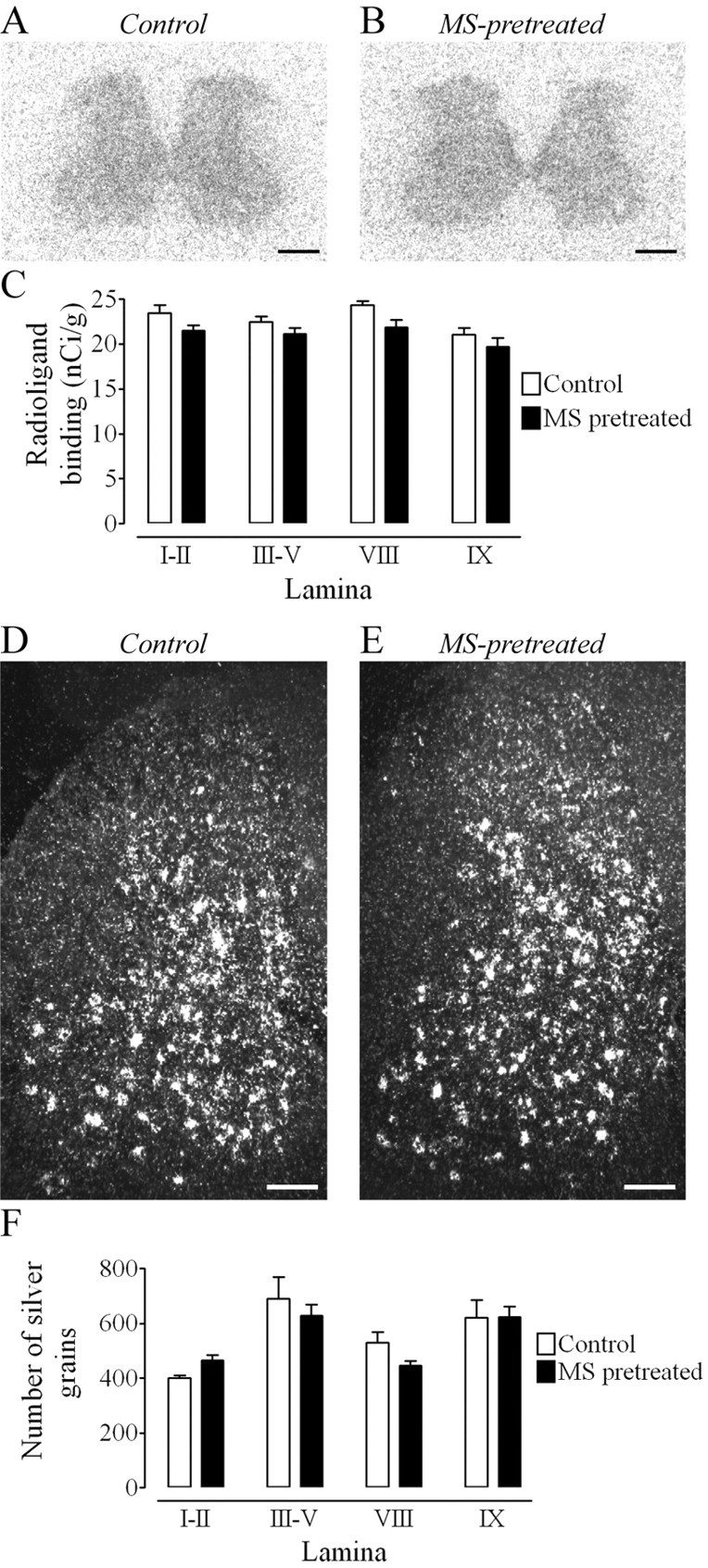
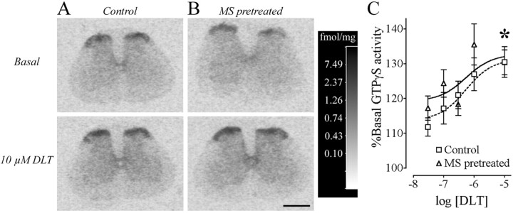
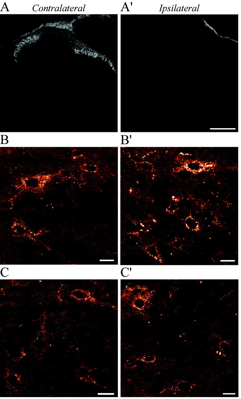
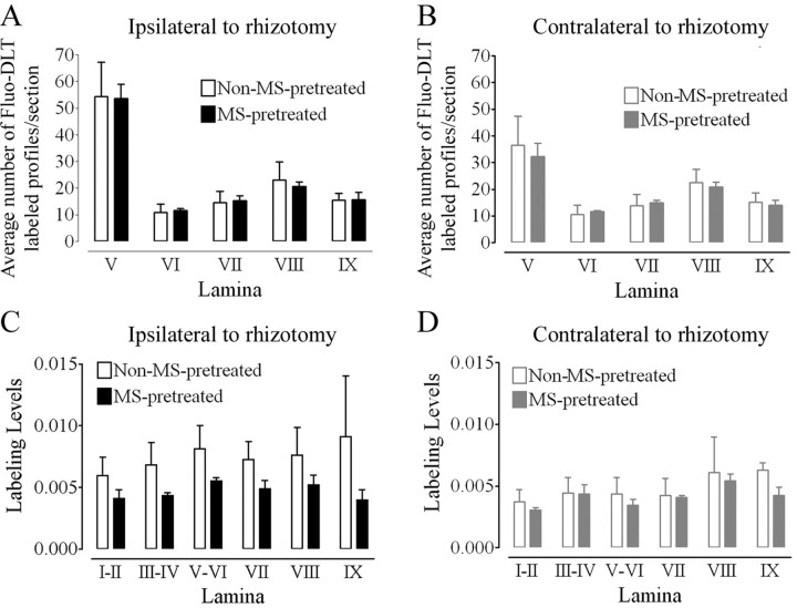
An in vivo fluorescent deltorphin (Fluo-DLT) internalization assay was used to assess the distribution and regulation of pharmacologically available delta opioid receptors (deltaORs) in the rat lumbar (L4-5) spinal cord. Under basal conditions, intrathecal injection of Fluo-DLT resulted in the labeling of numerous deltaOR-internalizing neurons throughout dorsal and ventral horns. The distribution and number of Fluo-DLT-labeled perikaryal profiles were consistent with that of deltaOR-expressing neurons, as revealed by in situ hybridization and immunohistochemistry, suggesting that a large proportion of these cells was responsive to intrathecally administered deltaOR agonists. Pretreatment of rats with morphine for 48 hr resulted in a selective increase in Fluo-DLT-labeled perikaryal profiles within the dorsal horn. These changes were not accompanied by corresponding augmentations in either deltaOR mRNA or (125)I-deltorphin-II binding levels, suggesting that they were attributable to higher densities of cell surface deltaOR available for internalization rather than to enhanced production of the receptor. Unilateral dorsal rhizotomy also resulted in increased Fluo-DLT internalization in the ipsilateral dorsal horn when compared with the side contralateral to the deafferentation or to non-deafferented controls, suggesting that deltaOR trafficking in dorsal horn neurons may be regulated by afferent inputs. Furthermore, morphine treatment no longer increased Fluo-DLT internalization on either side of the spinal cord after unilateral dorsal rhizotomy, indicating that microOR-induced changes in the cell surface availability of deltaOR depend on the integrity of primary afferent inputs. Together, these results suggest that regulation of deltaOR responsiveness through microOR activation in this region is linked to somatosensory information processing.


Figures

Figure 1.
Figure 1.
Antagonism of Fluo-DLT labeling by naloxone in the dorsal and ventral horn of the rat spinal cord. A-H, Anon-pretreated rat (control; A, C, E, G) and a rat pretreated with naloxone (B, D, F, H) were injected intrathecally with 0.8 nmol of Fluo-DLT. Internalized Fluo-DLT is observed by confocal microscopy in dorsal and ventral horn neurons of the lumbar (L4-5) spinal cord; typical images from laminas I-II (A, B), III-IV (C,D), VIII (E, F), and IX (G,H) are presented. Identical acquisition parameters were used to obtain the images for cells labeled in the presence or absence of naloxone. Images are displayed in glow-scale, where white represents the highest fluorescence intensity and red represents the lowest (black indicates the absence of fluorescent signal). Scale bar, 20 μm. I, Fluorescence labeling levels for Fluo-DLT intrathecally injected untreated (control) and naloxone-treated rats were determined as described in Materials and Methods. Each bar corresponds to the average fluorescence labeling levels of the naloxone-treated rat divided by the Fluo-DLT-labeling levels of the untreated rat calculated within each specified lamina (3 sections/animal) in one representative experiment.
Figure 2.
Figure 2.
High-magnification confocal microscopic images of Fluo-DLT-labeled neurons in dorsal and ventral horns of the rat spinal cord. Fluorescent puncta (denoted by arrows), corresponding to internalized Fluo-DLT, are evident throughout the perikaryal cytoplasm, sparing the nucleus (N). Images are displayed in glow-scale, where white represents the highest fluorescence intensity and red represents the lowest (black indicates the absence of fluorescent signal). Scale bar, 10 μm.
Figure 3.
Figure 3.
Comparison of the localization of cells labeled for δOR in the rat lumbar (L4-5) spinal cord. A, B, Immunohistochemical labeling of δOR (Fig. 5A) [Cahill et al. (2001a), reprinted with permission] (A) and δOR mRNA expression (B), as revealed by in situ hybridization, concur in demonstrating the presence of this receptor in neurons distributed throughout the gray matter of the spinal cord. C, Schematic representation of the distribution of Fluo-DLT-labeled perikaryal profiles in the lumbar spinal cord (L5) (Paxinos and Watson, 1986) of control, non-pretreated rats. The number of filled circles within each lamina represents the average number of Fluo-DLT-labeled cell body profiles per section. D, Comparison of the estimated number of cross-sectioned neurons counted by the three techniques in the dorsal (laminas III-VI) and ventral (VII-IX) horns. Fluo-DLT, immunuhistochemical (IHC)- and in situ hybridization (ISH)-labeled profiles were counted as described in Materials and Methods. Because these three techniques used sections of different thickness, counts were corrected to 20 μm according to the following formula: [(counts × 20)/(section thickness in micrometers)].
Figure 4.
Figure 4.
Cross-sectional area of Fluo-DLT-labeled perikaryal profiles in lamina III-V of the rat spinal cord before and after morphine pretreatment. Control and morphine-pretreated rats were injected intrathecally with 0.8 nmol of Fluo-DLT and subsequently processed for confocal microscopic imaging as described in Materials and Methods. Each bar represents the average cross-sectional area of labeled perikaryal profiles in AU for each animal (n = 3 animals per condition; 5 sections per animal). No statistically significant difference was noted between morphine-pretreated and control rats in either laminas III-IV or V. Statistical significance was determined by means of a repeated measures two-way ANOVA (drug and lamina as variables).
Figure 5.
Figure 5.
Morphine pretreatment produces a selective increase in the internalization of Fluo-DLT in the dorsal horn of the rat spinal cord. Control and morphine-pretreated rats were injected intrathecally with 0.8 nmol of Fluo-DLT and subsequently processed for visualization by confocal microscopy as described in Materials and Methods. Images of Fluo-DLT internalization from laminas III-IV (A) and V (B) are presented. Images are presented in red-white glow-scale. Scale bar, 10 μm. C, Comparison of the number of Fluo-DLT-labeled perikaryal profiles per section between control and morphine-pretreated rats in the L4-5 spinal cord segment. Each bar represents the average of three independent experiments. Morphine pretreatment produces a significant increase in the number of Fluo-DLT-labeled perikaryal profiles persection in laminas III-IV (denoted by one asterisk; p < 0.05) and in lamina V (denoted by two asterisks; p < 0.001) when compared with control rats. Statistical significance was determined using a two-way ANOVA with drug and lamina as variables. A Bonferroni's multiple comparison test (MCT) was used to determine which lamina, between control and morphine-pretreated animals, displayed a significant difference. D, Morphine pretreatment increased fluorescence-labeling levels when compared with control rats in dorsal horn neurons but not ventral horn neurons. Each bar represents the average of three independent experiments. For D, one asterisk corresponds to p < 0.01, whereas two asterisks representp < 0.001. Statistical significance was determined by means of a repeated measures two-way ANOVA (drug and lamina as variables) with a Bonferroni's MCT to determine which lamina between control and morphine-pretreated animals displayed a significant difference.
Figure 6.
Figure 6.
Morphine pretreatment produces no change in δOR agonist binding or mRNA expression. A, B, Binding of 125I-DLT-II in the lumbar (L4-5) spinal cord of control (A) and morphine-pretreated (B) rats. Scale bar, 0.5 mm. C, Densitometric quantification of autoradiographic signal in selected lamina. No significant difference, as assessed by means of a two-way ANOVA followed by a Bonferroni's MCT, was detected between control and morphine-pretreated rats in laminas I-II, III-V, VIII, or IX. Radioligand-binding values (nCi/gm) are based on a 14C series of calibration standards. Each bar represents the mean ± SEM for n = 4 rats per group. D, E, Comparison of δOR mRNA expression in the lumbar spinal cord of control (D) and morphine-pretreated (E) rats. Scale bar, 0.25 mm. F, Densitometric quantification of silver grains in selected lamina of the lumbar (L4-5) spinal cord. No significant difference was detected between control and morphine-pretreated rats in laminas I-II, III-V, VIII, or IX (two-way ANOVA followed by a Bonferroni's MCT). Silver grain counts are presented as means ± SEM for n = 4 rats per group.
Figure 7.
Figure 7.
[D-Ala2]-Deltorphin II-induced GTPγS binding in the lumbar spinal cord. Binding of GTγS in the absence of δOR agonist (basal) and in the presence of 10 μm DLT for control (n = 14; A) and morphine sulfate (MS)-pretreated (n = 13; B) rats in a representative experiment from the lumbar (L4-5) spinal cord is presented. The calibration scale (gray-scale converted to fmol/mg using 14C standards) for these four spinal cords taken from the same autoradiographic film is presented. Scale bar, 1.0 mm. C, Densitometric quantification of GTPγS binding in the lumbar spinal cord with increasing doses of deltorphin (expresssed as log10 of the concentration in mol/l) is expressed as a percentage of the basal binding observed in control and MS-pretreated rats. The asterisk denotes statistical significance between the 0.03 and 10 μm DLT (p < 0.05) for both control and MS-pretreated rats (unpaired t test followed by a Bonferroni's correction), demonstrating a dose-response effect.
Figure 8.
Figure 8.
A-C, IB4 immunostaining (A, A′) and Fluo-DLT labeling (B, B′,C,C′) in sections from the rat lumbar (L4-5) spinal cord 10 d after unilateral dorsal rhizotomy. Selective disappearance of IB4 immunostaining in the superficial layers of the dorsal horn on the side ipsilateral (A′) compared with the side contralateral (A) to the transected roots indicates complete degeneration of nonpeptidergic primary afferent fibers. In nonmorphine-pretreated rats (B, B′), increased internalization of Fluo-DLT is evident in neurons in lamina V on the side ipsilateral (B′) compared with contralateral (B) to the transected roots. In morphine-pretreated animals (C,C′), there is no apparent increase in the intensity of Fluo-DLT neuronal labeling on either the ipsilateral (compare C′ and B′) and contralateral (compare C and B) sides. Images B, B′, C, and C′ are displayed in red-white glow-scale. Scale bars: A, A′, 200 μm; B, B′, C, C′, 10 μm.
Figure 9.
Figure 9.
Effect of unilateral dorsal rhizotomy on Fluo-DLT labeling in the rat lumbar spinal cord. A, An increase in the number of Fluo-DLT-positive profiles is observed in laminas V-VI of the ipsilateral side of nonmorphine-pretreated rhizotomized rats when compared with either the side contralateral to the lesion (two-way ANOVA; Bonferroni's MCT; p < 0.001; denoted by #) or to control nonlesioned (two-way ANOVA; Bonferroni's MCT; p < 0.05; denoted by *) rats. B, Fluorescence labeling levels were significantly greater in nonmorphine-pretreated rhizotomized rats in laminas V-VI of the ipsilateral side compared with the contralateral side of lesioned animals (paired t test; p < 0.05; denoted by *), in agreement with the increase in the number of Fluo-DLT-labeled cells.
Figure 10.
Figure 10.
Effect of morphine pretreatment on Fluo-DLT labeling in the spinal cord of rats with a unilateral dorsal rhizotomy. A, B, Comparison of the number of Fluo-DLT-labeled perikaryal profiles in laminas V-IX on the contralateral (B) and ipsilateral side (A) reveals no significant difference between non-morphine sulfate (MS)-pretreated and MS-pretreated rats using two-way ANOVA. C, D, No statistically significant difference in Fluo-DLT-labeling levels between non-MS-pretreated and MS-pretreated animals was detected for either the side contralateral (D) or ipsilateral (C) to the transected roots in any of the lamina in the lumbar (L4-5) spinal cord using two-way ANOVA.

Similar articles

Cited by

References

    1. Abbadie C, Pan YX, Pasternak GW (2000) Differential distribution in rat brain of mu opioid receptor carboxy terminal splice variants MOR-1C-like and MOR-1-like immunoreactivity: evidence for region-specific processing. J Comp Neurol 419: 244-256. - PubMed
    1. Abbadie C, Pasternak GW, Aicher SA (2001) Presynaptic localization of the carboxy-terminus epitopes of the mu opioid receptor splice variants MOR-1C and MOR-1D in the superficial laminae of the rat spinal cord. Neuroscience 106: 833-842. - PubMed
    1. Abbadie C, Lombard MC, Besson JM, Trafton JA, Basbaum AI (2002) Mu and delta opioid receptor-like immunoreactivity in the cervical spinal cord of the rat after dorsal rhizotomy or neonatal capsaicin: an analysis of pre- and postsynaptic receptor distributions. Brain Res 930: 150-162. - PubMed
    1. Arvidsson U, Dado RJ, Riedl M, Lee JH, Law PY, Loh HH, Elde R, Wessendorf MW (1995) Delta-opioid receptor immunoreactivity: distribution in brainstem and spinal cord, and relationship to biogenic amines and enkephalin. J Neurosci 15: 1215-1235. - PMC - PubMed
    1. Bao L, Jin SX, Zhang C, Wang LH, Xu ZZ, Zhang FX, Wang LC, Ning FS, Cai HJ, Guan JS, Xiao HS, Xu ZQ, He C, Hokfelt T, Zhou Z, Zhang X (2003) Activation of delta opioid receptors induces receptor insertion and neuropeptide secretion. Neuron 37: 121-133. - PubMed

Publication types

MeSH terms

Grants and funding