. 2004 Jun 16;24(24):5632-42.
doi: 10.1523/JNEUROSCI.1248-04.2004.

Cone photoreceptors in bass retina use two connexins to mediate electrical coupling

Affiliations

Cone photoreceptors in bass retina use two connexins to mediate electrical coupling

John O'Brien et al. J Neurosci. .

Abstract

Electrical coupling via gap junctions is a common property of CNS neurons. In retinal photoreceptors, coupling plays important roles in noise filtering, intensity coding, and spatial processing. In many vertebrates, coupling is regulated during the course of light adaptation. To understand the mechanisms of this regulation, we studied photoreceptor gap junction proteins. We found that two connexins were expressed in bass cone photoreceptors. Connexin 35 (Cx35) mRNA was present in many cell types, including photoreceptors and amacrine, bipolar, and a few ganglion cells. Antibodies to Cx35 labeled abundant gap junctions in both the inner and outer plexiform layers. In the outer plexiform layer, numerous plaques colocalized with cone telodendria at crossing contacts and tip-to-tip contacts. Cx34.7 mRNA was found predominantly in the photoreceptor layer, primarily in cones. Cx34.7 immunolabeling was limited to small plaques immediately beneath cone pedicles and did not colocalize with Cx35. Cx34.7 plaques were associated with a dense complex of cone membrane beneath the pedicles, including apparent contacts between telodendria and cone pedicles. Tracer coupling studies of the connexins expressed in HeLa cells showed that coupling through Cx35 gap junctions was reduced by protein kinase A (PKA) activation and enhanced by PKA inhibition through a greater than fivefold activity range. Cx34.7 was too poorly expressed to study. PKA regulation suggests that coupling through Cx35 gap junctions can be controlled dynamically through dopamine receptor pathways during light adaptation. If Cx34.7 forms functional cell-cell channels between cones, it would provide a physically separate pathway for electrical coupling.


Figures
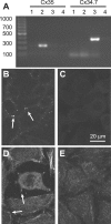
Figure 1.
Figure 1.
Expression of transfected connexins in HeLa cells. A, RT-PCR screening for expression of Cx35 and Cx34.7 in total RNA isolated from HeLa cell lines. For each primer set, lane 1 represents RNA from nontransfected HeLa cells; lane 2, RNA from Cx35-transfected cells; lane 3, RNA from Cx34.7-transfected cells; and lane 4, no template RT reaction control. Transfected HeLa cells showed specific expression of the connexin transfected. B, C, Immunostaining for Cx35 in Cx35-transfected cells (B) revealed punctate staining at cell-cell contacts (arrows) that was not present in nontransfected cells (C). Scale bar in C is the same for B-E. D, E, Immunostaining for Cx34.7 in Cx34.7-transfected cells (D) revealed punctate staining at cell contacts (arrows) and diffuse staining around the cell periphery. Nontransfected cells (E) lacked the diffuse staining and staining at contacts and showed only occasional speckles more consistent with nonspecific background.
Figure 2.
Figure 2.
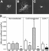
Cx35-mediated tracer coupling is regulated by protein kinase A. A, Neurobiotin injected into untransfected HeLa cells (control) spreads to a small number of cells through gap junctions composed of an endogenous connexin. B, Neurobiotin injection into cells stably transfected with Cx35 diffuses into a larger number of cells (for quantitative measure, see D). C, Bath application of the membrane-permeant cAMP analog Sp-8-cpt-cAMPS (8 μm) to Cx35-transfected HeLa cells 15 min before injection results in restriction of tracer diffusion to a few cells. D, Diffusion coefficients for Neurobiotin diffusion (for procedure and data analysis, see Materials and Methods) of HeLa cells either without transfection or stably transfected with Cx35 or Cx34.7. In control (cntrl) HeLa cells, application of 15 μm of the PKA activator Sp-8-cpt-cAMPS (cA+) or 15 μm of the PKA inhibitor Rp-8-cpt-cAMPS (cA-) caused no significant changes in diffusion coefficient (left plot, p > 0.05 vs no drug; no drug, n = 20; cA+, n = 10; cA-, n = 10). In Cx35-transfected cells, Sp-8-cpt-cAMPS caused a significant reduction in diffusion coefficient (center plot, p < 0.01 vs no drug; no drug, n = 26; cA+, n = 19), whereas Rp-8-cpt-cAMPS caused a significant increase in diffusion coefficient (p < 0.01 vs no drug; cA-, n = 10). Coupling in Cx34.7-transfected cells was not significantly different than in control HeLa cells (right plot, p > 0.05; n = 20). Coupling in all cell lines was significantly reduced to differing extent by 10 μm carbenoxolone (carb; nontransfected, n = 5; Cx35, n = 9; Cx34.7, n = 6). *Significant at the p < 0.05 level; **significant different at the p < 0.01 level.
Figure 5.
Figure 5.
Antibodies to Cx35 and Cx34.7 label connexins selectively. A, Western blot analysis of crude lysates of bacteria expressing a GST-Cx35 intracellular loop fusion protein (lanes 1, left and right panels) or a GST-Cx34.7 intracellular loop fusion protein (lanes 2, left and right panels). Both Cx35 monoclonal (left panel) and Cx34.7 polyclonal (right panel) antibodies are specific. B, Western blot analysis of hybrid bass tissues using anti-Cx35 monoclonal (left) and anti-Cx34.7 polyclonal (right) antibodies. Each lane contains 80 μg of crude protein from hybrid bass brain (lanes 1), retina (lanes 2), or liver (lanes 3). The Cx35 antibody recognizes a complex of bands at 30-33 kDa in brain and retina. A higher-mass band at ∼65 kDa may represent Cx35 dimers. The Cx34.7 antibody recognizes a single band of ∼36 kDa only in retina.
Figure 3.
Figure 3.
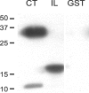
Cx35 cytoplasmic domains can be phosphorylated by PKA. Autoradiogram shows in vitro phosphorylation of bacterially expressed fusion proteins containing Cx35 intracellular domains. The C terminus of Cx35 (CT) was fused to glutathione-S-transferase, and the IL domain was fused to a 6×His tag. Both domains of Cx35 were phosphorylated by the PKA catalytic subunit, whereas GST alone was not.
Figure 4.
Figure 4.
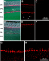
Cx35 and Cx34.7 mRNAs have different but overlapping distributions in the retina. A, In situ hybridization with a Cx35 intracellular loop antisense probe labeled three regions of the bass retina. Hybridization signal is visualized by Cy3 fluorescence in red overlaid on the Nomarski image of the section. Labeling is most prominent in the photoreceptor layer between the outer segments (OS) and ONL, within the INL, and in the GCL. Hybridization signal in the INL is restricted to the lower half, and the large, block-like horizontal cells (open arrow) are clearly not labeled. Scale bar in C applies to A-F. B, Confocal micrograph of a similar section showing faint labeling throughout the ONL. Cells within the GCL fall into two groups: scarce cells with large somata (filled arrow) presumably representing some types of ganglion cells and more common cells with small somata (arrowheads) that are more likely displaced amacrine cells. Image is a 4 μm confocal stack. C, Sections probed with the Cx35 sense strand show no labeling. Image is a 4 μm confocal stack. D, In situ hybridization with a Cx34.7 intracellular loop antisense probe (for details, see Materials and Methods) labels only one band in the photoreceptor layer strongly. Faint labeling was detected in the INL, but horizontal cells were again without label (open arrow). E, Confocal micrograph of a Cx34.7 antisense hybridization showing more clearly the labeling in the photoreceptor layer and very faint labeling in the INL. Image is a 4 μm confocal stack. F, Cx34.7 sense strand probe shows no labeling. Image is a 4 μm confocal stack. G, Confocal micrograph of the photoreceptor layer of Cx35-labeled section. Prominent areas of labeling are in the myoid regions of cone photoreceptors, which have large oval ellipsoids immediately above the labeled region. Image is a 4 μm confocal stack. H, Confocal micrograph of the photoreceptor layer of a Cx34.7-labeled section. Prominent labeling is in the same region of cone photoreceptors, as is Cx35 labeling. Image is a 4 μm confocal stack.
Figure 6.
Figure 6.
Cx35 gap junctions are widespread in bass retina. A, Immunolabeling for SV2 (blue) shows the synaptic layers, and PKCα (red) shows the rod-dominated Mb1 bipolar cell. Cx35 immunoreactivity (green) is abundant in both synaptic layers. Image is a 6 μm confocal stack. B, In the outer plexiform layer, Cx35 is predominantly located below the photoreceptor terminals labeled with SV2. Cx35 plaques vary from small plaques <0.5 μm in diameter to linear arrays up to 3 μm in length. There was little or no colocalization with dendritic processes of the Mb1 bipolar cells. Image is a 2 μm confocal stack. C, In the inner plexiform layer, Cx35 is abundant in both sublaminas. Plaques vary greatly in size and include large, flat plaques up to 3 × 8 μm in size. Telodendrial processes of the Mb1 bipolar cell axon terminals consistently colocalize with very small Cx35 plaques at points of constriction (arrows). These likely represent gap junctional coupling at tip-to-tip contacts of the telodendria. Image is an 8 μm confocal stack.
Figure 7.
Figure 7.
Cx34.7 protein is more limited in distribution. A, Double labeling for Cx34.7 (red) and Cx35 (green) shows distinctly different distributions of the two connexins. Cx34.7 immunoreactivity is restricted to the outer plexiform layer in regularly spaced clusters. Cx35 is also present at this location. Image is a 5 μm confocal stack. B, Labeling of the outer plexiform layer with SV2 (blue) shows that the Cx34.7 clusters (red) are located beneath cone photoreceptor pedicles, which appear as large trapezoids among the doughnut-shaped and much smaller rod terminals. Cx35 immunolabeling (green) in the same vicinity is not restricted to the cone pedicles. Image is a 5 μm confocal stack. C-E, Three views of Cx34.7 (red) and Cx35 (green) immunoreactivity in the OPL. Although the connexins are in very close proximity, the two do not occur in the same plaques. The limited overlap visible in some images was attributable to stacking of confocal slices from different depths. C is a 3 μm confocal stack of a whole-mount view; D, 4 μm confocal stack of a vertical section; E, 6 μm confocal stack of an oblique section. Scale bar in C applies to D, E.
Figure 8.
Figure 8.
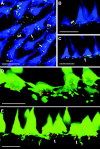
Gap junctions are located in the network of cone telodendria. A, Immunolabeling of double cones with FRet43 antibody (blue) reveals the network of telodendria connecting the cones. Cx35 immunostaining (green) is extensively colocalized with telodendria at linear plaques (triple arrowheads) and punctate spots indicative of crossing and tip-to-tip contacts (arrows). Cx34.7 (red) is predominantly localized to the cone pedicles. Image is a 2.5 μm confocal stack in whole-mount view. B, C, Vertical sections of cone terminals show telodendria that contact neighboring cones make gap junctions with Cx35 in linear arrays (triple arrowheads). Telodendria may also enter the central invagination of a neighboring cone, where Cx34.7 plaques are located (arrow). Both images are 1.5 μm confocal stacks. D, E, Double-labeled sections reveal Cx34.7 (red) association with cone telodendria matrix (green) more clearly. Cx34.7 plaques are scattered among the dense matrix of telodendria directly beneath each cone terminal. Small Cx34.7 plaques are located at the edges of elements of the matrix, whereas larger Cx34.7 plaques along telodendria appear to be located in areas from which the FRet43 antigen is excluded (arrows). D is a 4 μm confocal stack of an oblique section; E, 1.0 μm confocal stack of a vertical section. Scale bars, 10 μm.

Similar articles

Cited by

References

    1. Attwell D, Borges S, Wu SM, Wilson M (1987) Signal clipping by the rod output synapse. Nature 328: 522-524. - PubMed
    1. Bloomfield SA, Xin D, Osborne T (1997) Light-induced modulation of coupling between AII amacrine cells in the rabbit retina. Vis Neurosci 14: 565-576. - PubMed
    1. Cohen AI, Todd RD, Harmon S, O'Malley KL (1992) Photoreceptors of mouse retinas possess D4 receptors coupled to adenylate cyclase. Proc Natl Acad Sci USA 89: 12093-12097. - PMC - PubMed
    1. Condorelli DF, Parenti R, Spinella F, Salinaro AT, Belluardo N, Cardile V, Cicirata F (1998) Cloning of a new gap junction gene (Cx36) highly expressed in mammalian brain neurons. Eur J Neurosci 10: 1202-1208. - PubMed
    1. Cooper NG, McLaughlin BJ (1981) Gap junctions in the outer plexiform layer of the chick retina: thin section and freeze-fracture studies. J Neurocytol 10: 515-529. - PubMed

Publication types

MeSH terms