. 2004 Sep;15(9):4248-60.
doi: 10.1091/mbc.e03-11-0851. Epub 2004 Jun 16.

Nck-dependent activation of extracellular signal-regulated kinase-1 and regulation of cell survival during endoplasmic reticulum stress

Affiliations

Nck-dependent activation of extracellular signal-regulated kinase-1 and regulation of cell survival during endoplasmic reticulum stress

Duc Thang Nguyên et al. Mol Biol Cell. 2004 Sep.

Abstract

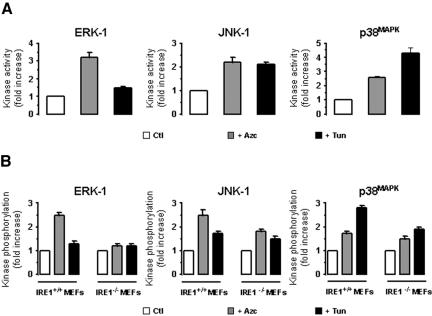
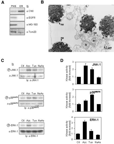
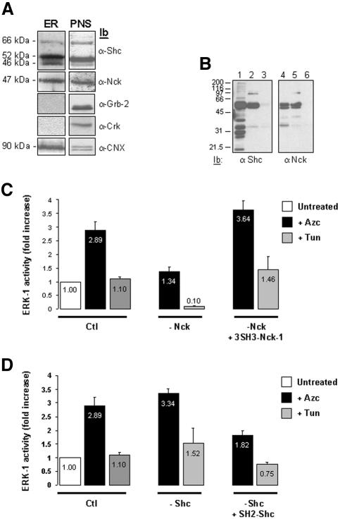
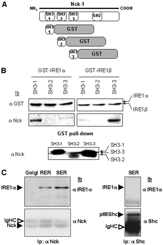
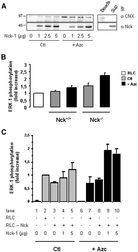
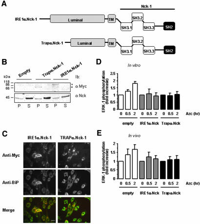
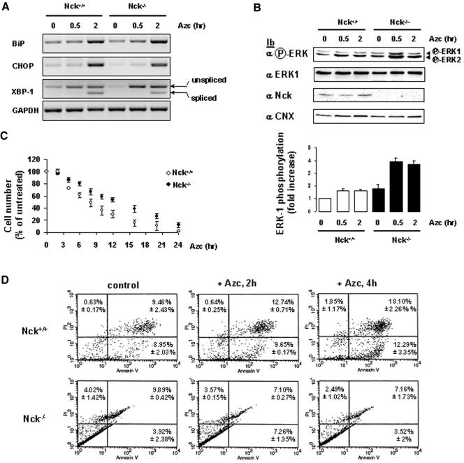
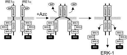
In response to stress, the endoplasmic reticulum (ER) signaling machinery triggers the inhibition of protein synthesis and up-regulation of genes whose products are involved in protein folding, cell cycle exit, and/or apoptosis. We demonstrate that the misfolding agents azetidine-2-carboxylic acid (Azc) and tunicamycin initiate signaling from the ER, resulting in the activation of Jun-N-terminal kinase, p44(MAPK)/extracellular signal-regulated kinase-1 (ERK-1), and p38(MAPK) through IRE1alpha-dependent mechanisms. To characterize the ER proximal signaling events involved, immuno-isolated ER membranes from rat fibroblasts treated with ER stress inducers were used to reconstitute the activation of the stress-activated protein kinase/mitogen-activate protein kinase (MAPK) pathways in vitro. This allowed us to demonstrate a role for the SH2/SH3 domain containing adaptor Nck in ERK-1 activation after Azc treatment. We also show both in vitro and in vivo that under basal conditions ER-associated Nck represses ERK-1 activation and that upon ER stress this pool of Nck dissociates from the ER membrane to allow ERK-1 activation. Moreover, under the same conditions, Nck-null cells elicit a stronger ERK-1 activation in response to Azc stress, thus, correlating with an enhanced survival phenotype. These data delineate a novel mechanism for the regulation of ER stress signaling to the MAPK pathway and demonstrate a critical role for Nck in ER stress and cell survival.


Figures

Figure 1.
Figure 1.
Effect of Azc and Tun on MAPK/SAPK activation. ERK-1, JNK-1 or p38MAPK activities/phosphorylation states were analyzed in FR3T3 cells, IRE1α+/+, or IRE1α–/– MEFs after 10-min treatment with 10 mM Azc or 10 μg/ml Tun. (A) JNK-1, p38MAPK, and ERK-1 were immunoprecipitated from clarified lysates obtained from FR3T3 cells, treated or not and immunoprecipitates were subjected to in vitro kinase assays with the following specific substrates: GST-Jun, GST-ATF2, and MBP respectively (n = 3, value ± SD). (B) MEF lysates were directly immunoblotted either with anti-phospho-ERK, anti-phospho-JNK-1 or anti-phospho-p38MAPK antibodies. Immunoblots were quantified by scanning densitometry (n = 2). JNK-1, p38MAPK, and ERK-1 activation are reported in A and B, respectively, as fold increase in either activity (for FR3T3 samples) or phosphorylation (for MEF samples) over control.
Figure 2.
Figure 2.
ER immuno-isolation and in vitro reconstitution of ER stress-activated signaling pathways. (A) Biochemical analysis of the immunopurified membranes by using CNX as the ER membrane trap. In each lane, total protein corresponding either to 10% of the PNS or to the total immunopurified membranes (ER) were immunoblotted with anti-CNX, anti-EGF receptor (plasma membrane marker), anti-MG-160 (Golgi marker), or anti-Tom20 (mitochondrial marker). (B) Morphological analysis of the immunopurified CNX compartment. Immunopurified membranes were treated as described under Materials and Methods. Rough membranes (RM), smooth membranes (SM), mitochondria (M), and magnetic beads (B). Bar, 0.5 μm. (C) Cells were treated with 10 mM Azc, 10 μg/ml Tun, or 50 μM NaAs for 10 min. CNX-enriched membranes were immunopurified as described under Materials and Methods. Each sample (∼1 mg of protein) was divided in three equal fractions (∼300 μg of protein each) and incubated with 3 mg of cytosol purified from rat liver. The phosphorylation level of ERK-1, JNK-1, or p38MAPK is shown (top gels); the amount of kinase was assessed by immunoblotting (bottom blots). (D) For each kinase, the ER stress-induced activity was quantified by the in vitro phosphorylation of GST-Jun, GST-ATF2 or MBP by immunoprecipitated JNK-1, p38MAPK, or ERK-1 respectively (n = 3, value ± SD).
Figure 3.
Figure 3.
Immunodepletion of cytosolic Nck alters the ER stress-mediated activation of ERK-1. (A) Immunoblotting of the ER immuno-isolated from FR3T3 cells with anti-Shc, anti-Nck, anti-Grb2, anti-Crk, or anti-CNX antibodies. A representative experiment is shown (n = 4). The same amount of total proteins corresponding either to total immuno-isolated ER membranes (ER) or 10% of the PNS were immunoblotted with anti-Shc, anti-Nck, anti-Grb-2, anti-Crk, and anti-CNX antibodies. (B) Shc and Nck immuno-depletion from cytosol. Lanes 1 and 2 and 4 and 5, subsequent immunoprecipitations of the total cytosol with anti-Shc or anti-Nck antibodies, followed by an immunoblot with respective antibodies. Lanes 3 and 6, Shc and Nck immunoblots of Shc- or Nck-immunodepleted cytosol. (C) ER microsomes immuno-isolated from nontreated, 10 mM Azc, or 10 μg/ml Tun treated cells were incubated with control RLC (Ctl), or RLC that had been previously Nck-immunodepleted (–Nck) either in the presence or absence of 10 μg of recombinant 3SH3-Nck-1 wt. ERK-1 activity is shown. Results are presented as fold increase over control (Azc, black bars; Tun, gray bars; n = 2, value ± 0.5 variation). Values in the graph bars indicate the relative ERK-1 activity. (D) Same experiment as in C except that ER microsomes were incubated with control RLC (Ctl), or RLC that had been previously Shc-immunodepleted (–Shc) either in the presence or absence of 10 μg of recombinant SH2-Shc wt. ERK-1 activity is shown. Results are presented as fold increase over control (Azc, black bars; Tun, gray bars; n = 2, value ± 0.5 variation). Values in the graph bars indicate the relative ERK-1 activity.
Figure 4.
Figure 4.
Distribution of BiP, Nck, and IRE1α in purified membrane fractions. Rough ER (RER), smooth ER (SER), ER-Golgi intermediate compartment (ERGIC) and Golgi were fractionated and purified from rat livers as reported previously (Lavoie et al., 2000). Each fraction was then immunoblotted with anti-BiP (middle), anti-Nck (bottom), or anti IRE1α (top) antibodies (n = 2).
Figure 5.
Figure 5.
In vitro association of Nck-1 SH3 domains with IRE1 cytosolic domain. (A) Representative scheme of the chimera constructed for these studies. (B) Individual SH3 domains of Nck-1, Nck-1-SH3(1), Nck-1-SH3(2), and Nck-1-SH3(3), fused to GST were cleaved with thrombin, GST and thrombin were depleted and the products were pulled down with GST-IRE1α or GST-IRE1β bound to glutathione-Sepharose beads. GST-associated proteins were immunoblotted with either anti-GST (top) or anti-Nck antibodies (middle). The imput of Nck-1-SH3(1), Nck-1-SH3(2), and Nck-1-SH3(3) domains was blotted with anti-Nck antibodies (bottom). (C) Rat liver Golgi or ER microsomes were solubilized, and clarified lysates (1 mg) were immunoprecipitated with anti-Nck antibodies. Immunoprecipitates were resolved by SDS-PAGE, transferred onto nitrocellulose membrane, and immunoblotted with either anti-IRE1α or anti-Nck antibodies (n = 3).
Figure 6.
Figure 6.
ER stress-mediated ERK-1 activation requires the association of Nck-1 with ER membranes. (A) ER immuno-isolated from Nck–/– MEFs treated or not with 10 mM Azc for 30 min was divided into four equal fractions and incubated in the absence or presence of 1, 2.5, or 5 μg of recombinant Nck-1(3SH3) wt. Magnetic beads were washed extensively and Nck-1 association to these membranes was assessed by immunoblotting by using anti-Nck (bottom left) and anti-CNX antibodies (top left). A representative immunoblot is shown (n = 4). Clarified lysate from untreated Nck–/– MEFs was incubated successively with anti-CNX antibodies and protein A-magnetic beads as described under Materials and Methods section for the immuno-isolation of ER enriched membranes, and the mixture was then incubated in the presence of 5 μg of recombinant Nck-1(3SH3) wt. After separation with a magnet, the supernatant was collected and the magnetic beads were washed extensively. The association of CNX and Nck-1 with the beads (Beads) or their presence in the incubation medium (Sup) was assessed by immunoblotting by using anti-CNX (top right) and anti-Nck (bottom right) antibodies. (B) RLC was incubated in the absence (white bar) or in the presence of ER purified from Nck+/+ MEFs or Nck–/– MEFs treated (black bars) or not (gray bars) with 10 mM Azc for 30 min. The phosphorylation state of ERK-1 was assessed as described under Materials and Methods section (n = 2, value ± 0.5 variation). (C) ER was purified from Nck–/– MEFs treated as in A. Each sample (∼1 mg of protein) was divided into five equal fractions and incubated in the absence of RLC (conditions 1 and 6), in the presence of RLC (2 and 7), in the presence of Nck-depleted RLC (conditions 3 and 8), or in the presence of 1 μg (conditions 4 and 9) or 5 μg (conditions 5 and 10) of recombinant Nck-1(3SH3) wt for 1h on ice before incubation with Nck-depleted RLC for 30 min at 30°C. Membrane and cytosolic fractions were separated with a magnet. Magnetic beads were washed extensively and immunoblotting was performed with anti-CNX and anti-Nck antibodies on the membrane fractions and with anti-ERK-1 and anti-phospho-ERK antibodies on the cytosolic fractions. Results are presented as fold increase of ERK-1 phosphorylation over ctl + RLC (n = 3, value ± SD).
Figure 7.
Figure 7.
Forced localization of Nck-1 at the ER membrane affects ER stress-mediated ERK-1 activation. (A) Representative scheme of the fusion proteins IRE1α.Nck-1 and Trapα.Nck-1 used. The luminal and transmembrane domains (gray) of IRE1α or Trapα were fused to the wild-type full-length Nck-1. These constructs were myc-tagged at the C terminus. (B) FR3T3 cells were transfected with pcDNA3, pcDNA3/IREα.Nck-1, or pcDNA3/Trapα.Nck-1, and expression of the respective recombinant protein was tested by immunoblot with anti-myc antibodies in the membrane fraction (P) and in the soluble fraction (S) (top blot). This was compared with the endogenous Nck content in both fractions by immunoblotting with anti-Nck antibodies (bottom blot). A representative immunoblot is shown (n = 3). (C) Immunofluorescence study of FR3T3 cells transfected with pcDNA3/IREα.Nck-1 or pcDNA3/Trapα.Nck-1. Cells were immuno-stained with anti-myc antibodies (top) and anti-BiP antibodies as an ER marker (middle). Costaining was detected by merging of both pictures (bottom). A representative picture is shown (n = 3). Bar, 20 μm. (D) In vitro reconstitution of ERK-1 activation in the presence of RLC was performed as described under Materials and Methods. These experiments were carried out using ER membranes purified from FR3T3 cells transfected with either pcDNA3 (empty) pcDNA3/IREα.Nck-1 or pcDNA3/Trapα.Nck-1 and treated with 10 mM Azc for 0.5 and 2 h. (n = 2, value ± 0.5 variation). (E) ERK-1 phosphorylation in FR3T3 cells transiently transfected either with pcDNA3 (empty) pcDNA3/IREα.Nck-1 or pcDNA3/Trapα.Nck-1 and treated with 10 mM Azc for 0.5 and 2 h (n = 2, value ± 0.5 variation). Lysates were directly immunoblotted with anti-phospho-ERK and anti-ERK-1 antibodies. Immunoblots were quantified by scanning densitometry (n = 2, value ± 0.5 variation).
Figure 8.
Figure 8.
Nck mediates Azc-induced apoptosis. (A) RT-PCR analysis of Bip, CHOP, XBP-1, and GAPDH mRNA expression in Nck+/+ and Nck–/– MEFs treated with 10 mM Azc for 0.5 to 2 h. A representative agarose gel of the RT-PCR assay is shown (n = 2). (B) ERK-1 phosphorylation was analyzed in Nck+/+ and Nck–/– MEFs after treatment with 10 mM Azc for 0.5 and 2 h. MEFs lysates were directly immunoblotted either with anti-phospho-ERK, anti-ERK-1, anti-Nck, and anti-CNX antibodies. Immunoblots were quantified by scanning densitometry. A representative immunoblot is shown (n = 5, value ± SD). (C) Nck+/+ and Nck–/– MEFs were treated with 10 mM Azc for 2 to 24 h. At each time point, the number of cells remaining after Azc treatment was compared with that of cells grown for the same time in Azc-free medium (n = 2, value ± 0.5 variation). (D) Nck+/+ and Nck–/– MEFs were untreated (control) or treated with 10 mM Azc for 2h (+Azc, 2 h) or 4 h (+Azc, 4 h) at which point apoptosis was determined by flow cytometry. A representative fluorescence-activated cell sorting analysis is shown (n = 2, value ± 0.5 variation).
Figure 9.
Figure 9.
Proposed model for the regulation of ERK-1 activation by Nck upon Azc stress. Under basal conditions IRE1 associates with BiP within the lumen of the ER and with Nck SH3 domains in the cytosol to prevent IRE1 signaling. On Azc stress, misfolded proteins accumulate in the lumen of the ER and as a consequence BiP dissociates from IRE1. This induces the oligomerization of IRE1 luminal domains that possibly leads to conformational change of IRE1 cytosolic domain leading to the release of IRE1-bound Nck and subsequent IRE1-mediated activation of ERK-1.

Similar articles

Cited by

References

    1. Aridor, M., and Balch, W.E. (1999). Integration of endoplasmic reticulum signaling in health and disease. Nat. Med. 5, 745–751. - PubMed
    1. Barbosa-Tessmann, I.P., Chen, C., Zhong, C., Schuster, S.M., Nick, H.S., and Kilberg, M.S. (1999). Activation of the unfolded protein response pathway induces human asparagine synthetase gene expression. J. Biol. Chem. 274, 31139–31144. - PubMed
    1. Bell, A.W., et al. (2001). Proteomics characterization of abundant Golgi membrane proteins. J. Biol. Chem. 276, 5152–5165. - PubMed
    1. Bertolotti, A., Zhang, Y., Hendershot, L.M., Harding, H.P., and Ron, D. (2000). Dynamic interaction of BiP and ER stress transducers in the unfolded-protein response. Nat. Cell Biol. 2, 326–332. - PubMed
    1. Bork, P., and Sander, C. (1993). A hybrid protein kinase-RNase in an interferon-induced pathway? FEBS Lett. 334, 149–152. - PubMed

Publication types

MeSH terms

LinkOut - more resources