Effect of thermal preconditioning before excimer laser photoablation
- PMID: 15201513
- PMCID: PMC2816848
- DOI: 10.3346/jkms.2004.19.3.437
Effect of thermal preconditioning before excimer laser photoablation
Abstract
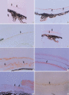
The purposes of this study were to assess the expression patterns of heat shock proteins (Hsps), after eyeball heating or cooling, and to elucidate their relationships with corneal wound healing and intraocular complications after excimer laser treatment. Experimental mice were grouped into three according to local pretreatment type: heating, cooling, and control groups. The preconditioning was to apply saline eyedrops onto the cornea prior to photoablation. Following photoablation, we evaluated corneal wound healing, corneal opacity and lens opacity. Hsp expression patterns were elucidated with Western blot and immunohistochemical staining. The heating and cooling groups recovered more rapidly, and showed less corneal and lens opacity than the control group. In the heating and cooling groups, there were more expressions of Hsps in the cornea and lens than in the control group. These results were confirmed in the Hsp 70.1 knockout mouse model. Our study showed that Hsps were induced by the heating or cooling preconditioning, and appeared to be a major factor in protecting the cornea against serious thermal damage. Induced Hsps also seemed to play an important role in rapid wound healing, and decreased corneal and lens opacity after excimer laser ablation.
Figures


Similar articles
-
The relationship between rabbit corneal opacity and immunohistochemical expression of heat shock protein 72/73 and c-fos after excimer laser photorefractive keratectomy.Ophthalmic Res. 1999;31(3):203-9. doi: 10.1159/000055533. Ophthalmic Res. 1999. PMID: 10224503
-
Excimer laser corneal surgery and free oxygen radicals.Jpn J Ophthalmol. 1996;40(2):154-7. Jpn J Ophthalmol. 1996. PMID: 8876381
-
Effect of basic fibroblast growth factor in transgenic mice: corneal epithelial healing process after excimer laser photoablation.Ophthalmologica. 2009;223(2):139-44. doi: 10.1159/000187686. Epub 2008 Dec 18. Ophthalmologica. 2009. PMID: 19092284
-
[Current status of infrared photoablation of the cornea].Klin Monbl Augenheilkd. 1999 Apr;214(4):195-202. doi: 10.1055/s-2008-1034776. Klin Monbl Augenheilkd. 1999. PMID: 10407800 Review. German.
-
Corneal wound healing relevance to wavefront guided laser treatments.Ophthalmol Clin North Am. 2004 Jun;17(2):225-31, vii. doi: 10.1016/j.ohc.2004.03.002. Ophthalmol Clin North Am. 2004. PMID: 15207564 Review.
Cited by
-
Centration axis in refractive surgery.Eye Vis (Lond). 2015 Feb 24;2:4. doi: 10.1186/s40662-015-0014-6. eCollection 2015. Eye Vis (Lond). 2015. PMID: 26605360 Free PMC article.
-
In vivo analysis of laser preconditioning in incisional wound healing of wild-type and HSP70 knockout mice with Raman spectroscopy.Lasers Surg Med. 2012 Mar;44(3):233-44. doi: 10.1002/lsm.22002. Epub 2012 Jan 24. Lasers Surg Med. 2012. PMID: 22275297 Free PMC article.
-
Thermal Injury Induces Small Heat Shock Protein in the Optic Nerve Head In Vivo.Korean J Ophthalmol. 2021 Dec;35(6):460-466. doi: 10.3341/kjo.2021.0027. Epub 2021 Oct 12. Korean J Ophthalmol. 2021. PMID: 34634865 Free PMC article.
-
Standard cross-linking versus photorefractive keratectomy combined with accelerated cross-linking for keratoconus management: a comparative study.Acta Ophthalmol. 2019 Jun;97(4):e623-e631. doi: 10.1111/aos.13986. Epub 2018 Nov 29. Acta Ophthalmol. 2019. PMID: 30499232 Free PMC article.
-
Shielding effect of the smoke plume by the ablation of excimer lasers.BMC Ophthalmol. 2018 Oct 23;18(1):273. doi: 10.1186/s12886-018-0942-8. BMC Ophthalmol. 2018. PMID: 30352572 Free PMC article.
References
-
- Marshall J, Trokel SL, Rothery S, Krueger RR. Long-term healing of the central cornea after photorefractive keratectomy using an excimer laser. Ophthalmology. 1988;95:1411–1421. - PubMed
-
- Langenbucher A, Seitz B, Kus MM, Nauman GO. Thermal effects in excimer laser trephination of the cornea. Graefes Arch for Clin Exp Ophthalmol. 1996;234(Suppl 1):S142–S148. - PubMed
-
- Bende T, Seiler T, Wollensak J. Corneal thermal gradients. Grafes Arch for Clin Exp Ophthalmol. 1988;226:277–280. - PubMed
-
- Tsubota K, Toda I, Itoh S. Reduction of subepithelial haze after photorefractive keratectomy by cooling the cornea. Am J Ophthalmol. 1993;115:820–821. - PubMed
-
- Park WC, Tseng SCG. Temperature cooling reduces keratocyte death in excimer laser ablated corneal and skin wounds. Invest Ophthmol Vis Sci. 1988;39(Suppl 1):2062.
Publication types
MeSH terms
Substances
LinkOut - more resources
Full Text Sources
Other Literature Sources

