

Intrapulmonary cystic lymphangioma in a 2-month-old infant
- PMID: 15201516
- PMCID: PMC2816851
- DOI: 10.3346/jkms.2004.19.3.458
Intrapulmonary cystic lymphangioma in a 2-month-old infant
Abstract
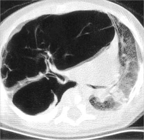
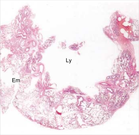
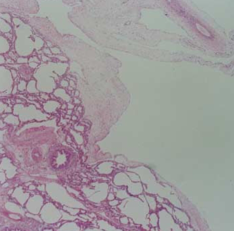
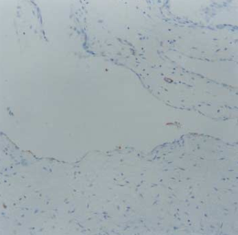
Lymphangioma is an abnormal collection of lymphatics that are developmentally isolated from the normal lymphatic system. Lymphangioma rarely presents as a solitary pulmonary lesion. We report a rare case of intrapulmonary cystic lymphangioma involving the upper lobe of the right lung, which presented with dyspnea in a 2-month-old infant. High-resolution computed tomography (HRCT) of the chest demonstrated a well-circumscribed, multiseptate, cystic lesion in the upper lobe of the right lung, mimicking the feature of type I congenital cystic adenomatoid mal-formation. The tumor was removed by bilobectomy of the upper and middle lobes of the right lung, and its pathologic examination confirmed the diagnosis of an intra-pulmonary cystic lymphangioma.
Figures
Similar articles
-
Solitary intrapulmonary cystic lymphangioma in an infant: a case report with literature review.Pathol Res Pract. 2010 Dec 15;206(12):851-6. doi: 10.1016/j.prp.2010.09.004. Epub 2010 Oct 16. Pathol Res Pract. 2010. PMID: 20952134 Review.
-
Cystic intrapulmonary lymphangioma: HRCT findings.Pediatr Radiol. 1995;25(3):206-7. doi: 10.1007/BF02021537. Pediatr Radiol. 1995. PMID: 7644305
-
Pulmonary lymphangioma.Ann Thorac Surg. 2008 Jan;85(1):336-9. doi: 10.1016/j.athoracsur.2007.07.033. Ann Thorac Surg. 2008. PMID: 18154844
-
Intrapulmonary cystic lymphangioma: report of a case.Surg Today. 1998;28(12):1310-2. doi: 10.1007/BF02482823. Surg Today. 1998. PMID: 9872557
-
[A case of intrapulmonary lymphangioma].Nihon Kokyuki Gakkai Zasshi. 1998 Feb;36(2):192-6. Nihon Kokyuki Gakkai Zasshi. 1998. PMID: 9617149 Review. Japanese.
Cited by
-
Diagnosis and treatment of diffuse pulmonary lymphangioma in children: A case report.Exp Ther Med. 2023 Mar 7;25(4):175. doi: 10.3892/etm.2023.11874. eCollection 2023 Apr. Exp Ther Med. 2023. PMID: 37006871 Free PMC article.
-
Managing pulmonary cystic Hygroma in adults: diagnostic and therapeutic considerations.Oxf Med Case Reports. 2025 Jun 27;2025(6):omaf074. doi: 10.1093/omcr/omaf074. eCollection 2025 Jun. Oxf Med Case Reports. 2025. PMID: 40585459 Free PMC article.
-
Pulmonary lymphangiectasis presenting as solitary pulmonary coin lesion.Indian J Thorac Cardiovasc Surg. 2019 Jan;35(1):81-84. doi: 10.1007/s12055-018-0722-3. Epub 2018 Aug 11. Indian J Thorac Cardiovasc Surg. 2019. PMID: 33060978 Free PMC article.
-
Experience of thoracoscopic extirpation of intrapulmonary lymphangioma.Jpn J Thorac Cardiovasc Surg. 2005 Jun;53(6):313-6. doi: 10.1007/s11748-005-0135-2. Jpn J Thorac Cardiovasc Surg. 2005. PMID: 15997754
References
-
- Faul JL, Berry GJ, Colby TV, Ruoss SJ, Walter MB, Rosen GD, Raffin TA. Thoracic lymphangiomas, lymphangiectasis, lymphangiomatosis, and lymphatic dysplasia syndrome. Am J Respir Crit Care Med. 2000;161:1037–1046. - PubMed
-
- Enzinger FM, Weiss SW. Tumors of lymph vessels. In: Gay SM, editor. Soft tissue tumors. St. Louis: Mosby; 1995. pp. 679–700.
-
- Tazelaar HD, Kerr D, Yousem SA, Saldana MJ, Langston C, Colby TV. Diffuse pulmonary lymphangiomatosis. Hum Pathol. 1993;24:1313–1322. - PubMed
-
- Shaffer K, Rosado-de-Christenson ML, Patz EF, Jr, Young S, Farver CF. Thoracic lymphangioma in adults: CT and MR imaging features. AJR Am J Roentgenol. 1994;162:283–289. - PubMed
-
- Wilson C, Askin FB, Heitmiller RF. Solitary pulmonary lymphangioma. Ann Thorac Surg. 2001;71:1337–1338. - PubMed
Publication types
MeSH terms
LinkOut - more resources
Full Text Sources
Medical
Research Materials
