. 2004 Jul 7;23(13):2674-83.
doi: 10.1038/sj.emboj.7600269. Epub 2004 Jun 17.

Mdc1 couples DNA double-strand break recognition by Nbs1 with its H2AX-dependent chromatin retention

Affiliations

Mdc1 couples DNA double-strand break recognition by Nbs1 with its H2AX-dependent chromatin retention

Claudia Lukas et al. EMBO J. .

Abstract

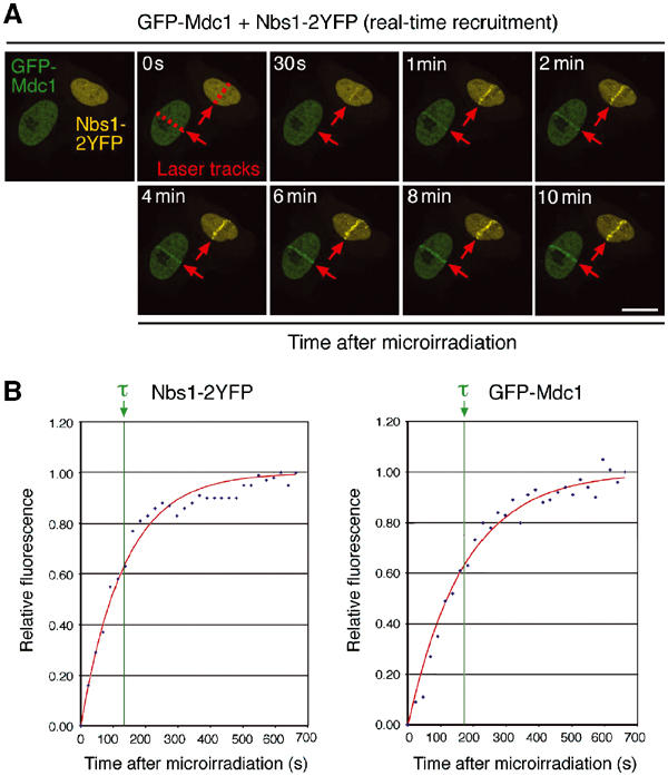
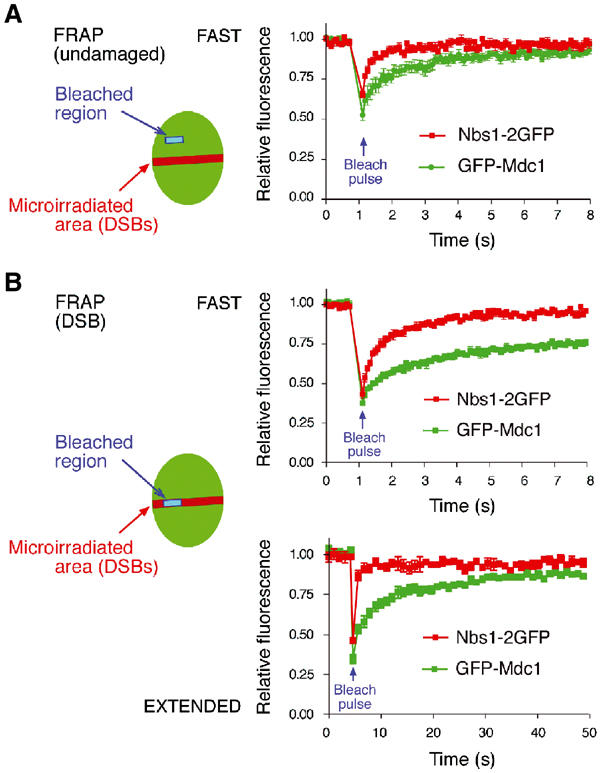
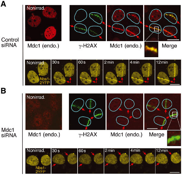
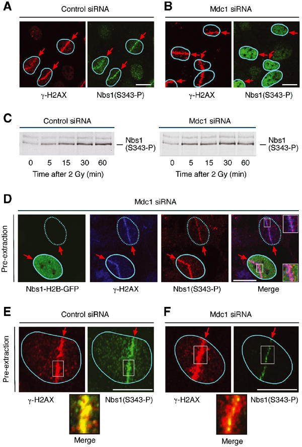
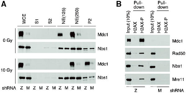
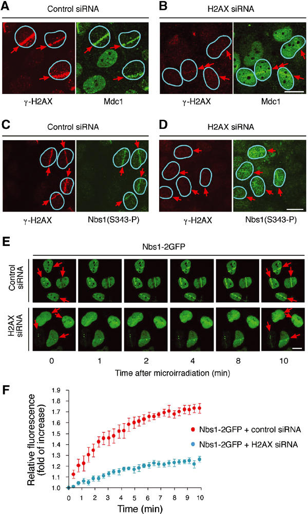
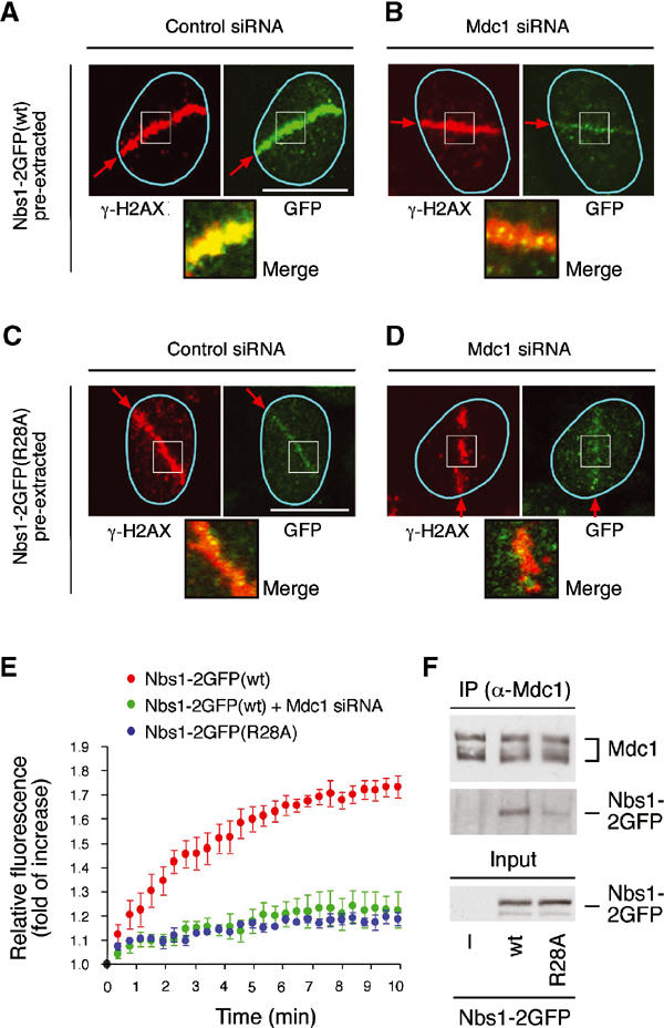
Mdc1/NFBD1 controls cellular responses to DNA damage, in part via interacting with the Mre11-Rad50-Nbs1 complex that is involved in the recognition, signalling, and repair of DNA double-strand breaks (DSBs). Here, we show that in live human cells, the transient interaction of Nbs1 with DSBs and its phosphorylation by ATM are Mdc1-independent. However, ablation of Mdc1 by siRNA or mutation of the Nbs1's FHA domain required for Mdc1 binding reduced the affinity of Nbs1 for DSB-flanking chromatin and caused aberrant pan-nuclear dispersal of Nbs1. This occurred despite normal phosphorylation of H2AX, indicating that lack of Mdc1 does not impair this DSB-induced chromatin change, but rather precludes the sustained engagement of Nbs1 with these regions. Mdc1 (but not Nbs1) became partially immobilized to chromatin after DSB generation, and siRNA-mediated depletion of H2AX prevented such relocalization of Mdc1 and uncoupled Nbs1 from DSB-flanking chromatin. Our data suggest that Mdc1 functions as an H2AX-dependent interaction platform enabling a switch from transient, Mdc1-independent recruitment of Nbs1 to DSBs towards sustained, Mdc1-dependent interactions with the surrounding chromosomal microenvironment.


Figures

Figure 1
Figure 1
Real-time recruitment of Nbs1 and Mdc1 to the microlaser-generated sites of DNA damage. (A) U-2-OS cells expressing Nbs1-2YFP or GFP-Mdc1 were mixed (1:1) and co-cultivated for 24 h. The YFP and GFP signals were unmixed through the spectral analyser to discriminate the Nbs1- and Mdc1-expressing cells. The cells were microirradiated with a laser beam along 0.5–1-μm-wide tracks spanning the entire nuclear diameter, and the redistribution of the respective fluorophore-tagged proteins was recorded by a repeated scanning of the same field at 20 s intervals. Selected time points covering the earliest signs of Nbs1 and/or Mdc1 recruitment up to their stabilized interaction with DSB are shown. The arrows indicate the laser movement during microirradiation. Scale bars=10 μm. (B) Kinetic curves of Nbs1 and Mdc1 recruitment to laser-generated, DSB-containing nuclear compartments. The data show examples of normalized data from one cell expressing Nbs1-2YFP and GFP-Mdc1, fitted to a model for the first-order response to a step change (step=DSB generation; Supplementary information 1). The value of the calculated recruitment time constant (τ) is indicated by a green line.
Figure 2
Figure 2
Distinct intranuclear movement of Nbs1 and Mdc1 before and after DNA damage. U-2-OS cells expressing Nbs1-2GFP or GFP-Mdc1 were microirradiated and 10 min later subjected to the FRAP analysis (Supplementary information 1). The positions of the microirradiated (red stripes) and the bleached regions (blue boxes) in undamaged nucleoplasm (A) and in the DSB-containing nuclear compartments (B) are schematically indicated. The total duration of image recording in the postbleached period is indicated by FAST (10 s) and EXTENDED (60 s). The latter protocol was used to achieve the maximum recovery of both Nbs1 and Mdc1 in the microirradiated nuclear compartments. The data integrate measurements form 10 independent cells for each nuclear location.
Figure 3
Figure 3
Decreased affinity of Nbs1 to the DSB-containing nuclear compartments in cells lacking Mdc1. U-2-OS cells were treated with control (A, top panel) and the Mdc1-targeting (B, top panel) siRNA oligonucleotides for 96 h. The nuclei were then subjected to local microirradiation, fixed 10 min later, and immunostained with antibodies to γ-H2AX (green) and total Mdc1 (red). The insets in (A, B) show a higher magnification of the respective microirradiated areas. In parallel, U-2-OS cells stably expressing Nbs1-2YFP were treated with control siRNA (A, bottom panel) or Mdc1-targeting siRNA (B, bottom panel) and analysed by the in vivo real-time recruitment assay as described in Figure 1. The arrows indicate the laser movement during microirradiation. Scale bars=10 μm.
Figure 4
Figure 4
Impaired redistribution of ATM-phosphorylated Nbs1 in Mdc1-depleted cells. (A, B) U-2-OS cells were treated with control (A) and Mdc1-specific (B) siRNA oligonucleotides for 96 h, microirradiated, incubated for 10 min, fixed, and co-immunostained with phospho-specific antibodies to γ-H2AX and Nbs1(S343). (C) U-2-OS cells were transfected with control or Mdc1-specific siRNA duplexes for 96 h and exposed to IR (2 Gy). At the indicated time points, the cell lysates were analysed by immunoblotting with the phospho-specific antibody to Nbs1(S343). (D) U-2-OS cells were treated with Mdc1-specific siRNA oligonucleotides for 72 h, microinjected with the Nbs1-H2B-GFP expression plasmid (10 μg/ml), incubated for additional 36 h, pre-extracted to remove the bulk of endogenous Nbs1 (Materials and methods), fixed, and immunostained with the indicated antibodies. Note the stronger, DSB-restricted Nbs1-S343 phosphorylation in the cell nucleus expressing the immobile form of Nbs1 (marked by solid circle) compared to the neighbouring control nucleus (dashed circle). The insets show enlargements of the respective segments in the merged image. (E, F) U-2-OS cells were treated with control (E) or Mdc1-targeting (F) siRNA oligonucleotides, and microirradiated as in (A, B). After 10 min, the cells were pre-extracted for 5 min (Materials and methods), fixed, and co-immunostained with the indicated phospho-specific antibodies. The insets show magnified microirradiated fields. The arrows indicate the laser movement during microirradiation. Scale bars=10 μm.
Figure 5
Figure 5
(A) Mdc1 but not Nbs1 chromatin binding increases upon induction of DSBs. HeLa cells stably expressing Mdc1-targeting (M) or a LacZ (Z) control shRNA were exposed to 10 Gy of IR. After 1 h, the cell lysates were fractionated (Materials and methods) to separate cytoplasmic (S1) and soluble nuclear (S2) proteins from chromatin. The chromatin pellet was subsequently washed with increasing salt concentrations (125 and 250 mM, respectively), and the remaining pellet (P2) was resuspended in SDS sample buffer and briefly sonicated to release the chromatin-bound proteins. All fractions were analysed by immunoblotting with antibodies to the indicated proteins. (B) Binding of MRN to γ-H2AX requires Mdc1. Nuclear extracts from HeLa cells stably expressing Mdc1-targeting (M) or a LacZ (Z) control shRNA were incubated with phosphorylated (H2AX-P) or nonphosphorylated (H2AX) peptides (Materials and methods). The bound proteins were analysed by immunoblotting with the indicated antibodies.
Figure 6
Figure 6
Transient ablation of H2AX impairs Mdc1 recruitment to DSBs and attenuates sustained interaction of Nbs1 with the damaged nuclear compartments. (A, B) U-2-OS cells were treated with control (A) and the H2AX-targeting (B) siRNA oligonucleotides for 96 h, microirradiated, and after additional 10 min fixed and co-immunostained with phospho-specific antibodies to γ-H2AX and total Mdc1. (C, D) U-2-OS cells were treated as in (A, B), and co-immunostained with phospho-specific antibodies to γ-H2AX and Nbs1(S343). (E) U-2-OS cells stably expressing Nbs1-2GFP were treated with control siRNA or H2AX-targeting siRNA as indicated and analysed by the in vivo real-time recruitment assay (Figure 1). The arrows indicate the laser movement during microirradiation. Scale bars=10 μm. (F) Quantification of the real-time recruitment data obtained in (E). The graph integrates the data from 10 cells for each setting and shows the fold of increase of relative GFP-associated fluorescence in the microirradiated areas during the first 10 min after the laser treatment.
Figure 7
Figure 7
Disruption of the FHA domain uncouples Nbs1 from the γ-H2AX-modified chromatin regions. (A, B) U-2-OS cells stably expressing the wild-type (wt) form of Nbs1-2GFP were treated with control (A) or Mdc1-targeting (B) siRNA for 96 h, microirradiated, pre-extracted fixed after 10 min, and immunostained with phospho-specific antibodies to γ-H2AX. (C, D) U-2OS cells expressing the FHA-deficient (R28A) form of Nbs1-2GFP were treated with siRNA oligonucleotides as indicated and processed as in (A, B). (E) U-2-OS cells expressing the indicated forms of Nbs1-2GFP were microirradiated and subjected to the kinetic measurement of their DSB recruitment as described in Figure 1. Where indicated, endogenous Mdc1 was depleted by the siRNA oligonucleotides for 96 h before microirradiation. The graph integrates the data from 10 cells for each setting and shows the fold of increase of relative GFP-associated fluorescence in the microirradiated areas during the first 10 min after the laser treatment. (F) U-2-OS cells were transfected with expression plasmids coding for wild-type or R28A forms of Nbs1-2GFP. At 24 h after transfection, the cells were exposed to 2 Gy of ionizing radiation and cultured for additional 1 h. The cell lysates were immunoprecipitated with anti-Mdc1 antibody and immunoblotted with antibodies to Mdc1 or Nbs1 as indicated. The arrows indicate the laser movement during microirradiation. Scale bars=10 μm.

Similar articles

Cited by

References

    1. Bakkenist CJ, Kastan MB (2003) DNA damage activates ATM through intermolecular autophosphorylation and dimer dissociation. Nature 421: 499–506 - PubMed
    1. Brummelkamp TR, Bernards R, Agami R (2002) A system for stable expression of short interfering RNAs in mammalian cells. Science 296: 550–553 - PubMed
    1. Carson CT, Schwartz RA, Stracker TH, Lilley CE, Lee DV, Weitzman MD (2003) The Mre11 complex is required for ATM activation and the G(2)/M checkpoint. EMBO J 22: 6610–6620 - PMC - PubMed
    1. Celeste A, Fernandez-Capetillo O, Kruhlak MJ, Pilch DR, Staudt DW, Lee A, Bonner RF, Bonner WM, Nussenzweig A (2003) Histone H2AX phosphorylation is dispensable for the initial recognition of DNA breaks. Nat Cell Biol 5: 675–679 - PubMed
    1. Cerosaletti KM, Concannon P (2003) Nibrin forkhead-associated domain and breast cancer C-terminal domain are both required for nuclear focus formation and phosphorylation. J Biol Chem 278: 21944–21951 - PubMed

Publication types

MeSH terms