. 2004 Jul 7;23(13):2608-19.
doi: 10.1038/sj.emboj.7600275. Epub 2004 Jun 17.

Binding of the 7SK snRNA turns the HEXIM1 protein into a P-TEFb (CDK9/cyclin T) inhibitor

Affiliations

Binding of the 7SK snRNA turns the HEXIM1 protein into a P-TEFb (CDK9/cyclin T) inhibitor

Annemieke A Michels et al. EMBO J. .

Abstract

The positive transcription elongation factor b (P-TEFb) plays a pivotal role in productive elongation of nascent RNA molecules by RNA polymerase II. Core active P-TEFb is composed of CDK9 and cyclin T. In addition, mammalian cell extracts contain an inactive P-TEFb complex composed of four components, CDK9, cyclin T, the 7SK snRNA and the MAQ1/HEXIM1 protein. We now report an in vitro reconstitution of 7SK-dependent HEXIM1 association to purified P-TEFb and subsequent CDK9 inhibition. Yeast three-hybrid tests and gel-shift assays indicated that HEXIM1 binds 7SK snRNA directly and a 7SK snRNA-recognition motif was identified in the central part of HEXIM1 (amino acids (aa) 152-155). Data from yeast two-hybrid and pull-down assay on GST fusion proteins converge to a direct binding of P-TEFb to the HEXIM1 C-terminal domain (aa 181-359). Consistently, point mutations in an evolutionarily conserved motif (aa 202-205) were found to suppress P-TEFb binding and inhibition without affecting 7SK recognition. We propose that the RNA-binding domain of HEXIM1 mediates its association with 7SK and that P-TEFb then enters the complex through association with HEXIM1.


Figures
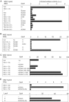
Figure 1
Figure 1
7SK snRNA interacts with HEXIM1 in yeast three-hybrid assays. β-Galactosidase activities in yeast three-hybrid (A, C, D), modified three-hybrid (B) and two-hybrid (D) assays. Proteins fused to the Gal4 activation or LexA DNA-binding domains and RNA fused to the MS2-binding sites are indicated in the left panels. Pools of more than 10 transformed yeast colonies grown on selective medium were assayed for β-galactosidase activities and quantified in arbitrary units (a.u.). Interaction between HIV TAR RNA and Tat was used as a positive control. (A) Three-hybrid assay testing truncated 7SK RNAs for interaction with full-length CDK9, cyclin T1 or HEXIM1. (B) Modified three-hybrid testing the capacity of HEXIM1 to link 7SK RNA to CDK9 or cyclin T1. The additional genuine protein is indicated in the central column. (C) Three-hybrid assay testing 7SK(1–175) for interaction with HEXIM1 deletion mutants. (D) Three-hybrid and two-hybrid assays testing 7SK(1–175) and cyclin T1 respectively for interaction with HEXIM1(ILAA).
Figure 2
Figure 2
In vitro binding of HEXIM1 to P-TEFb requires 7SK snRNA. Proteins were detected by Western blot with anti-CDK9, anti-cyclin T1 or anti-HEXIM1 antibodies. (A) Left: Glutathione beads coated with GST-HEXIM1(1–359) were incubated with cell extracts in the presence of increasing concentrations of 7SK (0–80 nM) or U2 RNA (0–160 nM). Right: RNase A was added (+) or not (−) to HEXIM1/7SK/P-TEFb complexes preformed on the beads. Inputs (I), supernatants (S) and beads (B) were probed for cyclin T1 and CDK9. (B) An extract of actinomycin-treated HeLa cells was used to immunoprecipitate P-TEFb with anti-cyclin T1, which was incubated with purified His10-HEXIM1 and increasing concentrations of 7SK (nM) or U2 (nM) RNA.
Figure 3
Figure 3
The KHRR motif is involved in in vitro and in vivo formation of the P-TEFb/HEXIM1/7SK RNA complex. (A) Pull-down assay of GST, GST-HEXIM1 WT (WT) and GST-HEXIM1(ILAA) with (+) or without (−) addition of 7SK RNA (80 nM). GST (fusion) proteins bound to glutathione beads were probed with anti-GST. (B) HeLa cells transiently transfected with Flag-HEXIM1(WT), Flag-HEXIM1(ILAA), Flag-HEXIM1(150–359) or Flag-HEXIM1(156–359) were processed for immunofluorescence with anti-Flag antibodies. Nuclei were stained with DAPI. (C) HeLa cells transfected with Flag-HEXIM1(WT), Flag-HEXIM1(ILAA), Flag-HEXIM1(150–359) or Flag-HEXIM1(156–359) or an empty vector (control) were treated (+) or not (−) with actinomycin D (ActD), lysed and immunoprecipitated with anti-Flag antibodies. Proteins in the extracts (inputs) or immunoprecipitated (beads) were probed with anti-Flag, anti-cyclin T1 and anti-CDK9 antibodies. 7SK RNA was detected by Northern blot.
Figure 4
Figure 4
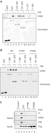
Involvement of the PYNT motif in HEXIM1 C-terminal domain in in vivo and in vitro binding to P-TEFb. (A) Glutathione beads coated with GST-HEXIM1 full-length (WT) or truncated proteins were incubated with cell extracts with (+) or without (−) 80 nM 7SK RNA. GST-HEXIM1 proteins bound to the beads were detected by Coomassie blue staining. (B) Full-length (1–359) or truncated (181–359) GST-HEXIM proteins with or without the PYND or PDND mutation were tested for binding to P-TEFb in the presence (+) and absence (−) of 80 nM 7SK. (C) Cells transfected with empty vector (control) or Flag-HEXIM1 with or without the PYND or PDND mutations were treated (+) or not (−) with actinomycin D (ActD), lysed and immunoprecipitated with anti-Flag. Beads were probed for cyclin T1, CDK9 and 7SK. Extracts were probed with anti-Flag (inputs).
Figure 5
Figure 5
Binding of P-TEFb to GST-HEXIM1 represses its kinase activity. P-TEFb from a cell extract was retained on glutathione beads coated with WT (WT) or truncated (181–359) GST-HEXIM1 with (−) or without (+) 80 nM 7SK. As a control, P-TEFb was immunoprecipitated from a cell extract with anti-cyclin T1 (IP). (A) P-TEFb retained on beads was analyzed using anti-cyclin T1 and CDK9 antibodies. For an accurate comparison, increasing amounts of material (μl) were loaded on the gels. (B) The reactions were analyzed by SDS–PAGE using autoradiography. (C) 32P incorporation into the CTD4 peptide was quantified in arbitrary units (a.u.) and plotted versus time (min). This experiment was performed three times with similar results. A typical experiment is shown.
Figure 6
Figure 6
Binding of HEXIM1 to P-TEFb represses its kinase activity. P-TEFb immunoprecipitated with anti-cyclin T1 was incubated with buffer (control), or recombinant histidine-tagged HEXIM1, or T7-transcribed 7SK RNA (80 nM), or a combination of both for 1 h at 21°C. Kinase activity was assayed on the beads as in Figure 5. When indicated, RNase A was added to the kinase assay. This experiment was performed twice with similar results. A typical experiment is shown.
Figure 7
Figure 7
Analysis of P-TEFb inhibition by 7SK and HEXIM1 in a defined system. Kinase assays were performed with recombinant P-TEFb and HEXIM1 proteins, and T7-transcribed 7SK RNA. The reactions were analyzed by SDS–PAGE using autoradiography (inset) and quantified. (A) Using DSIF as substrate, the kinase reactions contained increasing amounts of a mixture of HEXIM1 and 7SK (8:1 molar ratio) and P-TEFb (containing cyclin T1 or T2a). (B) Using RNAPII as substrate, kinase assays were performed with cyclin T2a-containing P-TEFb and the indicated amounts of HEXIM1 and/or 7SK. When HEXIM1 and 7SK are both present, they are in an equal molar ratio with the indicated amounts of each individually. (C) Using RNAPII as substrate, kinase assays with cyclin T2a-containing P-TEFb and the indicated HEXIM1 proteins and/or 7SK were performed. HEXIM1 WT and mutant proteins (and 7SK) were present at either 5 or 2 pmol per reaction.
Figure 8
Figure 8
EMSA analysis of HEXIM1, P-TEFb and 7SK interactions. HEXIM1 alone or when combined with P-TEFb shifts radioactive 7SK RNA. (A) Reactions contained indicated amounts of WT HEXIM1, about 1.5 pmol of P-TEFb, 150 ng of anti-CDK9 or affinity-purified anti-HEXIM1. (B) Indicated amounts of cold 7SK were added either during (D, lanes 2–7) or after (A, lanes 8–13) the formation of the complexes. Radioactive 7SK/HEXIM1 complexes were allowed to form first, and cold 7SK was added next with P-TEFb (asterisk, lanes 14–16). (C) Comparison between WT (WT) HEXIM1 or mutant proteins. Reactions contained 0.5 pmol of WT (WT) HEXIM1 or mutant proteins, about 1.5 pmol of P-TEFb. (D) DTT (5 mM) was added where indicated (lanes 13–17). Reactions contained 0.5 pmol of WT (WT) HEXIM1 or mutant proteins, about 1.5 pmol of P-TEFb, 150 ng of anti-CDK9 and 5 mM DTT as indicated (lanes 13–17).
Figure 9
Figure 9
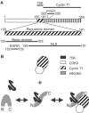
(A) HEXIM1 functional domains. Positions of amino acids are indicated. (B) Model: HEXIM1 binds 7SK RNA first and subsequently associates and inactivates P-TEFb. These processes are regulated in vivo.

Similar articles

Cited by

References

    1. Bieniasz PD, Grdina TA, Bogerd HP, Cullen BR (1998) Recruitment of a protein complex containing Tat and cyclin T1 to TAR governs the species specificity of HIV-1 Tat. EMBO J 17: 7056–7065 - PMC - PubMed
    1. Chen R, Yang Z, Zhou Q (2004) Phosphorylated P-TEFb is tagged for inhibition through association with 7SK snRNA. J Biol Chem 279: 4153–4160 - PubMed
    1. Clemens MJ (1997) PKR—a protein kinase regulated by double-stranded RNA. Int J Biochem Cell Biol 29: 945–949 - PubMed
    1. Deleault NR, Lucassen RW, Supattapone S (2003) RNA molecules stimulate prion protein conversion. Nature 425: 717–720 - PubMed
    1. Fraldi A, Licciardo P, Majello B, Giordano A, Lania L (2001) Distinct regions of cyclinT1 are required for binding to CDK9 and for recruitment to the HIV-1 Tat/TAR complex. J Cell Biochem 36: 247–253 - PubMed

Publication types

MeSH terms