Visual recognition of faces, objects, and words using degraded stimuli: where and when it occurs
- PMID: 15202108
- PMCID: PMC6872030
- DOI: 10.1002/hbm.20039
Visual recognition of faces, objects, and words using degraded stimuli: where and when it occurs
Abstract
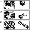
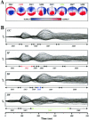
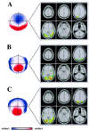
We studied time course and cerebral localisation of word, object, and face recognition using event-related potentials (ERPs) and source localisation techniques. To compare activation rates of these three categories, we used degraded images that easily pop out without any change in the physical features of the stimuli, once the meaning is revealed. Comparisons before and after identification show additional periods of activation beginning at 100 msec for faces and at around 200 msec for objects and words. For faces, this activation occurs predominantly in right temporal areas, whereas for objects, the specific time period gives rise to bilateral posterior but right dominant foci. Finally, words show a maximum area of activation in the left temporooccipital area at their specific time period. These results provide unequivocal evidence that when effects of low-level visual features are circumvented, faces, objects, and words are not only distinct in terms of their anatomic routes, but also in terms of their times of processing.
Copyright 2004 Wiley-Liss, Inc.
Figures


Similar articles
-
Early lateralization and orientation tuning for face, word, and object processing in the visual cortex.Neuroimage. 2003 Nov;20(3):1609-24. doi: 10.1016/j.neuroimage.2003.07.010. Neuroimage. 2003. PMID: 14642472 Clinical Trial.
-
Women are better at seeing faces where there are none: an ERP study of face pareidolia.Soc Cogn Affect Neurosci. 2016 Sep;11(9):1501-12. doi: 10.1093/scan/nsw064. Epub 2016 May 5. Soc Cogn Affect Neurosci. 2016. PMID: 27217120 Free PMC article.
-
Source analysis of the N170 to faces and objects.Neuroreport. 2004 Jun 7;15(8):1261-5. doi: 10.1097/01.wnr.0000127827.73576.d8. Neuroreport. 2004. PMID: 15167545
-
Words in the brain's language.Behav Brain Sci. 1999 Apr;22(2):253-79; discussion 280-336. Behav Brain Sci. 1999. PMID: 11301524 Review.
-
Domain Specificity vs. Domain Generality: The Case of Faces and Words.Vision (Basel). 2023 Dec 21;8(1):1. doi: 10.3390/vision8010001. Vision (Basel). 2023. PMID: 38535755 Free PMC article. Review.
Cited by
-
Temporal Tuning of Word- and Face-selective Cortex.J Cogn Neurosci. 2016 Nov;28(11):1820-1827. doi: 10.1162/jocn_a_01002. Epub 2016 Jul 5. J Cogn Neurosci. 2016. PMID: 27378330 Free PMC article.
-
Early and late cortical responses to directly gazing faces are task dependent.Cogn Affect Behav Neurosci. 2018 Aug;18(4):796-809. doi: 10.3758/s13415-018-0605-5. Cogn Affect Behav Neurosci. 2018. PMID: 29736681
-
Developmental changes in visual object recognition between 18 and 24 months of age.Dev Sci. 2009 Jan;12(1):67-80. doi: 10.1111/j.1467-7687.2008.00747.x. Dev Sci. 2009. PMID: 19120414 Free PMC article.
-
Stimulus dependency of object-evoked responses in human visual cortex: an inverse problem for category specificity.PLoS One. 2012;7(2):e30727. doi: 10.1371/journal.pone.0030727. Epub 2012 Feb 17. PLoS One. 2012. PMID: 22363479 Free PMC article.
-
Changes in visual object recognition precede the shape bias in early noun learning.Front Psychol. 2012 Dec 3;3:533. doi: 10.3389/fpsyg.2012.00533. eCollection 2012. Front Psychol. 2012. PMID: 23227015 Free PMC article.
References
-
- Allison T, McCarthy G, Nobre A, Puce A, Belger A (1994): Human extrastriate visual cortex and the perception of faces, words, numbers, and colors. Cereb Cortex 4: 544–554. - PubMed
-
- Allison T, Puce A, Spencer DD, McCarthy G (1999): Electrophysiological studies of human face perception. I: Potentials generated in occipitotemporal cortex by face and non‐face stimuli. Cereb Cortex 9: 415–430. - PubMed
-
- Bar M, Tootell RB, Schacter DL, Greve DN, Fischl B, Mendola JD, Rosen BR, Dale AM (2001): Cortical mechanisms specific to explicit visual object recognition. Neuron 29: 529–535. - PubMed
-
- Beauchamp MS, Lee KE, Haxby JV, Martin A (2002): Parallel visual motion processing streams for manipulable objects and human movements. Neuron 34: 149–159. - PubMed
Publication types
MeSH terms
LinkOut - more resources
Full Text Sources

