. 2004 Sep 15;382(Pt 3):803-10.
doi: 10.1042/BJ20040122.

Exchange protein directly activated by cAMP (EPAC) interacts with the light chain (LC) 2 of MAP1A

Affiliations

Exchange protein directly activated by cAMP (EPAC) interacts with the light chain (LC) 2 of MAP1A

Maria M Magiera et al. Biochem J. .

Abstract

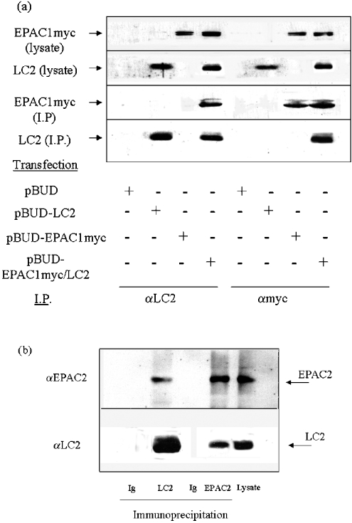
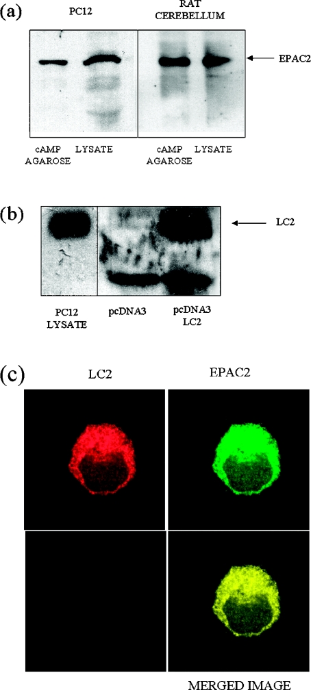
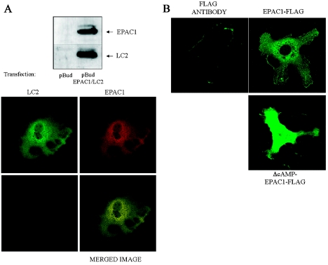
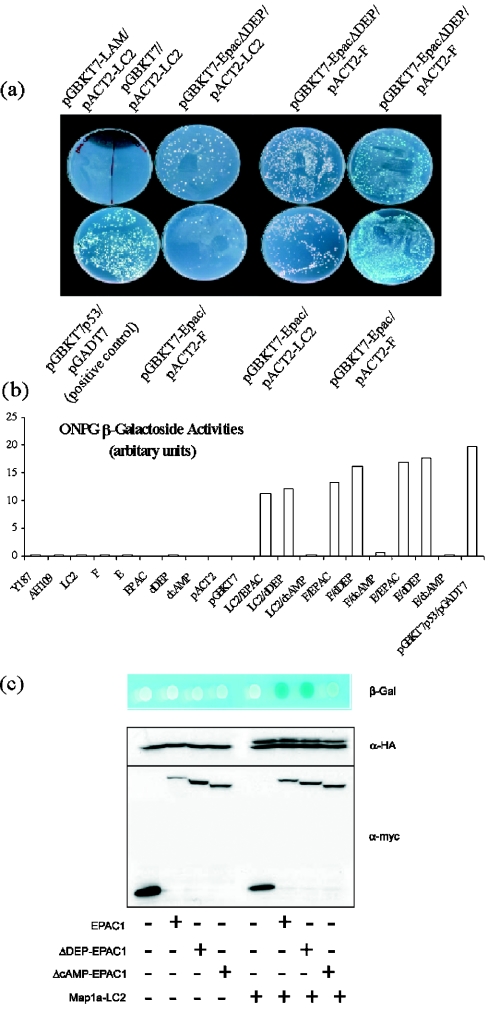
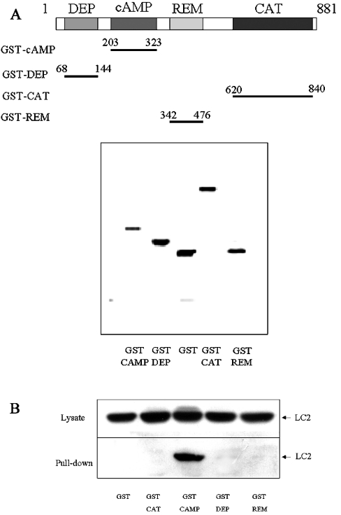
Using EPAC1 (exchange protein directly activated by cAMP 1) as bait in two-hybrid screens of foetal and adult human brain libraries, we identified the LC2 (light chain 2) of MAP1A (microtubule-associated protein 1A) as a protein capable of interaction with EPAC1. We applied an immunoprecipitation assay to demonstrate protein interaction between EPAC1 and LC2 in co-transfected human embryonic kidney 293 cells. EPAC2 also co-immunoprecipitated with LC2 from extracts of rat cerebellum. Immunolocalization in co-transfected human embryonic kidney 293 cells revealed that EPAC1 co-localizes with LC2 throughout the cell body. We found that endogenous EPAC2 is also immunolocalized with LC2 in PC12 cells. Immunolocalization of EPAC1 in transfected COS1 cells showed that EPAC1 is associated with the perinuclear region surrounding the nucleus and filamentous structures throughout the cell. Removal of the cAMP-binding domain of EPAC1 (DeltacAMP-EPAC1) appeared to disrupt targeting of EPAC1 in cells resulting in a more dispersed staining pattern. Using two-hybrid assay, we tested the ability of LC2 to interact with DeltacAMP-EPAC1 and DeltaDEP-EPAC1, which lacks a DEP domain (dishevelled, Egl-10 and pleckstrin homology domain). We found that deletion of the cAMP-binding domain inhibited interaction between EPAC1 and LC2 in a two-hybrid assay, but removal of the DEP domain had little effect. LC2 was found to interact with a glutathione-S-transferase-fusion protein of the cAMP-binding domain of EPAC1 in a pull-down assay, but not the DEP, REM (Ras exchange motif) or CAT (catalytic) domains. Together with our two-hybrid results, this suggests that the cAMP-binding domain of EPAC1 mediates interaction with LC2.


Figures

Figure 1
Figure 1. Co-immunoprecipitation of LC2 with EPAC1 and EPAC2
(a) HEK-293 cells were transfected with pBUD, pBUD-LC2, pBUD-EPAC1 or pBUD-EPAC1/LC2 vector, which allows co-expression of myc-tagged EPAC1 and LC2. Cell extracts were prepared and immunoprecipitated with IgG, LC2 or myc antibodies, as indicated. Immunoprecipitates and samples of cell extract were then separated by SDS/PAGE and immunoblotted. The upper half of immunoblots was probed with anti-myc antibody and the lower half with anti-LC2. EPAC1 and LC2 were found to precipitate with both myc and LC2 antibodies. (b) Rat cerebellum lysate was subjected to immunoprecipitation with IgG, the EPAC2 antibody or the LC2 antibody. Samples of rat cerebellum lysate and immunoprecipitates were then subjected to SDS/PAGE. The upper half of the gel was immunoblotted with the EPAC2-specific antibody. The lower half of the gel was immunoblotted with the LC2-specific antibody, and the position of the 25 kDa LC2 species is indicated. EPAC2 and LC2 were found to precipitate with both the EPAC2 and LC2 antibodies.
Figure 2
Figure 2. Expression and immunolocalization of EPAC2 and LC2 in PC12 cells
(a) Lysates were prepared from rat cerebellum (right panel) and PC12 cells (left panel) and then precipitated with cAMP-agarose beads. Samples of cell lysate and cAMP-agarose bead precipitates were separated by SDS/PAGE and probed with an anti-EPAC2 polyclonal antibody. Immunoblotting revealed the presence a major immunoreactive species of approx. 120 kDa (EPAC2) within PC12 and rat cerebellum cell lysates and associated with the cAMP-agarose beads. (b) Recombinant LC2 was synthesized from pcDNA3-LC2 by coupled, in vitro transcription/translation (see the Experimental section) and separated by SDS/PAGE together with PC12 cell lysate. A control transcription/translation reaction was also subjected to SDS/PAGE, using pcDNA3 as a template. Immunoblots were probed with the LC2 antibody, demonstrating the presence of LC2 in PC12 lysates, which co-migrated with recombinant LC2 at 25 kDa. (c) Laser scans of confocal optical sections were taken through PC12 cells that had been probed with an LC2-specific polyclonal antibody (detected with an anti-rabbit IgG Texas Red conjugate) and an EPAC2-specific polyclonal antibody (detected with an anti-goat FITC conjugate). The intracellular distributions of EPAC2 and LC2 were very similar, as indicated by the yellow colour of overlaid images.
Figure 3
Figure 3. Localization of EPAC1 in COS1 cells and immunolocalization of EPAC1 with LC2 in HEK-293 cells
(A) HEK-293 cells were transfected with pBUD-EPAC1/LC2 vector, which allows co-expression of myc-tagged EPAC1, which can be detected by an anti-myc antibody, and LC2, detected with the LC2 antibody, as shown in the upper panel. Individual cells transfected with pBUD-EPAC1/LC2 were probed with a myc-specific monoclonal antibody to detect transfected EPAC1 (detected with an anti-mouse IgG Texas Red conjugate) and the LC2-specific polyclonal antibody to detect transfected LC2 (detected with an anti-goat FITC conjugate), and then analysed with an immunofluorescent microscope (lower panel). The LC2 and EPAC1 frames are given together as a superimposed image shown in the panel on the bottom right. In all the treatments applied, the immunofluorescent staining patterns of both LC2 and EPAC1 were very similar in all the images taken. In each case, this yielded a uniform yellow image indicative of a high degree of co-localization seen in the three different transfection studies. Analyses were repeated using different combinations of fluorescent-labelled antisera with similar results. (B) COS1 cells were transfected with myc-EPAC1-FLAG or constitutively active myc-ΔcAMP-EPAC1-FLAG (which lacks the cAMP-binding domain, amino acids 203–323) and then probed with an anti-Flag polyclonal antibody, secondary labelled with an anti-rabbit FITC conjugate, to detect myc-EPAC1-FLAG. A series of 0.25 μm optical sections were then captured using a laser scanning confocal microscope. The green-coloured immunofluorescent-staining pattern of myc-EPAC1-FLAG or constitutively active myc-ΔcAMP-EPAC1-FLAG was different in that myc-EPAC1-FLAG displayed a more diffuse staining pattern within the cell, demonstrating interaction with cytoskeletal components. The localization of myc-ΔcAMP-EPAC1-FLAG was more widespread within the cell and no longer seemed to be associated with cytoskeletal components.
Figure 4
Figure 4. LC2 interacts with EPAC1 and EPAC1-ΔDEP, but not with EPAC1-ΔcAMP
(a) AH109 yeast were transformed with the bait vector pGBKT7 or pGBKT7–EPAC1, pGBKT7–EPAC1–ΔDEP or pGBKT7-LAM (negative control), which express the full-length EPAC1 cDNA, EPAC1 cDNA lacking the DEP domain (amino acids 68–144; pGBKT7–EPAC1–ΔDEP) or laminin cDNA (pGBKT7-LAM) respectively, as an in-frame fusion with the Gal4 DNA-binding domain (BD). These were then mated with Y187 yeast that had been transformed with the library vector pACT2 or pACT2 containing either rat LC2 cDNA or clone F3 (F in the Figure) as in-frame fusions with the Gal4 activation domain (AD) in the combinations indicated in the Figure. As a positive control, AH109 yeast containing pGBKT7-p53 was mated with Y187 yeast containing the pGADT7 vector (positive control). After 2–3 days of incubation at 30 °C, the mated yeast was then plated on -Ade/-His/-Leu/-Trp plates to test for positive interaction between EPAC1-fusion protein (EPAC1-BD or EPAC1ΔDEP-BD) and the DNA activation domain (LC2-AD or F-AD). Growth of mated yeast on -Ade/-His/-Leu/-Trp plates indicates positive BD–AD protein interaction. In some cases, X-Gal was added to plates (+X-Gal) to check for expression and activation of α-galactosidase, a further determinant of BD–AD protein interaction, indicated by blue colour formation by mated colonies. (b) Yeast cells derived from the indicated mating pairs (pGBKT7 or pGBKT7–EPAC1, pGBKT7–EPAC1–ΔDEP or pGBKT7–EPAC1–ΔcAMP mated with pACT2 or pACT2-LC2 or pACT2-E4 or pACT2-F3) were grown overnight. Next day, cells were collected by centrifugation, resuspended in assay buffer and A600 measured. Following this, β-galactosidase substrate was added and the A values were measured at 420 and 550 nm. (c) AH109 cells were co-transformed with the empty bait vector, pGBKT7 or pGBKT7 containing EPAC1, EPAC1-ΔDEP or EPAC1-ΔcAMP and the empty ‘library vector’, pACT2 or pACT2-LC2. After 2–3 days, cells were spotted on to -Ade/-His/-Leu/-Trp agar plates containing X-Gal to check for activation of β-galactosidase expression. To check that the ratio of expression levels of EPAC1-BD, EPAC1-ΔDEP-BD and EPAC1-ΔcAMP-BD to E4-AD was equal, protein extracts were extracted from co-transformed yeast. Protein extracts were separated by SDS/PAGE and immunoblotted with an anti-HA-epitope monoclonal antibody (to detect E4-AD) and an anti-myc-epitope monoclonal antibody (to detect EPAC1-BD, EPAC1-ΔDEP-BD and EPAC1-ΔcAMP-BD). In all experiments, EPAC1-ΔcAMP-BD showed reduced interaction with E4.
Figure 5
Figure 5. LC2 interacts with the cAMP-binding domain of EPAC1
(A) Individual EPAC1 DEP (amino acids 68–144), REM (amino acids 342–476), cAMP binding (amino acids 203–323) and CAT (amino acids 620–840) domains were subcloned into pGEX6X, which expresses each domain as an in-frame fusion with GST, thereby facilitating purification of recombinant protein chimaeras from bacteria using glutathione (GSH)–Sepharose. To check the purity of isolated recombinant chimaeras, GST-DEP, GST-REM, GST-cAMP and GST-CAT were separated by SDS/PAGE and stained with Coomassie Blue (lower panel). The position of the domains in the primary structure of EPAC1 is shown diagrammatically in the upper panel. (B) To check for the ability of GST-fusion proteins to interact with LC2, PC12 Triton X-100 lysates were used as a source of soluble LC2 protein. PC12 lysates were precipitated with GST-DEP, GST-REM, GST-cAMP or GST-CAT. Cell lysates and precipitates were then separated by SDS/PAGE, followed by immunoblotting with the LC2 polyclonal antibody. In all experiments, LC2 was found to interact with GST-cAMP but not with GST-DEP, GST-REM or GST-CAT.

Similar articles

Cited by

References

    1. Kopperud R., Krakstad C., Selheim F., Doskeland S. O. cAMP effector mechanisms. Novel twists for an ‘old’ signaling system. FEBS Lett. 2003;546:121–126. - PubMed
    1. Houslay M. D. Adaptation in cyclic AMP signalling processes: a central role for cyclic AMP phosphodiesterases. Semin. Cell Dev. Biol. 1998;9:161–167. - PubMed
    1. Engh R. A., Bossemeyer D. The protein kinase activity modulation sites: mechanisms for cellular regulation – targets for therapeutic intervention. Adv. Enzyme Regul. 2001;41:121–149. - PubMed
    1. Medina D. L., Santisteban P. Thyrotropin-dependent proliferation of in vitro rat thyroid cell systems. Eur. J. Endocrinol. 2000;143:161–178. - PubMed
    1. Stork P. J., Schmitt J. M. Crosstalk between cAMP and MAP kinase signaling in the regulation of cell proliferation. Trends Cell Biol. 2002;12:258–266. - PubMed

Publication types

LinkOut - more resources