

Type-selective muscular degeneration promotes infiltrative growth of intramuscular lipoma
- PMID: 15202946
- PMCID: PMC441390
- DOI: 10.1186/1471-2474-5-20
Type-selective muscular degeneration promotes infiltrative growth of intramuscular lipoma
Abstract
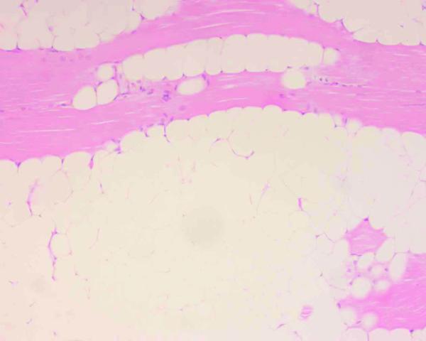
Background: Intramuscular lipoma is a relatively common benign neoplasm that is occasionally described as an infiltrating lipoma. Typical benign tumors show a clear margin, however, the infiltrative growth pattern of this lipoma mimics that of a malignant tumor. Although its growth has an effect on muscle bundles and it is known to never metastasize, the mechanism of infiltrative growth is not well understood. Previously, little attention has been paid to pathogenic features of muscle fibers around an intramuscular lipoma.
Methods: In the present study, we focused on pathologic changes of the surrounding skeletal muscles especially to the degenerative features of involving muscular types, and evaluate the role of type-selective muscular degeneration for the infiltrative growth of intramuscular lipomas. Following a review of the medical records in our institute, 17 lesions containing muscle tissues in their specimens (15 infiltrating lipomas, 2 well-circumscribed lipomas) were analyzed immunohistochemically. The tumor from the most recent case was also subjected to ultrastructural analysis. Two cases of the traumatic muscle damage were also evaluated as the control experiments.
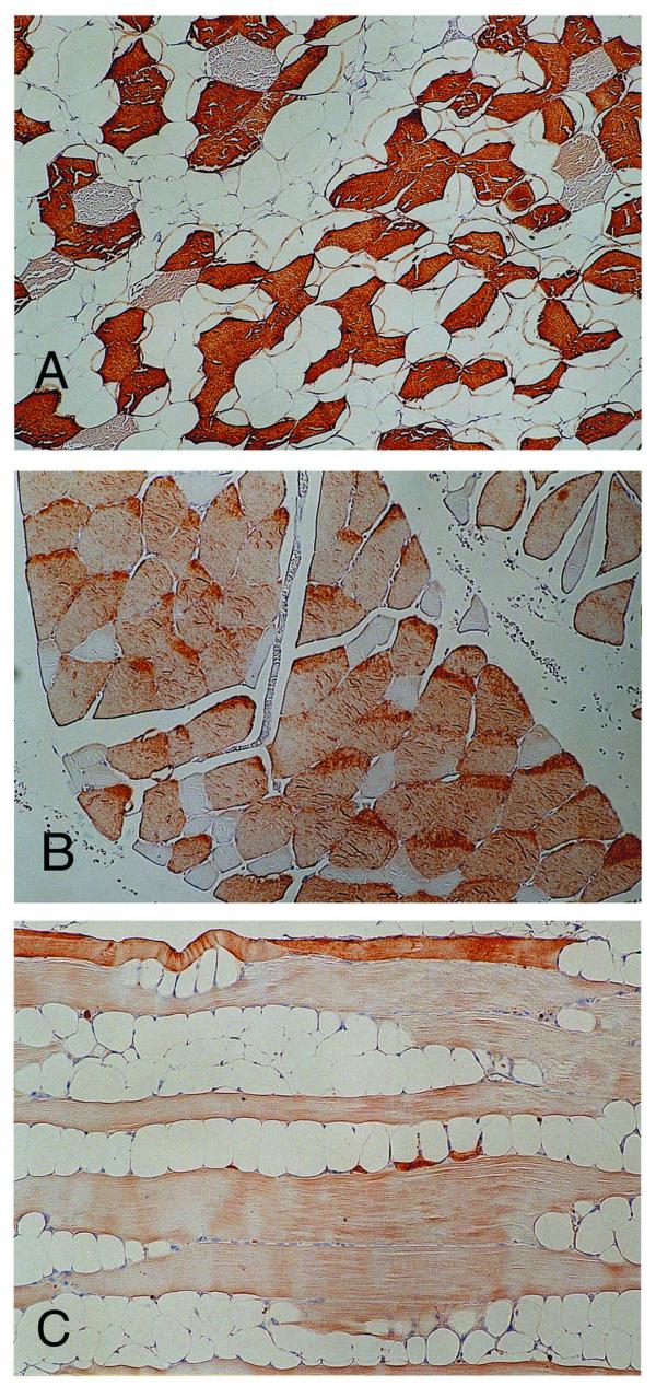
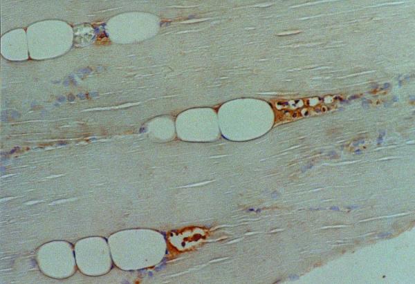
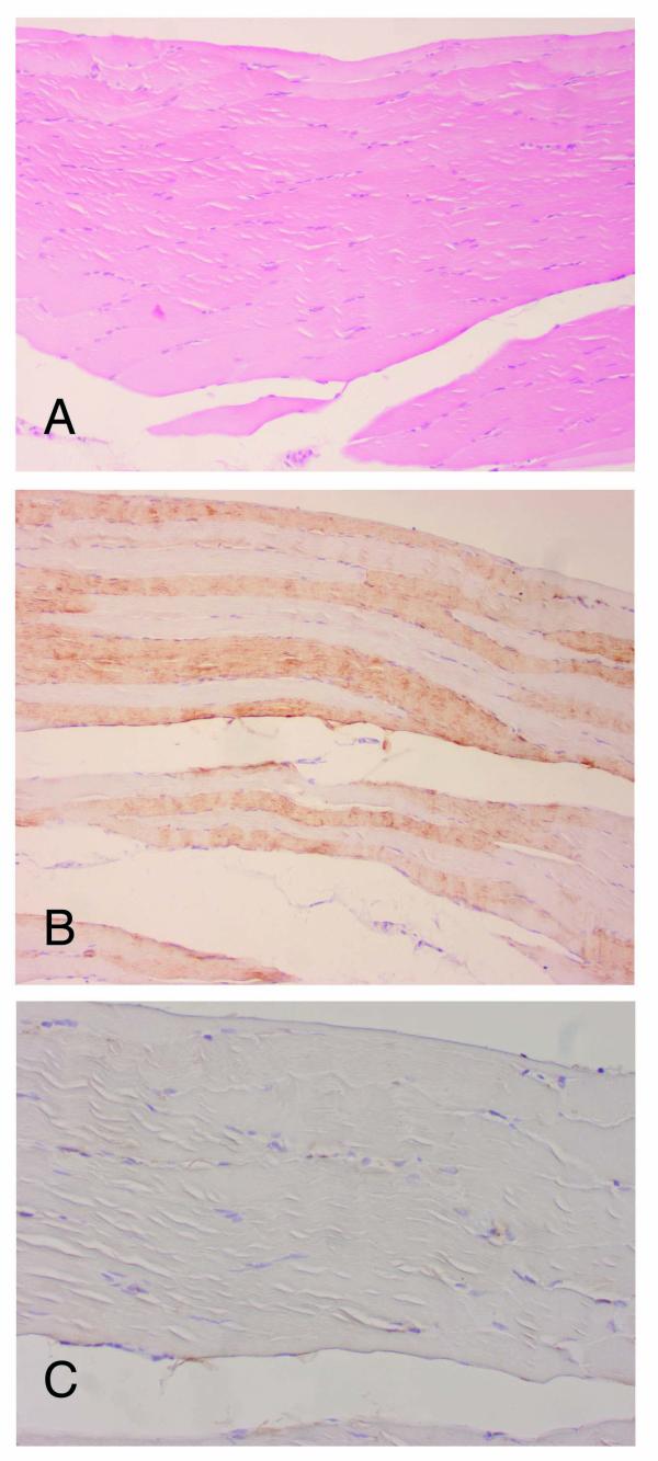
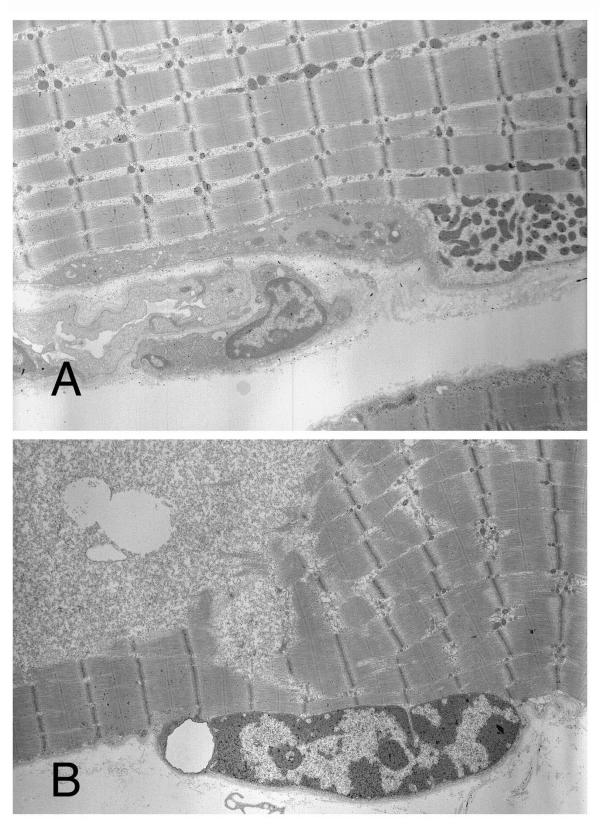
Results: These analyses revealed type-selective muscle involution in 11 of 17 intramuscular lipomas and in 10 of 11 of the infiltrative type, with an involving pattern that resembled that of a neurogenic or myogenic disorder. Immunoreactivity to cathepsin-D, a lysosomal catabolic enzyme, was increased in the involved muscle fibers. Subsarcolemmal vacuoles in the muscle fibers of the peripheral areas were also positive for cathepsin-D, while degenerative findings were not visually apparent in these areas. Ultrastructural analysis revealed degenerative changes in those fibers. Neither positive staining for cathepsin-D nor type-selective atrophy was detected in the sections of traumatic muscle damage.
Conclusions: Our findings suggest that type-selective muscular degeneration and endomysial fatty growth as a result of atrophy may modulate the infiltrating growth characteristic of intramuscular lipoma.
Figures
Similar articles
-
Intramuscular lipoma of the superior oblique muscle.Orbit. 2006 Sep;25(3):227-33. doi: 10.1080/01676830600575519. Orbit. 2006. PMID: 16987771
-
Light-microscopic study of the beta 1 integrin subunit in human skeletal muscle.Clin Neuropathol. 1997 Nov-Dec;16(6):319-27. Clin Neuropathol. 1997. PMID: 9401799
-
Intramuscular lipoma of the sternocleidomastoid muscle.J Craniofac Surg. 2010 Nov;21(6):1976-8. doi: 10.1097/SCS.0b013e3181f502cd. J Craniofac Surg. 2010. PMID: 21119474 Review.
-
Morphologic changes in the vastus medialis muscle in patients with osteoarthritis of the knee.Arthritis Rheum. 2007 Nov;56(11):3626-33. doi: 10.1002/art.22960. Arthritis Rheum. 2007. PMID: 17968889
-
Well-circumscribed intramuscular lipoma of the sternocleidomastoid muscle.Auris Nasus Larynx. 2004 Sep;31(3):283-5. doi: 10.1016/j.anl.2004.03.017. Auris Nasus Larynx. 2004. PMID: 15364365 Review.
Cited by
-
Lipoma in the pronator quadratus: A case report.Medicine (Baltimore). 2020 May 22;99(21):e20248. doi: 10.1097/MD.0000000000020248. Medicine (Baltimore). 2020. PMID: 32481302 Free PMC article.
-
Rapidly growing intramuscular lipoma: a unique entity of benign lipomas in children.BMJ Case Rep. 2024 Feb 27;17(2):e253408. doi: 10.1136/bcr-2022-253408. BMJ Case Rep. 2024. PMID: 38417947
-
Intramuscular Lipoma of the Sternocleidomastoid Muscle: A Rare Entity Revisited.Clin Pathol. 2024 Jun 10;17:2632010X241260200. doi: 10.1177/2632010X241260200. eCollection 2024 Jan-Dec. Clin Pathol. 2024. PMID: 38864025 Free PMC article.
-
The Lipid Enigma: A Case Report Highlighting Diagnostic Challenges in Adipocytic Tumors.Cureus. 2024 Oct 10;16(10):e71233. doi: 10.7759/cureus.71233. eCollection 2024 Oct. Cureus. 2024. PMID: 39525228 Free PMC article.
-
Large intramuscular lipoma of the tongue.Radiol Case Rep. 2018 Feb 3;13(2):361-364. doi: 10.1016/j.radcr.2018.01.014. eCollection 2018 Apr. Radiol Case Rep. 2018. PMID: 29904473 Free PMC article.
References
-
- Tallini G, Dal Cin P, Rhoden KJ, Chiapetta G, Manfioletti G, Giancotti V, Fusco A, Van den Berghe H, Sciot R. Expression of HMGI-C and HMGI (Y) in ordinary lipoma and atypical lipomatous tumors: immunohistochemical reactivity correlates with karyotypic alterations. Am J Pathol. 1997;151:37–43. - PMC - PubMed
-
- Tkachenko A, Ashar HR, Meloni AM, Sandberg AA, Chada KK. Misexpression of disrupted HMGI architectural factors activates alternative pathways of tumorigenesis. Cancer Res. 1997;57:2276–2280. - PubMed
-
- Cullen MJ, Mastaglia FL. Pathological reactions of skeletal muscle. In: Mastaglia FL, Walton SJ, editor. In Skeletal muscle pathology. New York: Churchill Livingstone; 1982. pp. 88–139.
-
- Dubowitz V. Definition of pathological changes seen in muscle biopsies. In: Dubowitz V, editor. In Muscle biopsy-a practical approach. England: Bailliere Tindall; 1985. pp. 82–128.
-
- Bird IM, Dhoot GK, Wilkinson JM. Identification of multiple variants of fast muscle troponin T in the chicken using monoclonal antibodies. Eur J Biochem. 1985;150:517–525. - PubMed
Publication types
MeSH terms
Substances
LinkOut - more resources
Full Text Sources
