

ERP evidence of impaired central nervous system function in virally suppressed HIV patients on antiretroviral therapy
- PMID: 15203059
- PMCID: PMC2367143
- DOI: 10.1016/j.clinph.2004.02.015
ERP evidence of impaired central nervous system function in virally suppressed HIV patients on antiretroviral therapy
Abstract
Objective: To examine the effects of human immunodeficiency virus (HIV) on central nervous system (CNS) function in patients receiving antiretroviral therapy (ART) who have suppressed viral loads.
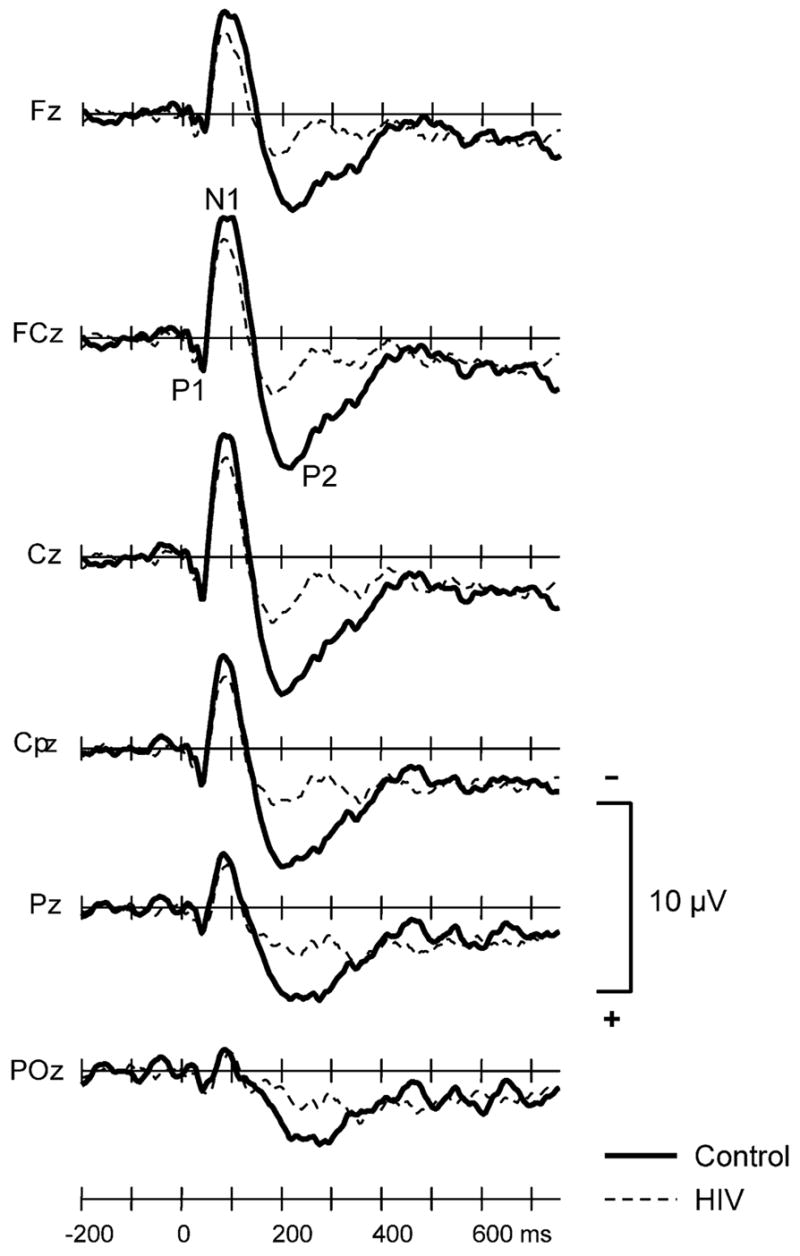
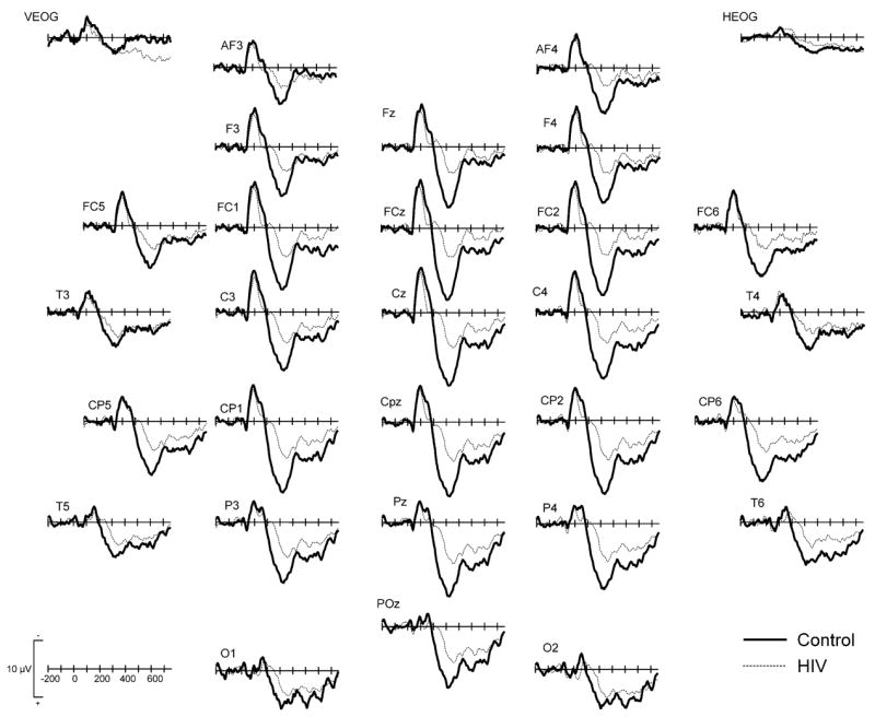
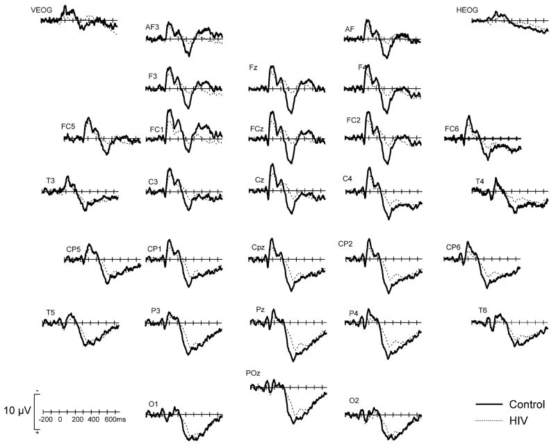
Methods: Event-related brain potentials (ERPs) were recorded from 15 virally suppressed HIV patients and 15 age-, sex-, and education-matched controls while they performed a 3-stimulus auditory oddball task. The amplitude and latency of the P3a, P3b, and early auditory components were examined in HIV patients and controls.
Results: Virally suppressed HIV patients on ART were more depressed than controls, as determined by the Beck Depression Inventory (BDI). After controlling for the effects of depression, HIV patients had smaller P2, P3a, and P3b amplitudes and longer P3a and P3b latency than control subjects. BDI scores correlated positively with N1 latency in HIV patients and negatively with P3b amplitude in all subjects.
Conclusions: These electrophysiological results suggest that, even in the absence of detectable levels of HIV in the peripheral blood, viral replication persists in the CNS and continues to cause disease in HIV patients on ART.
Figures
Similar articles
-
P3a and P3b auditory ERPs in HIV patients receiving anti-viral medication.Clin Electroencephalogr. 2002 Jul;33(3):97-101. doi: 10.1177/155005940203300305. Clin Electroencephalogr. 2002. PMID: 12192663 Clinical Trial.
-
Auditory P3 in antidepressant pharmacotherapy treatment responders, non-responders and controls.Eur Neuropsychopharmacol. 2013 Nov;23(11):1561-9. doi: 10.1016/j.euroneuro.2013.03.003. Epub 2013 May 9. Eur Neuropsychopharmacol. 2013. PMID: 23664712 Free PMC article. Clinical Trial.
-
A longitudinal study of event related potentials and correlations with psychosocial functioning and clinical features in first episode psychosis patients.Int J Psychophysiol. 2019 Nov;145:48-56. doi: 10.1016/j.ijpsycho.2019.05.007. Epub 2019 May 17. Int J Psychophysiol. 2019. PMID: 31108121 Free PMC article.
-
Brain macrophages harbor latent, infectious simian immunodeficiency virus.AIDS. 2019 Dec 1;33 Suppl 2(Suppl 2):S181-S188. doi: 10.1097/QAD.0000000000002269. AIDS. 2019. PMID: 31789817 Free PMC article. Review.
-
HIV-1 persistence in the central nervous system: viral and host determinants during antiretroviral therapy.Curr Opin Virol. 2019 Oct;38:54-62. doi: 10.1016/j.coviro.2019.06.004. Epub 2019 Aug 4. Curr Opin Virol. 2019. PMID: 31390580 Review.
Cited by
-
HIV/AIDS and an overweight body mass are associated with excessive intra-individual variability in response preparation.J Neurovirol. 2018 Oct;24(5):577-586. doi: 10.1007/s13365-018-0644-2. Epub 2018 May 17. J Neurovirol. 2018. PMID: 29777461 Free PMC article.
-
A family history of psychopathology modifies the decrement in cognitive control among patients with HIV/AIDS.Brain Cogn. 2008 Jun;67(1):103-14. doi: 10.1016/j.bandc.2007.12.004. Epub 2008 Jan 28. Brain Cogn. 2008. PMID: 18226846 Free PMC article.
-
Brainstem Auditory Evoked Potential in HIV-Positive Adults.Med Sci Monit. 2015 Oct 20;21:3172-8. doi: 10.12659/msm.894958. Med Sci Monit. 2015. PMID: 26485202 Free PMC article.
-
A Gap in Time: Extending our Knowledge of Temporal Processing Deficits in the HIV-1 Transgenic Rat.J Neuroimmune Pharmacol. 2017 Mar;12(1):171-179. doi: 10.1007/s11481-016-9711-8. Epub 2016 Oct 3. J Neuroimmune Pharmacol. 2017. PMID: 27699630 Free PMC article.
-
Selective Estrogen Receptor β Agonists: a Therapeutic Approach for HIV-1 Associated Neurocognitive Disorders.J Neuroimmune Pharmacol. 2020 Jun;15(2):264-279. doi: 10.1007/s11481-019-09900-y. Epub 2019 Dec 19. J Neuroimmune Pharmacol. 2020. PMID: 31858373 Free PMC article.
References
-
- Aylward EH, Brettschneider PD, McArthur JC, Harris GJ, Schlaepfer TE, Henderer JD, Barta PE, Tien AY, Pearlson GD. Magnetic resonance imaging measurement of gray matter volume reductions in HIV dementia. Am J Psychiatry. 1995;152:987–94. - PubMed
-
- Barker PB, Lee RR, McArthur JC. AIDS dementia complex: evaluation with proton MR spectroscopic imaging. Radiology. 1995;195:58–64. - PubMed
-
- Beck AJ. Depression: clinical, experimental and theoretical aspects. New York: Harper & Row; 1967.
-
- Beck AT, Ward CH, Mendelson M, Erbaugh JK. An inventory for measuring depression. Arch Gen Psychiatry. 1961;4:567–71. - PubMed
-
- Becker JT, Sanchez J, Dew MA, Lopez OL, Dorst SK, Banks G. Neuropsychological abnormalities among HIV-infected individuals in a community-based sample. Neuropsychology. 1997;11:592–601. - PubMed
Publication types
MeSH terms
Substances
Grants and funding
LinkOut - more resources
Full Text Sources
Medical
