. 2004 Jun;10(3):141-51.
doi: 10.1080/13550280490441103.

Apoptotic death of striatal neurons induced by human immunodeficiency virus-1 Tat and gp120: Differential involvement of caspase-3 and endonuclease G

Affiliations

Apoptotic death of striatal neurons induced by human immunodeficiency virus-1 Tat and gp120: Differential involvement of caspase-3 and endonuclease G

Indrapal N Singh et al. J Neurovirol. 2004 Jun.

Abstract

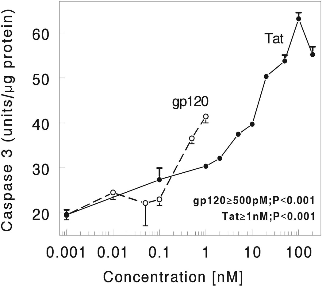
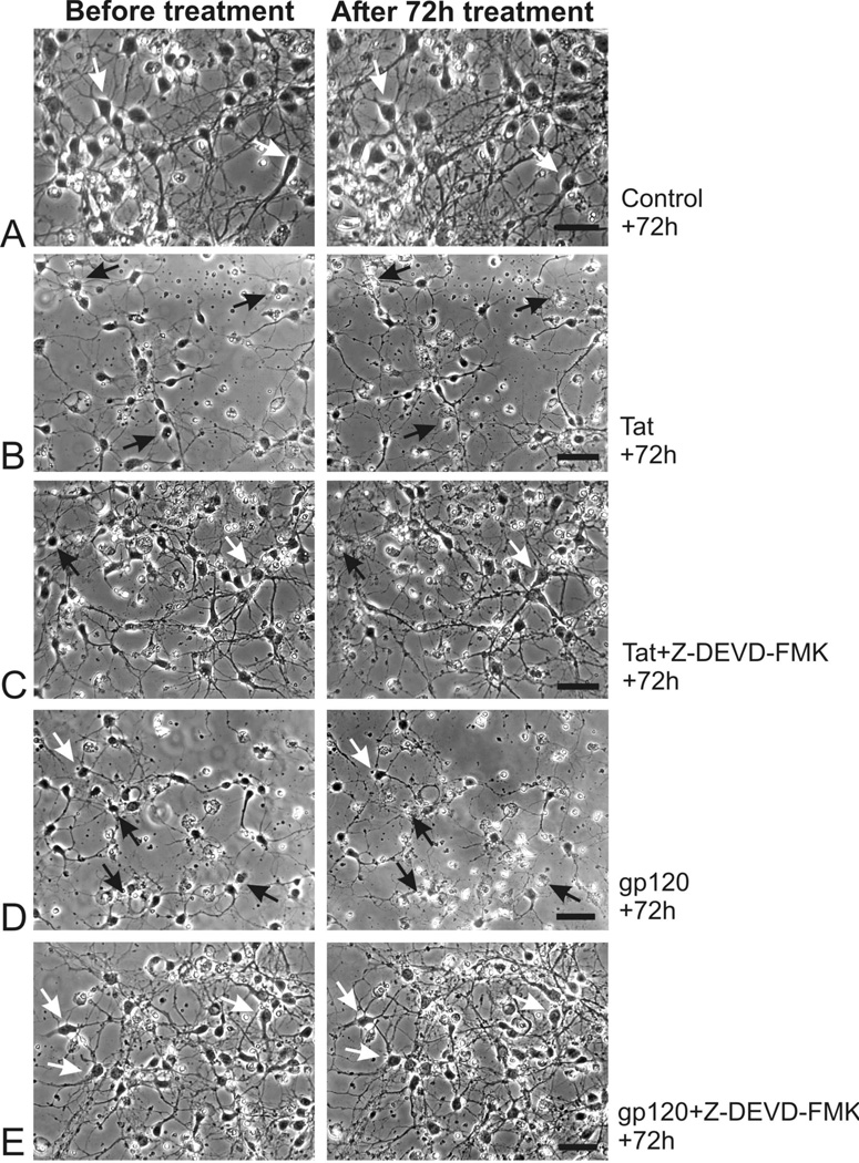
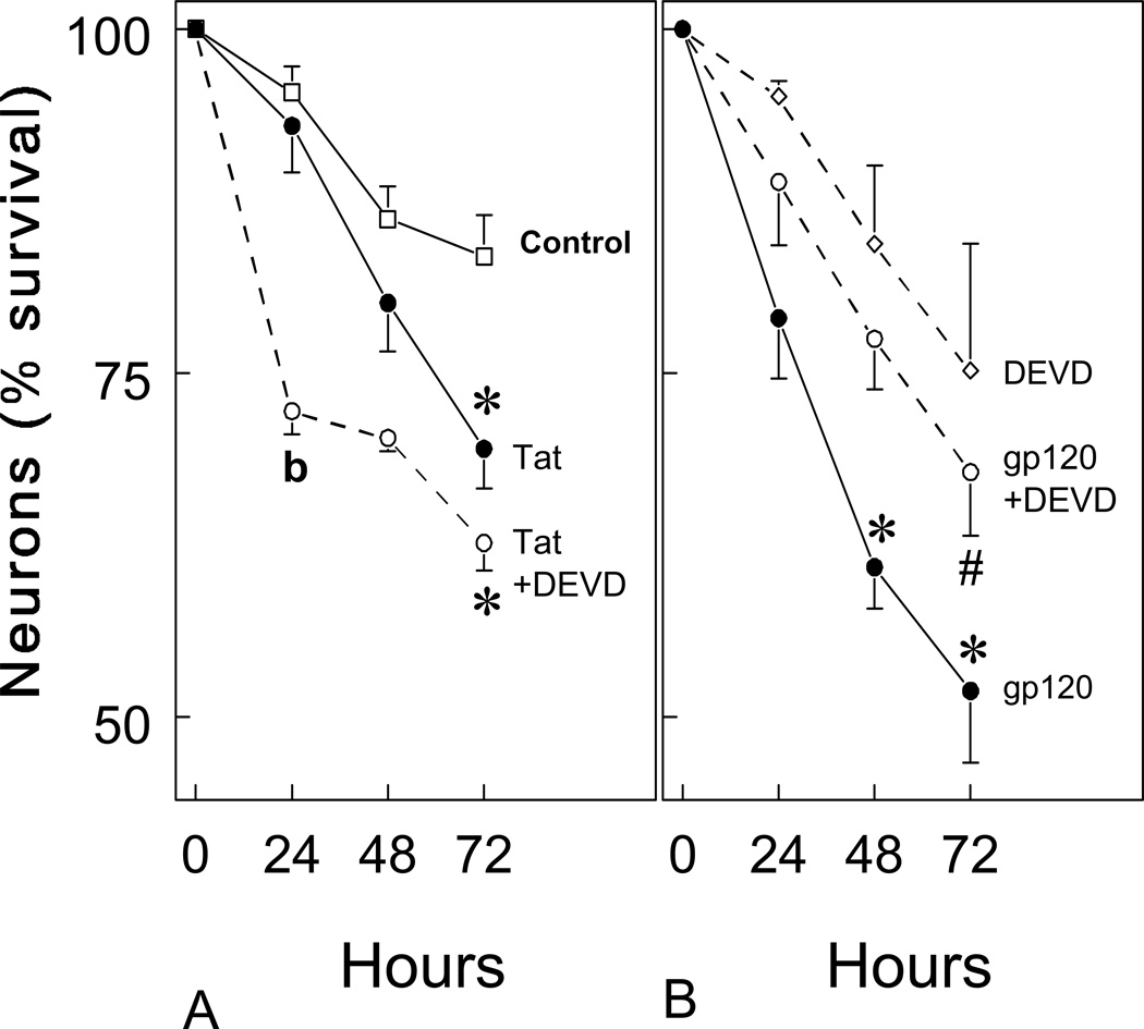
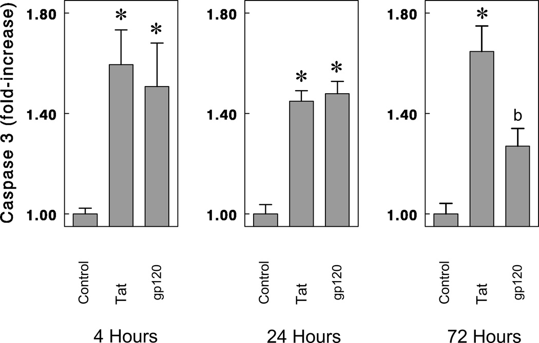
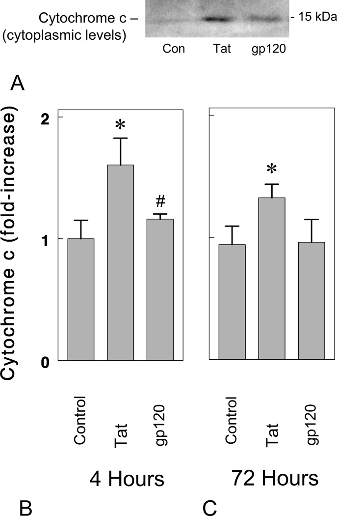
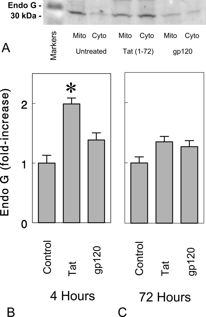
Human immunodeficiency virus-1 (HIV-1) infection affects the striatum, resulting in gliosis and neuronal losses. To determine whether HIV-1 proteins induce striatal neurotoxicity through an apoptotic mechanism, mouse striatal neurons isolated on embryonic day 15 and the effects of HIV-1 Tat(1-72) and gp120 on survival were assessed in vitro. Mitochondrial release of cytochrome c, caspase-3 activation, and neuron survival, as well as an alternative apoptotic pathway involving endonuclease G (endo G), were assessed at 4 h, 24 h, 48 h, and/or 72 h using enzyme assays and immunoblotting. Both HIV-1 Tat and gp120 significantly increased caspase-3 activation in a concentration-dependent manner in striatal neurons at 4 h following continuous exposure in vitro. Tat(1-72) and gp120 caused significant neuronal losses at 48 h and/or 72 h. Tat(1-72) increased cytochrome c release, and caspase-3 and endo G activation at 4 h, 24 h, and/or 72 h. By contrast, gp120 increased caspase-3 activation, but failed to increase cytochrome c or endo G levels in the cytoplasm at 4 h, 24 h, and/or 72 h. The cell permeant caspase inhibitor Z-DEVD-FMK significantly attenuated gp120-induced, but not Tat(1-72)-induced, neuronal death, suggesting that gp120 acts in large part through the activation of caspase(s), whereas Tat(1-72)-induced neurotoxicity was accompanied by activating an alternative pathway involving endo G. Thus, although Tat(1-72) and gp120 induced significant neurotoxicity, the nature of the apoptotic events preceding death differed. Collectively, our findings suggest that HIV-1 proteins are intrinsically toxic to striatal neurons and the pathogenesis is mediated through separate actions involving both caspase-3 and endo G.


Figures

Figure 1
Figure 1
HIV-1 Tat and gp120 caused concentration-dependent increases in caspase-3 activation in striatal neurons at 4 h following continuous exposure in vitro. Striatal neurons were grown for 7 days in culture and incubated with varied concentrations of HIV-1 Tat (closed circles with solid line) and gp120 (open circles with dotted lines) for 4 h. Caspase-3 activity was measured as described in the Methods section. Results are the mean ± the S.E.M from n=4 experiments.
Figure 2
Figure 2
Time-lapse digital photomicrographs showing the effects of HIV-1 Tat1-72 or gp120 on striatal neuron survival prior to (Before treatment; left column) and at 72 h following exposure (right column) in vitro (A–F). The effects of Tat or gp120 were additionally assessed in the presence or absence of Z-DEVD-FMK (30 µM) (DEVD) a caspase inhibitor applied 4 hours prior to viral protein exposure. Tat1-72 (100 nM) (B) and gp120 (500 pM) (D) were neurotoxic compared to treatment with media alone (A). However, only gp120 (E), but not Tat (C), induced neuronal losses were significantly attenuated by DEVD. Black arrows represent dying neurons, white arrows show viable neurons; scale bars = 25 µm.
Figure 3
Figure 3
HIV-1 Tat1-72 and gp120 reduced striatal neuron survival at 24, 48, and 72 hours in vitro. Both Tat (100 nM) (A) and gp120 (500 pM) (B) significantly increased the proportion of dying neurons at 48 and/or 72 h (*P < 0.05 versus vehicle-treated controls). The cytotoxic effects of gp120, but not Tat1-72, were significantly attenuated by the caspase inhibitor Z-DEVD-FMK (30 µM; #P < 0.01 versus gp120 treatment alone). Interestingly, Z-DEVD-FMK exposure appeared to enhance Tat toxicity at 24 h (bP < 0.05 versus Tat-treated or vehicle-treated cultures). About 50–75 neurons were arbitrarily sampled per culture. At least 4–6 separate cultures, each consisting of cells isolated and maintained from separate mice were assessed per experimental group. Rates of neuronal death in DEVD-FMK treated cultures did not differ significantly from vehicle-treated controls; DEVD = Z-DEVD-FMK.
Figure 4
Figure 4
Fluorescent photomicrographs showing the co-localization neuronal nuclear (NeuN) marker and ethidium monoazide (EMA) in striatal neuron cultures at 24 h following exposure to medium alone (A), gp120 (500 pM) with or without Z-DEVD-FMK (DEVD) (50 µM) (B,C), or DEVD alone (50 µM) (D). Cell cultures were incubated with DEVD for 4 h prior to exposure to gp120. gp120 treatment increased the proportion of dying neurons and the toxicity was prevented by co-administering DEVD (see Table 1). Viable NeuN immunofluorescence neurons (arrowheads) appear green and exclude EMA (red fluorescence) from their nuclei. Non-viable neurons (arrows) fail to exclude EMA (red) and their nuclei appear yellow. Tat (100 nM) was also assayed but showed no effect at 24 h (see Table 1). The large photomicrographs (right-side) are composite images of NeuN reactivity (upper left insets) and EMA (lower left insets); scale bar = 20 µm.
Figure 5
Figure 5
Effects of Tat1-72 and gp120 on caspase-3 activation in striatal neurons at 4 h, 24 h, and 72 h following continuous exposure in vitro. Both Tat (100 nM) and gp120 (500 pM) significantly increased caspase-3 activity at 4 h, 24 h, and 72 h. Caspase-3 activity was expressed as fluorescence units per µg of cytosolic protein and averaged from triplicate determinations each from at least n = 4 separate experiments. Mean caspase-3 activity in vehicle-treated (control) cultures was 69.71 units/µg protein (*P < 0.015 versus vehicle-treated cultures; ANOVA, post hoc Duncan’s test; bP< 0.05 versus vehicle-treated or Tat-treated cultures; ANOVA, post hoc Duncan’s test).
Figure 6
Figure 6
HIV-1 Tat1-72 increases cytoplasmic levels of cytochrome c in striatal neurons in vitro. Tat1-72 (100 nM), but not gp120 (500 pM), significantly increased levels of cytochrome c in the cytoplasm at 4 (A & B) and 72 hours (C) (*P < 0.05 versus vehicle-treated control (Con) cultures; #P < 0.05 versus Tat-treated neurons; n = 4 experiments).
Figure 7
Figure 7
Effects of Tat and gp120 on endonuclease G (endo G) in the cytoplasm at 4 and 72 h following continuous treatment. HIV-1 Tat1-72 (100 nM) increased cytoplasmic levels of in striatal neurons at 4 hours in vitro (*P < 0.05 versus vehicle-treated control cultures), while gp120 (500 pM) failed to increase endo G levels in the cytoplasm (Cyto) at 4 h or 72 h. Although cytoplasmic changes in endo G are shown (B,C), mitochondrial (Mito) levels were also assessed and varied inversely to cytoplasmic levels in control and Tat-treated neurons as expected (A). The results show the mean ± SEM of n = 4 experiments.

References

    1. Adamson DC, Wildemann B, Sasaki M, Glass JD, McArthur JC, Christov VI, Dawson TM, Dawson VL. Immunologic NO synthase: elevation in severe AIDS dementia and induction by HIV-1 gp41. Science. 1996;274:1917–1921. - PubMed
    1. Adle-Biassette H, Levy Y, Colombel M, Poron F, Natchev S, Keohane C, Gray F. Neuronal apoptosis in HIV infection in adults. Neuropathol Appl Neurobiol. 1995;21:218–227. - PubMed
    1. Albini A, Benelli R, Presta M, Rusnati M, Ziche M, Rubartelli A, Paglialunga G, Bussolino F, Noonan D. HIV-1 Tat protein is a heparin-binding angiogenic growth factor. Oncogene. 1996a;12:289–297. - PubMed
    1. Albini A, Soldi R, Giunciuglio D, Giraudo E, Benelli R, Primo L, Noonan D, Salio M, Famussi G, Rockl W, Bussolino F. The angiogenesis induced by HIV-1 Tat protein is mediated by the Flk-1/KDR receptor on vascular endothelial cells. NatMedl: 1996b:1371–1375. - PubMed
    1. Bagetta G, Corasaniti MT, Aloe L, Berliocchi L, Costa N, Finazzi-Agro A, Nistico G. Intracerebral injection of HIV-1 coat protein gp120 differentially affects the expression of NGF and nitric oxide synthase in the hippocampus of rat. Proc Natl Acad Sci USA. 1996a;93:928–933. - PMC - PubMed

Publication types

MeSH terms