

Reversible MR changes in the cat brain after cerebral fat embolism induced by triolein emulsion
- PMID: 15205130
- PMCID: PMC7975688
Reversible MR changes in the cat brain after cerebral fat embolism induced by triolein emulsion
Erratum in
- AJNR Am J Neuroradiol. 2004 Aug;25(7):1301
Abstract
Background and purpose: Clinical cerebral-fat embolism shows both reversible and irreversible changes. We used MR imaging to investigate the reversibility of embolized lesions induced with a fat-emulsion technique and to evaluate the histologic findings.
Methods: A fat emulsion was made with 0.05 mL of triolein and 20 mL of normal saline and vigorous to-and-fro movement through a three-way stopcock. In 50 cats, the internal carotid artery was infused with the fat emulsion. Cats were divided into six groups on the basis of time delay after embolization: 1 hour; 1 and 4 days; and 1, 2, and 3 weeks. MR imaging and histologic examination were performed at these times.
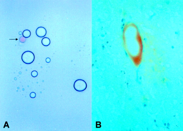
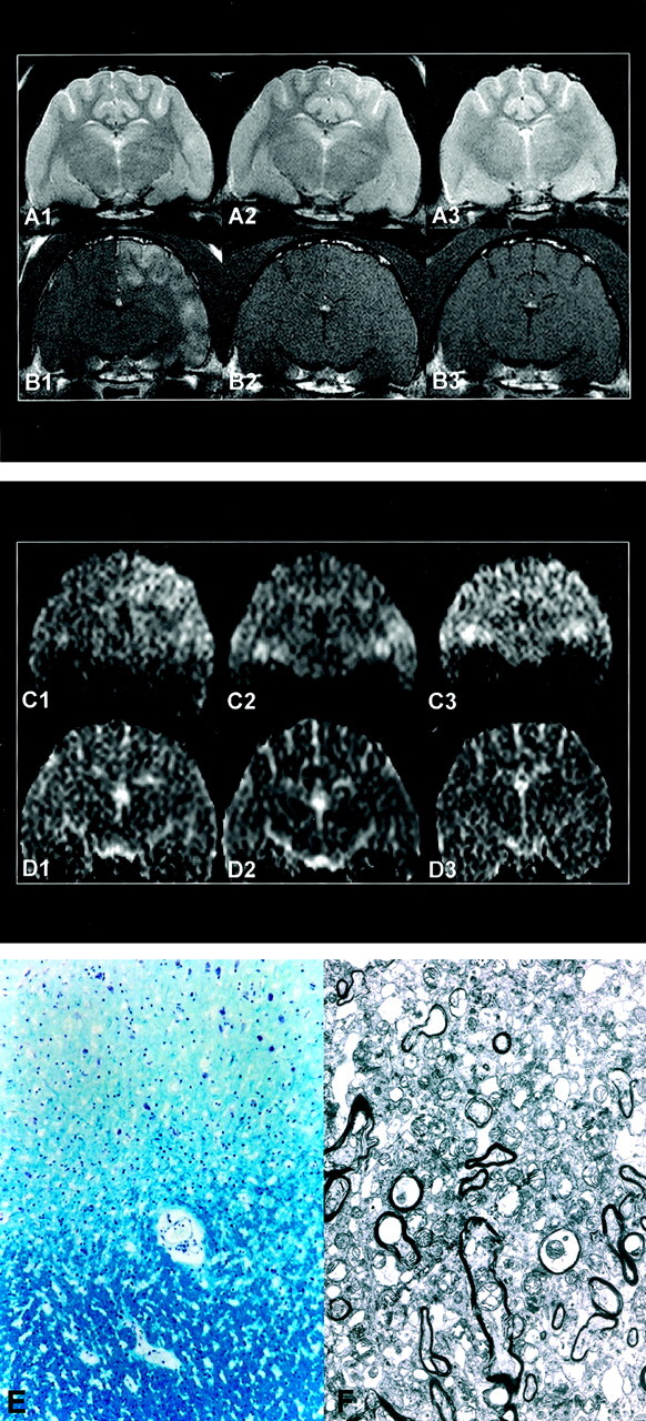
Results: Embolized lesions were hyperintense on T2-weighted images, isointense or mildly hyperintense on diffusion-weighted images, isointense on apparent diffusion coefficient maps, and enhancing on gadolinium-enhanced T1-weighted images at 1 hour. These MR imaging findings were less evident at day 1 and reverted to normal after day 4 (isointense on all images). Electron microscopy showed minimal findings in the cortical lesion in groups 1 and 2 (group 1 at 1 hour and group 2 at 1 hour and 1 day). Light microscopic findings revealed evidence of necrosis-small focal gliosis and demyelination in the periventricular white matter-in only one cat. The number of intravascular fat globules was not significantly different between groups, as visualized by oil red O staining.
Conclusion: Cerebral-fat embolism induced by a triolein emulsion revealed reversible MR findings and minimal histologic findings.
Figures
Similar articles
-
Experimental cerebral fat embolism: embolic effects of triolein and oleic acid depicted by MR imaging and electron microscopy.AJNR Am J Neuroradiol. 2002 Oct;23(9):1516-23. AJNR Am J Neuroradiol. 2002. PMID: 12372741 Free PMC article.
-
Magnetic resonance imaging and histologic findings of experimental cerebral fat embolism.Invest Radiol. 2003 Oct;38(10):625-34. doi: 10.1097/01.rli.0000077055.48406.e2. Invest Radiol. 2003. PMID: 14501490
-
The study of cerebral hemodynamics in the hyperacute stage of fat embolism induced by triolein emulsion.AJNR Am J Neuroradiol. 2006 Feb;27(2):398-401. AJNR Am J Neuroradiol. 2006. PMID: 16484418 Free PMC article.
-
Massive cerebral involvement in fat embolism syndrome and intracranial pressure management.J Neurosurg. 2013 Nov;119(5):1263-70. doi: 10.3171/2013.7.JNS13363. Epub 2013 Aug 16. J Neurosurg. 2013. PMID: 23952720 Review.
-
Diffusion-weighted MR of the brain: methodology and clinical application.Radiol Med. 2005 Mar;109(3):155-97. Radiol Med. 2005. PMID: 15775887 Review. English, Italian.
Cited by
-
Gradient-echo MRI in defining the severity of cerebral fat embolism.J Clin Neurol. 2008 Dec;4(4):164-6. doi: 10.3988/jcn.2008.4.4.164. Epub 2008 Dec 31. J Clin Neurol. 2008. PMID: 19513292 Free PMC article.
-
Dynamic MR imaging patterns of cerebral fat embolism: a systematic review with illustrative cases.AJNR Am J Neuroradiol. 2014 Jun;35(6):1052-7. doi: 10.3174/ajnr.A3605. Epub 2013 May 2. AJNR Am J Neuroradiol. 2014. PMID: 23639561 Free PMC article.
-
Triolein emulsion infusion into the hepatic artery increases vascular permeability to doxorubicin in rabbit liver.World J Gastroenterol. 2021 Jan 14;27(2):152-161. doi: 10.3748/wjg.v27.i2.152. World J Gastroenterol. 2021. PMID: 33510556 Free PMC article.
-
The steroid effect on the blood-ocular barrier change induced by triolein emulsion as seen on contrast-enhanced MR images.Korean J Radiol. 2008 May-Jun;9(3):205-11. doi: 10.3348/kjr.2008.9.3.205. Korean J Radiol. 2008. PMID: 18525222 Free PMC article.
-
Fat embolism syndrome with cerebral fat embolism through a patent foramen ovale: A case report.Medicine (Baltimore). 2020 Jun 12;99(24):e20569. doi: 10.1097/MD.0000000000020569. Medicine (Baltimore). 2020. PMID: 32541485 Free PMC article.
References
-
- Sevitt S. The significance and pathology of fat embolism. Ann Clin Res 1977;9:173–180 - PubMed
-
- Chrysikopoulos H, Maniatis V, Pappas J, Filalithis P, Gogali C, Sfyras D. Case report: post-traumatic cerebral fat embolism: CT and MR findings—report of two cases and review of the literature. Clin Radiol 1996;51:728–732 - PubMed
-
- Saito A, Meguro K, Matsumura A, Komatsu Y, Oohashi N. Magnetic resonance imaging of a fat embolism of the brain: case report. Neurosurgery 1990;26:882–885 - PubMed
-
- Kim HJ, Lee CH, Lee SH, Moon TY. Magnetic resonance imaging and histologic findings of experimental cerebral fat embolism. Invest Radiol 2003;38:625–634 - PubMed
Publication types
MeSH terms
Substances
LinkOut - more resources
Full Text Sources
Medical
Miscellaneous