

Case Reports
Prenatal MR findings of the middle interhemispheric variant of holoprosencephaly
Affiliations
- PMID: 15205143
- PMCID: PMC7975649
Item in Clipboard
Case Reports
Prenatal MR findings of the middle interhemispheric variant of holoprosencephaly
AJNR Am J Neuroradiol.
2004 Jun-Jul.
Abstract
We report a case of the middle interhemispheric variant of holoprosencephaly (MIH) with noncleavage of the posterior portion of the frontal lobes and the parietal regions in a fetus at 22 weeks' gestation. To our knowledge, this is the first case of the rare MIH variant to be diagnosed in utero by use of ultrafast MR imaging and one of the few such reports to document gross and microscopic pathologic findings. Neuroimaging results correlated with those of gross and microscopic pathologic specimens obtained from the stillborn child. We conclude that ultrafast MR imaging can accurately distinguish holoprosencephaly subtypes in utero, which may affect counseling of parents.
Figures
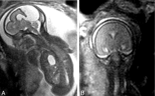
Fetal MR images obtained at 22 weeks’ gestation. A, Sagittal half-Fourier single-shot turbo spin-echo (HASTE) (TR/TE/NA, 1000/95/1) image obtained through the midline reveals the dorsal cyst and cleft formed by communication of the sylvian fissures over the vertex. Herniation of the stomach into the chest is also apparent. B, Coronal HASTE image obtained through the midportion of the brain depicts the lack of separation of the hemispheres and absent interhemispheric fissure more dorsally.
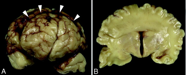
Gross specimens obtained from the stillborn female neonate. A, Frontal view of the brain shows a clear interhemispheric fissure extending to the rostral aspect of the frontal lobes. An abnormal transverse fissure can be seen extending into the dorsal midline of both hemispheres (arrowheads). B, Coronal section at the level of the third ventricle demonstrates bilaterally symmetric, separated basal forebrain structures (hypothalamus, basal ganglia, and anterior thalamus). In contrast, the cerebral hemispheres are continuous across the dorsal midline with a mass of white matter and cerebral cortex crossing the dorsal midline with no clear corpus callosum.
References
-
- Girard N, Raybaud C, Gambarelli D, Figarella-Branger D. Fetal brain MR imaging. Magn Reson Imaging Clin North Am 2001;9:19–56 - PubMed
-
- Lewis AJ, Simon EM, Barkovich AJ, et al. Middle interhemispheric variant of holoprosencephaly: a distinct cliniconeuroradiolgic subtype. Neurology 2002;59:1860–1865 - PubMed
-
- Bernard P, Drummond CL, Zaarour P, et al. A new clue to the prenatal diagnosis of lobar holoprosencephaly: the abnormal pathway of the anterior cerebral artery crawling under the skull. Ultrasound Obstet Gynecol 2002;19:605–607 - PubMed
Publication types
MeSH terms
LinkOut - more resources
Full Text Sources
Medical