

Intraobserver variability in the MR determination of tumor volume in squamous cell carcinoma of the pharynx
- PMID: 15205156
- PMCID: PMC7975672
Intraobserver variability in the MR determination of tumor volume in squamous cell carcinoma of the pharynx
Abstract
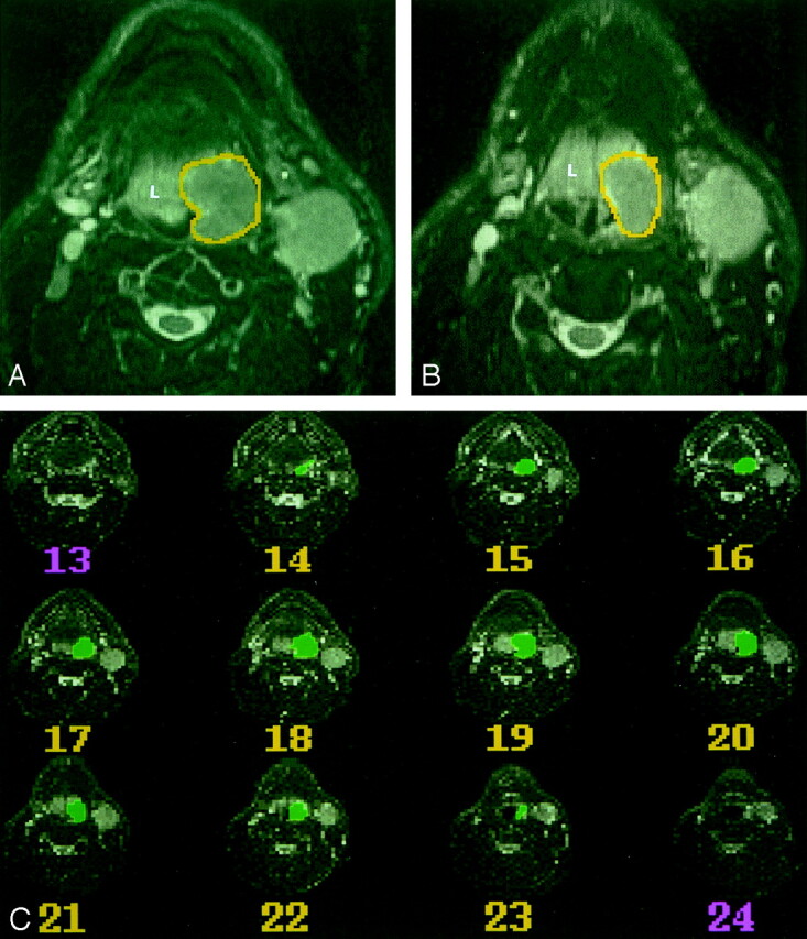
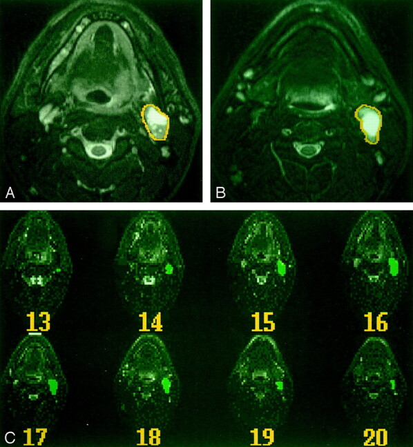
Background and purpose: If tumor volumes are to be used for evaluating responses to treatment and long-term outcomes of patients with primary pharyngeal carcinomas, the reproducibility of these measurements must be established. We determined the intraobserver variability of MR imaging-based volume measurements of these cancers and their regional metastases.
Methods: We used an interactive computer program (IDL) that enables the extraction of tumor volumes from 3D MR data to obtain 202 volume measurements in 17 patients with pharyngeal carcinoma (two to five time points each). The primary cancer and largest nodal mass were manually outlined on every T2-weighted image of each MR study. The same neuroradiologist reanalyzed this MR dataset 2-41 weeks later. Measurement error and percentage measurement error (intraobserver variability) were determined. Differences in intraobserver variability between primary lesions and nodes, as well as between stages of treatment were tested with a Wilcoxon rank sum test.
Results: The mean and median percentage measurement errors, respectively, were 13% and 12% (range, 0-53%; 95% CI: 10%, 16%) for primary tumors and 9% and 7% (range, 0-37%; 95% CI: 7%, 12%) for nodal metastases. The difference in the percentage measurement error between primary lesions and cervical nodes approached statistical significance (P =.07). Differences in the variation of volume measurements based on the stage of therapy were significant (P =.01).
Conclusion: Our results suggest that MR imaging-based tumor volumes are reliably reproducible. Such measurements may be important in predicting patient outcome, determining appropriate therapy, and conducting patient follow-up.
Figures
Similar articles
-
Intraobserver and interobserver variability of MR imaging- and CT-derived prostate volumes after transperineal interstitial permanent prostate brachytherapy.Radiology. 1998 Jun;207(3):785-9. doi: 10.1148/radiology.207.3.9609905. Radiology. 1998. PMID: 9609905 Clinical Trial.
-
Are pancreatic IPMN volumes measured on MRI images more reproducible than diameters? An assessment in a large single-institution cohort.Eur Radiol. 2018 Jul;28(7):2790-2800. doi: 10.1007/s00330-017-5268-z. Epub 2018 Feb 5. Eur Radiol. 2018. PMID: 29404774
-
Accuracy of magnetic resonance imaging in predicting absence of fixation of head and neck cancer to the prevertebral space.Head Neck. 2005 Feb;27(2):95-100. doi: 10.1002/hed.20128. Head Neck. 2005. PMID: 15627263
-
Neoplastic fixation to the prevertebral compartment by squamous cell carcinoma of the head and neck.AJR Am J Roentgenol. 1998 May;170(5):1389-94. doi: 10.2214/ajr.170.5.9574622. AJR Am J Roentgenol. 1998. PMID: 9574622
-
Squamous cell carcinoma of the upper aerodigestive tract: a review.Radiol Clin North Am. 2015 Jan;53(1):81-97. doi: 10.1016/j.rcl.2014.09.013. Radiol Clin North Am. 2015. PMID: 25476174 Review.
Cited by
-
Volumetric MR imaging of oral, maxillary sinus, oropharyngeal, and hypopharyngeal cancers: correlation between tumor volume and lymph node metastasis.AJNR Am J Neuroradiol. 2005 Oct;26(9):2384-9. AJNR Am J Neuroradiol. 2005. PMID: 16219850 Free PMC article.
-
Imaging in head and neck cancer.Curr Treat Options Oncol. 2006 Jan;7(1):23-34. doi: 10.1007/s11864-006-0029-2. Curr Treat Options Oncol. 2006. PMID: 16343366 Review.
-
Efficacy of diffusion-weighted imaging for the differentiation between lymphomas and carcinomas of the nasopharynx and oropharynx: correlations of apparent diffusion coefficients and histologic features.AJNR Am J Neuroradiol. 2012 Apr;33(4):761-6. doi: 10.3174/ajnr.A2834. Epub 2011 Dec 15. AJNR Am J Neuroradiol. 2012. PMID: 22173765 Free PMC article.
-
Clinical efficacy of primary tumor volume measurements: comparison of different primary sites.Clin Exp Otorhinolaryngol. 2009 Jun;2(2):78-84. doi: 10.3342/ceo.2009.2.2.78. Epub 2009 Jun 27. Clin Exp Otorhinolaryngol. 2009. PMID: 19565032 Free PMC article.
-
Development and implementation of optimized endogenous contrast sequences for delineation in adaptive radiotherapy on a 1.5T MR-linear-accelerator: a prospective R-IDEAL stage 0-2a quantitative/qualitative evaluation of in vivo site-specific quality-assurance using a 3D T2 fat-suppressed platform for head and neck cancer.J Med Imaging (Bellingham). 2023 Nov;10(6):065501. doi: 10.1117/1.JMI.10.6.065501. Epub 2023 Nov 6. J Med Imaging (Bellingham). 2023. PMID: 37937259 Free PMC article.
References
-
- National Cancer Institute 2001 FactBook. Bethesda: National Cancer Institute,2001
-
- Gilbert RW, Birt D, Shulman H, et al. Correlation of tumor volume with local control in laryngeal carcinoma treated by radiotherapy. Ann Otol Rhinol Laryngol 1987;96:514–518 - PubMed
-
- Freeman DE, Mancuso AA, Parsons JT, Mendenhall WM, Million RR. Irradiation alone for supraglottic larynx carcinoma: can CT findings predict treatment results? Int J Rad Oncol Biol Phys 1990;19:485–490 - PubMed
-
- Lee WR, Mancuso AA, Saleh EM, Mendenhall WM, Parsons JT, Million RR. Can pretreatment computed tomography findings predict local control in T3 squamous cell carcinoma of the glottic larynx treated with radiotherapy alone? Int J Rad Oncol Biol Phys 1993;25:683–687 - PubMed
-
- Castelijns JA, van den Brekel MWM, Smit EMT, et al. Predictive value of MR imaging-dependent and non-MR imaging-dependent parameters for recurrence of laryngeal cancer after radiation therapy. Radiology 1995;196:735–739 - PubMed
Publication types
MeSH terms
Grants and funding
LinkOut - more resources
Full Text Sources
Medical