. 2004 Jul;186(13):4110-23.
doi: 10.1128/JB.186.13.4110-4123.2004.

The YopD translocator of Yersinia pseudotuberculosis is a multifunctional protein comprised of discrete domains

Affiliations

The YopD translocator of Yersinia pseudotuberculosis is a multifunctional protein comprised of discrete domains

Jan Olsson et al. J Bacteriol. 2004 Jul.

Abstract

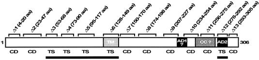
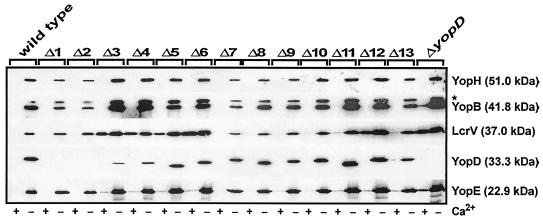
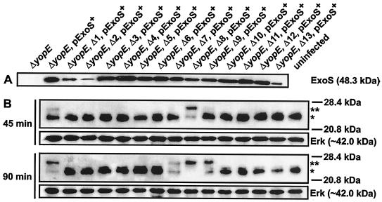
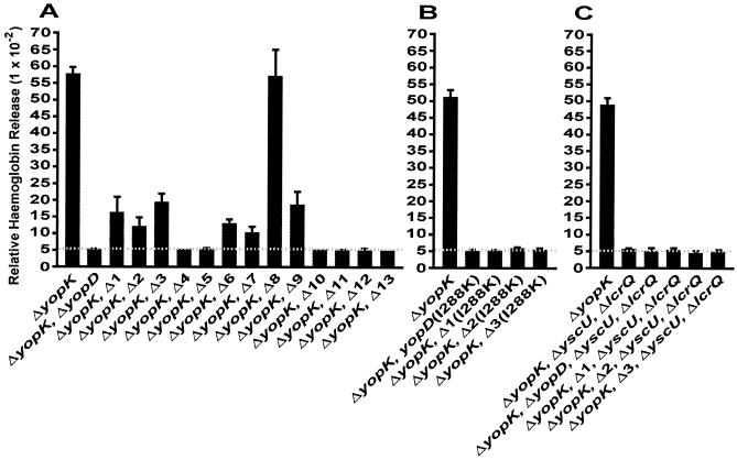
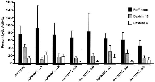
To establish an infection, Yersinia pseudotuberculosis utilizes a plasmid-encoded type III translocon to microinject several anti-host Yop effectors into the cytosol of target eukaryotic cells. YopD has been implicated in several key steps during Yop effector translocation, including maintenance of yop regulatory control and pore formation in the target cell membrane through which effectors traverse. These functions are mediated, in part, by an interaction with the cognate chaperone, LcrH. To gain insight into the complex molecular mechanisms of YopD function, we performed a systematic mutagenesis study to search for discrete functional domains. We highlighted amino acids beyond the first three N-terminal residues that are dispensable for YopD secretion and confirmed that an interaction between YopD and LcrH is essential for maintenance of yop regulatory control. In addition, discrete domains within YopD that are essential for both pore formation and translocation of Yop effectors were identified. Significantly, other domains were found to be important for effector microinjection but not for pore formation. Therefore, YopD is clearly essential for several discrete steps during efficient Yop effector translocation. Recognition of this modular YopD domain structure provides important insights into the function of YopD.


Figures

FIG. 1.
FIG. 1.
Schematic diagram of the 306-amino-acid YopD protein of pathogenic Yersinia spp. The structural features of YopD include the putative transmembrane domain (TM) predicted by using the TMPRED web server (http://www.ch.embnet.org/software/TMPRED_form.html). Two amphipathic α-helices, one located internally (AD1) and the other a biologically relevant domain at the C terminus (AD2) (22, 62), were both identified by helical wheel projection (Antheprot, version 3.2; G. Deleage, Lyon, France). A predicted coiled coil region (CC) was identified by using the COILS web server (http://www.ch.embnet.org/software/COILS_form.html). A question mark indicates a domain that was predicted only when low-stringency parameters were used. Also shown are the locations of the following in-frame sequential ΔYopD deletions used in this study: Δ1, YPIII/pIB625 (Δ4-20 aa [deletion of amino acids 4 to 20]); Δ2, YPIII/pIB605 (Δ23-47 aa); Δ3, YPIII/pIB626 (Δ53-68 aa); Δ4, YPIII/pIB627 (Δ73-90 aa); Δ5, YPIII/pIB628 (Δ95-117 aa); Δ6, YPIII/pIB623 (Δ128-149 aa); Δ7, YPIII/pIB629 (Δ150-170 aa); Δ8, YPIII/pIB630 (Δ174-198 aa); Δ9, YPIII/pIB631 (Δ207-227 aa); Δ10, YPIII/pIB632 (Δ234-254 aa); Δ11, YPIII/pIB633 (Δ256-275 aa); Δ12, YPIII/pIB622 (Δ278-292 aa); and Δ13, YPIII/pIB624 (Δ293-305 aa). The regulatory status of individual mutants, as determined by MOX analysis (5, 23) (see Materials and Methods), is indicated. CD reflects wild-type regulatory control of Yop synthesis, and TS reflects defective regulatory control in which Yop synthesis is constitutive. YopD domains important for regulatory control (indicated by a solid line) correspond to identical domains required for binding the dedicated chaperone LcrH (20, 21).
FIG. 2.
FIG. 2.
Analysis of Yop secretion from Y. pseudotuberculosis strains grown in BHI broth either with (+) or without (−) Ca2+. Secreted Yops (a mixture of Yops present only in cleared culture supernatants) were separated by SDS-PAGE and identified by immunoblot analysis by using polyclonal rabbit anti-YopH, anti-YopB, anti-LcrV, anti-YopD, and anti-YopE antisera. The asterisk indicates a nonspecific cross-reactive band detected by using anti-YopB antiserum. The molecular masses indicated in parentheses were deduced from the primary sequences.
FIG. 3.
FIG. 3.
Infection of HeLa cells by Y. pseudotuberculosis. Strains were allowed to infect a monolayer of growing HeLa cells, and at 2 h postinfection the effect of the bacteria on the HeLa cells was determined by phase-contrast microscopy. Note the extensive rounding of the YopE-dependent cytotoxically affected HeLa cells (B and I to K). HeLa cells infected with strains carrying a yopD null mutation or the in-frame Δ1 to Δ6 and Δ10 to Δ13 YopD deletions had normal uninfected cell morphology (compare panel A with panels C to H and L to P). Prolonged infections (up to 5 h) did not alter the experimental outcome (data not shown).
FIG. 4.
FIG. 4.
(A) Analysis of secreted ExoS produced in trans by a ΔyopE null mutant (YPIII/pIB522) of Y. pseudotuberculosis harboring sequential deletions of YopD and grown under inducing conditions (without Ca2+). Secreted protein from cleared bacterial supernatants was separated by SDS-PAGE and identified by immunoblot analysis by using polyclonal rabbit anti-ExoS antisera. ExoS, together with its dedicated chaperone Orf1, is encoded on the high-copy-number plasmid pTS103-Gm (pExoS+) under control of the native promoter (10). (B) Ras modification in HeLa cells infected with bacteria expressing ExoS. HeLa cells were harvested after infection at 45 and 90 min and dissolved in sample buffer. Proteins in the HeLa cell lysates were fractionated by SDS-PAGE, and this was followed by immunoblotting with anti-Ras monoclonal antibody. As a loading control, the same filters were also probed with a monoclonal antibody directed against the eukaryotic cytosolic marker protein Erk. One asterisk indicates the position of unmodified Ras, while two asterisks indicate the position of the more slowly migrating modified version of Ras. No further Ras modification was observed at later times (data not shown).
FIG. 5.
FIG. 5.
Conformational analysis of YopD internal deletion mutants. (A and B) Intrabacterial stability of YopD proteins produced by Y. pseudotuberculosis grown at 37°C in the presence of 5 mM CaCl2. At time zero, chloramphenicol was added in order to stop new protein synthesis. Aliquots were taken at different times, and the amounts of proteins were determined by Western blot analysis (A) and by a densitometry analysis in which images were first acquired with a Fluor-S MultiImager (Bio-Rad) and after inversion the intensity of each band was quantified by using the Quantity One quantitation software (version 4.2.3; Bio-Rad) (B). (C and D) Immunoblots of secreted YopD prepared from cleared culture supernatants that had been incubated with (+) or without (−) chymotrypsin for 30 min prior to trichloroacetic acid precipitation. YopD was identified by using polyclonal rabbit anti-YopD antiserum (α-YopD) in combination with enhanced chemiluminescence detection prior to normal exposure (A and C) and overexposure (D) to X-ray film. For reference, the asterisk identifies identical bands in panels C and D.
FIG. 6.
FIG. 6.
Lysis of erythrocytes by a ΔyopK null mutant (YPIII/pIB155) of Y. pseudotuberculosis also having a sequential in-frame deletion of YopD (A) in combination with the site mutation I288K located in the C-terminal amphipathic α-helix (B) or with a defective type III secretion apparatus (ΔyscU ΔlcrQ) (C). Sheep erythrocytes were infected with the different strains of Y. pseudotuberculosis for 3 h. After this the amount of released hemoglobin was determined spectrophotometrically. The values are the average lytic activities ± standard deviations (as determined with Microsoft Excel 2000) for at least four individual experiments performed in quadruplicate.
FIG. 7.
FIG. 7.
Osmoprotection of infected erythrocytes by different carbohydrates. Sheep erythrocytes were infected with the different strains of Y. pseudotuberculosis for 3 h in the presence of carbohydrates having different diameters, including raffinose (diameter, 1.2 to 1.4 nm), dextrin 15 (diameter, 2.2 nm), and dextran 4 (diameter, 3 to 3.5 nm), after which the amounts of released hemoglobin were determined spectrophotometrically. The lytic activity is expressed as a percentage of the lysis in the absence of sugars. The bars and error bars indicate the means and standard errors of the means (as calculated by using Mathsoft Axum software, version 7.0), respectively, for three independent experiments.
FIG. 8.
FIG. 8.
Analysis of YopD synthesis and secretion from Y. pseudotuberculosis strains grown in BHI broth without Ca2+. YopD in the total fractions (mixtures of proteins in intact bacteria secreted into the culture medium) (A) and YopD in the secreted fractions (mixtures of trichloroacetic acid-precipitated proteins in cleared culture supernatants) (B) were separated by SDS-PAGE and identified by immunoblot analysis by using polyclonal rabbit anti-YopD antiserum (α-YopD).

Similar articles

Cited by

References

    1. Aepfelbacher, M., and J. Heesemann. 2001. Modulation of Rho GTPases and the actin cytoskeleton by Yersinia outer proteins (Yops). Int. J. Med. Microbiol. 291:269-276. - PubMed
    1. Anderson, D. M., K. S. Ramamurthi, C. Tam, and O. Schneewind. 2002. YopD and LcrH regulate expression of Yersinia enterocolitica YopQ by a posttranscriptional mechanism and bind to yopQ RNA. J. Bacteriol. 184:1287-1295. - PMC - PubMed
    1. Anderson, D. M., and O. Schneewind. 1999. Type III machines of Gram-negative pathogens: injecting virulence factors into host cells and more. Curr. Opin. Microbiol. 2:18-24. - PubMed
    1. Barbieri, J. T. 2000. Pseudomonas aeruginosa exoenzyme S, a bifunctional type-III secreted cytotoxin. Int. J. Med. Microbiol. 290:381-387. - PubMed
    1. Bergman, T., S. Håkansson, Å. Forsberg, L. Norlander, A. Macellaro, A. Bäckman, I. Bölin, and H. Wolf-Watz. 1991. Analysis of the V antigen lcrGVH-yopBD operon of Yersinia pseudotuberculosis: evidence for a regulatory role of LcrH and LcrV. J. Bacteriol. 173:1607-1616. - PMC - PubMed

Publication types

LinkOut - more resources