Energetics of gliding motility in Mycoplasma mobile
- PMID: 15205428
- PMCID: PMC421629
- DOI: 10.1128/JB.186.13.4254-4261.2004
Energetics of gliding motility in Mycoplasma mobile
Abstract
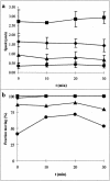
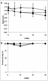
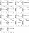
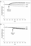
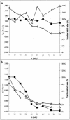
Mycoplasma mobile glides on surfaces at up to 7 microm/s by an unknown mechanism. We studied the energetics that power gliding by using a novel, growth medium-free system. We found that cells could glide in defined media if the glass substrate is preconditioned by exposure to horse serum. The active component that potentiates gliding is sensitive to proteinase K treatment. We used the defined medium system to test the effect of various inhibitors, ionophores, and poisons on motility of M. mobile. Valinomycin, carbonyl cyanide 4-(trifluoromethoxy)phenylhydrazone (FCCP), N,N'-dicyclohexylcarbodiimide, phenamil, amiloride, rifampin, and puromycin had no short-term effects on gliding. We also confirmed that we were able to modulate the membrane potential with valinomycin and FCCP by using a potential-sensitive dye. Shifting the pH likewise had no effect on motility. These results rule out the use of conventional ion motive forces to power gliding. Arsenate had a dramatic inhibitory effect on gliding, and both the speed and the fraction of cells moving tracked ATP levels. Sodium orthovanadate had a slight but significant inhibitory effect on gliding. Taken together, these results suggest that the motor system of M. mobile is likely an ATPase or is directly coupled to an ATPase.
Figures
Similar articles
-
Effects of energization on membrane organization in mycoplasma.Biochim Biophys Acta. 1982 May 7;687(2):281-90. doi: 10.1016/0005-2736(82)90556-9. Biochim Biophys Acta. 1982. PMID: 7093258
-
Behaviors and Energy Source of Mycoplasma gallisepticum Gliding.J Bacteriol. 2019 Sep 6;201(19):e00397-19. doi: 10.1128/JB.00397-19. Print 2019 Oct 1. J Bacteriol. 2019. PMID: 31308069 Free PMC article.
-
Source of energy for gliding motility in Flexibacter polymorphus: effects of metabolic and respiratory inhibitors on gliding movement.J Bacteriol. 1977 Aug;131(2):544-56. doi: 10.1128/jb.131.2.544-556.1977. J Bacteriol. 1977. PMID: 885839 Free PMC article.
-
Prospects for the gliding mechanism of Mycoplasma mobile.Curr Opin Microbiol. 2016 Feb;29:15-21. doi: 10.1016/j.mib.2015.08.010. Epub 2015 Oct 21. Curr Opin Microbiol. 2016. PMID: 26500189 Review.
-
Unique centipede mechanism of Mycoplasma gliding.Annu Rev Microbiol. 2010;64:519-37. doi: 10.1146/annurev.micro.112408.134116. Annu Rev Microbiol. 2010. PMID: 20533876 Review.
Cited by
-
Involvement of P1 adhesin in gliding motility of Mycoplasma pneumoniae as revealed by the inhibitory effects of antibody under optimized gliding conditions.J Bacteriol. 2005 Mar;187(5):1875-7. doi: 10.1128/JB.187.5.1875-1877.2005. J Bacteriol. 2005. PMID: 15716461 Free PMC article.
-
Gliding Direction of Mycoplasma mobile.J Bacteriol. 2015 Oct 26;198(2):283-90. doi: 10.1128/JB.00499-15. Print 2016 Jan 15. J Bacteriol. 2015. PMID: 26503848 Free PMC article.
-
Directed Binding of Gliding Bacterium, Mycoplasma mobile, Shown by Detachment Force and Bond Lifetime.mBio. 2016 Jun 28;7(3):e00455-16. doi: 10.1128/mBio.00455-16. mBio. 2016. PMID: 27353751 Free PMC article.
-
Sequence analysis of the gliding protein Gli349 in Mycoplasma mobile.Biophysics (Nagoya-shi). 2005 May 25;1:33-43. doi: 10.2142/biophysics.1.33. eCollection 2005. Biophysics (Nagoya-shi). 2005. PMID: 27857551 Free PMC article.
-
Gliding ghosts of Mycoplasma mobile.Proc Natl Acad Sci U S A. 2005 Sep 6;102(36):12754-8. doi: 10.1073/pnas.0506114102. Epub 2005 Aug 26. Proc Natl Acad Sci U S A. 2005. PMID: 16126895 Free PMC article.
References
-
- Aluotto, B. B., R. G. Wittler, C. O. Williams, and J. E. Faber. 1970. Standardized bacteriologic techniques for the characterization of Mycoplasma species. Int. J. Syst. Bacteriol. 20:35-58.
-
- Atsumi, T., Y. Maekawa, H. Tokuda, and Y. Imae. 1992. Amiloride at pH 7.0 inhibits the Na(+)-driven flagellar motors of Vibrio alginolyticus but allows cell growth. FEBS Lett. 314:114-116. - PubMed
-
- Atsumi, T., L. McCarter, and Y. Imae. 1992. Polar and lateral flagellar motors of marine Vibrio are driven by different ion-motive forces. Nature 355:182-184. - PubMed
-
- Berg, H. C., and S. M. Block. 1984. A miniature flow cell designed for rapid exchange of media under high-power microscope objectives. J. Gen. Microbiol. 130:2915-2920. - PubMed
Publication types
MeSH terms
Substances
Grants and funding
LinkOut - more resources
Full Text Sources
Molecular Biology Databases

