. 2004 Jul;186(13):4262-75.
doi: 10.1128/JB.186.13.4262-4275.2004.

Autoinduction of Bacillus subtilis phoPR operon transcription results from enhanced transcription from EsigmaA- and EsigmaE-responsive promoters by phosphorylated PhoP

Affiliations

Autoinduction of Bacillus subtilis phoPR operon transcription results from enhanced transcription from EsigmaA- and EsigmaE-responsive promoters by phosphorylated PhoP

Salbi Paul et al. J Bacteriol. 2004 Jul.

Abstract

The phoPR operon encodes a response regulator, PhoP, and a histidine kinase, PhoR, which activate or repress genes of the Bacillus subtilis Pho regulon in response to an extracellular phosphate deficiency. Induction of phoPR upon phosphate starvation required activity of both PhoP and PhoR, suggesting autoregulation of the operon, a suggestion that is supported here by PhoP footprinting on the phoPR promoter. Primer extension analyses, using RNA from JH642 or isogenic sigE or sigB mutants isolated at different stages of growth and/or under different growth conditions, suggested that expression of the phoPR operon represents the sum of five promoters, each responding to a specific growth phase and environmental controls. The temporal expression of the phoPR promoters was investigated using in vitro transcription assays with RNA polymerase holoenzyme isolated at different stages of Pho induction, from JH642 or isogenic sigE or sigB mutants. In vitro transcription studies using reconstituted EsigmaA, EsigmaB, and EsigmaE holoenzymes identified PA4 and PA3 as EsigmaA promoters and PE2 as an EsigmaE promoter. Phosphorylated PhoP (PhoP approximately P) enhanced transcription from each of these promoters. EsigmaB was sufficient for in vitro transcription of the PB1 promoter. P5 was active only in a sigB mutant strain. These studies are the first to report a role for PhoP approximately P in activation of promoters that also have activity in the absence of Pho regulon induction and an activation role for PhoP approximately P at an EsigmaE promoter. Information concerning PB1 and P5 creates a basis for further exploration of the regulatory coordination or overlap of the PhoPR and SigB regulons during phosphate starvation.


Figures
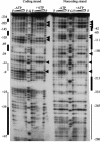
FIG. 1.
FIG. 1.
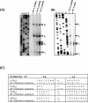
DNase I footprint analysis of the phoPR promoter bound by PhoP and PhoP∼P. Various amounts of PhoP, incubated with *PhoR (1.4 μg) in the presence or absence of 4 mM ATP, were mixed with the 407-bp phoPR promoter labeled on either the coding or noncoding strand and treated with DNase I. The concentration of PhoP used in each reaction mixture was, from left to right, 0 nM, 55 nM, 275 nM, 1.38 μM, and 6.7 μM. Lanes with PhoP∼P are labeled +ATP, and those with unphosphorylated PhoP are labeled −ATP. Lanes F, PhoP-free lanes; lane G, the G-sequencing reaction lane used as a reference. The thick black vertical lines represent the PhoP and PhoP∼P binding regions, while the thin lines represent sites bound only by PhoP∼P. The hypersensitive sites are marked with a dark arrowhead. Base pairs are numbered relative to the translation start site (as +1).
FIG. 2.
FIG. 2.
Transcription start sites and PhoP binding sites on the phoPR promoter sequence and 5′ PhoP coding sequence. Gray shading identifies sequence protected by both PhoP and PhoP∼P. Stippled shading identifies sequence protected only by PhoP∼P. Transcriptional start sites for PB1, PE2, PA3, PA4, or P5 are indicated by bold sequence base pairs that are identified by a bent arrow followed by the promoter number. The −10 consensus sequence for each promoter is underlined by a slender rectangle marked −10 followed by the promoter number. The −35 consensus sequence for each promoter is overlined by a slender rectangle marked −35 followed by the promoter number. The consensus repeats for PhoP dimer binding [TT(A/C/T)A(C/T)A] are underlined with the sequence in bold print. The translational start codon, ATG, is boxed and identified by a bent arrow marked +1. Sequence numbering is relative to the A of ATG as +1. Arrows with half arrowheads identify primers used to amplify sequences in the two promoter fusion constructs analyzed below in Fig. 3. The asterisk identifies the transcription site of PBX1.
FIG. 3.
FIG. 3.
The roles of PhoP or PhoP binding regions in phoPR transcription differ during phosphate-limited and phosphate-replete growth. (A) Growth and phoPR expression in LPDM. An arrow marks the induction of APase in phoP+ strains MH5562 and MH5559. (B) Growth and phoPR expression in SSG. Filled symbols represent growth; open symbols represent expression of various phoP-lacZ promoter fusions. Circle, MH5562 (JH642 strain; phoP-lacZ); square, MH5565 (phoP strain; phoP-lacZ); triangle, MH5559 (JH642 strain; phoP-lacZ fusion containing a deletion of bp +25 to +92 that removed the 3′ PhoP binding site, phoPΔ25-92-lacZ); inverted triangle, MH5567 (Δ phoP; phoPΔ25-92-lacZ); diamond, MH5580 (sigE phoP-lacZ).
FIG. 4.
FIG. 4.
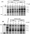
Primer extension identified four mRNA 5′ ends in the phoPR promoter region. (A) Primer extension analysis of the phoPR promoter region. The end-labeled primer FMH079 was annealed to RNA from transition or post-exponential-stage cultures. (A) Lane 1, RNA isolated from LPDM-grown cells during early induction (T1); lanes 2 and 3, RNA isolated from LPDM-grown cells during later Pho induction (T3 and T4); lanes T, C, G, and A, sequencing ladders generated by annealing the same end-labeled primer to a plasmid (pSB5) containing the full-length promoter region of the phoPR operon and extending it with Sequenase (U.S. Biochemical Corp.). Arrowheads labeled PB1, PE2, PA3, and PA4 identify the mRNA 5′ ends. (B) Comparison of phoP 5′ ends in RNA isolated from postexponential cells grown under phosphate starvation or phosphate-replete sporulation conditions. Lane 1, RNA isolated from LPDM-grown cells during late Pho induction, T4; lane 2, DNA isolated from SSG-grown cells at sporulation stage T4. (Labeling is as in panel A.) (C) Putative promoter −10 and −35 consensus regions for PB1, PE2, PA3, and PA4 compared to sigma factor consensus sequences (10). Bold letters in the PB1, PE2, PA3, and PA4 sequences represent matches to the sigma binding consensus sequence. In the consensus sequence, capital letters indicate highly conserved positions and lowercase letters indicate less-conserved positions. R = A or G; W = A or T.
FIG. 5.
FIG. 5.
Growth stage-specific RNAP shows temporal expression of in vitro phoPR promoter transcripts and the absence of PE2 with RNAP from a sigE mutant strain. (A) In vitro transcription of the phoPR promoter with RNAP isolated from stage T0, T3, and T4 cells grown in LPDM. The in vitro transcription reactions were carried out as described in Materials and Methods. M, RNA marker. In vitro transcripts were generated using RNAP from LPDM-grown MH5636 cells harvested at T0 (lane 1), T3 (lane 2), or T4 (lane 3) or from MH5654 (sigE) cells at T4 (lane 4). All reaction mixtures contained 5 pmol each of PhoP and *PhoR plus 1 mM ATP. (B) In vitro transcription products identified by primer extension. Markings and procedures were the same as for Fig. 4A, except the mRNA was generated by in vitro transcription (panel A, lanes 3 and 4).
FIG. 6.
FIG. 6.
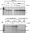
PhoP∼P enhances transcription from PE2 and PA4. (A) PhoP phosphorylation affects RNAP T4 phoPR promoter transcript PE2. Symbols are the same as for Fig. 5. Lanes 1 and 5 contain no PhoP; lanes 2 to 4 and lanes 6 to 8 contain increasing concentrations of PhoP (1, 2.5, and 5 pmol). For phosphorylation of PhoP, equal molar concentrations of PhoP and *PhoR were in reaction mixtures applied to lanes 6 to 8. (B) T4 RNAP from a sigE strain yielded PB1, PA3, and PA4 transcripts but no PE2 transcript; PhoP∼P enhanced PA4 transcription. Lane 1 contains an in vitro transcription reaction identical to lane 8 in panel A for a direct comparison between reactions using T4 RNAP from JH642 and from a mutant strain. Lanes 2 and 5 contain no PhoP; lanes 3 and 4 and lanes 6 and 7 contain increasing concentrations of PhoP (2.5 and 5 pmol, respectively). Lanes 6 and 7 each contain equal molar amounts of PhoP and *PhoR plus ATP for phosphorylation of PhoP.
FIG. 7.
FIG. 7.
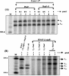
PhoP∼P activates transcription from PA4 and PE2 by using different forms of RNAP. (A) Expression of PA3 and PA4 requires EσA. Lane M contains the 200-nucleotide marker. Lanes 1 to 5 and 7 to 13 contain reconstituted EσA. Lane 6 contains core RNAP with no sigma factor added. Lanes 1 and 7 contain no PhoP or PhoP∼P. Lanes 2 to 5 contain increasing amounts of PhoP (0.1 to 5.0 pmol). Lane 6 contains 5.0 pmol of PhoP∼P. Lanes 8 to 13 contain increasing amounts of PhoP∼P (0.1 to 5 pmol). Lanes 8 to 13 contain equal molar amounts of PhoP and *PhoR plus ATP for phosphorylation of PhoP. (B) PhoP∼P specifically activates the PE2E promoter of the phoPR operon. The 100-nucleotide marker is in the lane marked M. Lanes 1 to 8 contain reconstituted EσE plus the phoPR template. Lanes 2 to 4 contain increasing amounts (0.5, 1, and 5 pmol) of PhoP. Lanes 5 to 8 contain increasing amounts (0.25, 0.5, 1, and 5 pmol) of PhoP∼P. Lanes 9 to 13 contain reconstituted EσE plus the spoIIID template. Lanes 10 and 11 contain 1 and 5 pmol of PhoP, respectively. Lanes 12 and 13 contain 1 and 5 pmol of PhoP∼P, respectively.
FIG. 8.
FIG. 8.
Core plus SigB is sufficient to transcribe from PB1 or P(x1); RNAP from a sigB mutant strain cannot transcribe from PB1 or P(x1). (A) EσB transcription from the PB1 or P(x1) promoter does not require PhoP or PhoP∼P. Lane M contains the 100-nucleotide marker. Reaction mixtures in lanes 1 to 9 contained the phoPR promoter template and reconstituted EσB. Lanes 2 to 5 and 6 to 9 contained increasing amounts (0.1, 0.5, 1, and 5 pmol) of PhoP or PhoP∼P, respectively. (B) Neither T0 RNAP nor T4 RNAP from a sigB mutant strain can transcribe from PB1. Lanes 1 to 7 contain the phoPR template. The reaction mixture in lane 1 contained T0 sigE RNAP plus PhoP∼P and identified the migration positions of P(BX1), PB1, PA3, and PA4. Lane 2 contains core RNAP plus σB as in lane 1 of panel A. Reaction mixtures in lanes 3 and 4 contained T0 RNAP, and those in lanes 5 to 7 contained T4 RNAP from a sigB mutant strain. Reaction mixtures in lanes 3 and 5 contained PhoP, and lanes 4, 6, and 7 contained PhoP∼P.
FIG. 9.
FIG. 9.
RNA from a sigE mutant strain contains no PE2 transcript, and RNA from a sigB mutant strain shows temporal regulation of phoPR promoter transcripts and identifies a 5′ mRNA terminus upstream of PA4. Primer extension and generation of sequencing ladders were the same as described in the legend for Fig. 4. WT lanes used RNA for primer extension studies that was isolated from the parental strain JH642 at T0, T2, or T4. σE lanes used RNA for primer extension studies that was isolated from the sigE mutant strain EU8701 at T0, T2, or T4. σB lanes used RNA for primer extension studies that was isolated from sigB mutant strain PB344 at T0, T1, T2, T3, and T4.

Similar articles

Cited by

References

    1. Antelmann, H., C. Scharf, and M. Hecker. 2000. Phosphate starvation-inducible proteins of Bacillus subtilis: proteomics approach and transcriptional analysis. J. Bacteriol. 182:4478-4490. - PMC - PubMed
    1. Archibald, A. R., I. C. Hancock, and C. R. Harwood. 1993. Cell wall structure, synthesis and turnover, p. 381-410. In J. A. Hoch, R. Losick, and A. L. Sonenshein (ed.), Bacillus subtilis and other gram-positive bacteria: biochemistry, physiology, and molecular biology. American Society for Microbiology, Washington, D.C.
    1. Blencke, H. M., G. Homuth, H. Ludwig, U. Mader, M. Hecker, and J. Stulke. 2003. Transcriptional profiling of gene expression in response to glucose in Bacillus subtilis: regulation of the central metabolic pathways. Metab. Eng. 5:133-149. - PubMed
    1. Chen, Y., C. Birck, J. P. Samama, and F. M. Hulett. 2003. Residue R113 is essential for PhoP dimerization and function: a residue buried in the asymmetric PhoP dimer interface determined in the PhoPN three-dimensional crystal structure. J. Bacteriol. 185:262-273. - PMC - PubMed
    1. Chesnut, R. S., C. Bookstein, and F. M. Hulett. 1991. Separate promoters direct expression of phoAIII, a member of the Bacillus subtilis alkaline phosphatase multigene family, during phosphate starvation and sporulation. Mol. Microbiol. 5:2181-2190. - PubMed

Publication types

LinkOut - more resources