

Primary de novo malignant giant cell tumor of kidney: a case report
- PMID: 15207006
- PMCID: PMC446202
- DOI: 10.1186/1471-2490-4-7
Primary de novo malignant giant cell tumor of kidney: a case report
Abstract
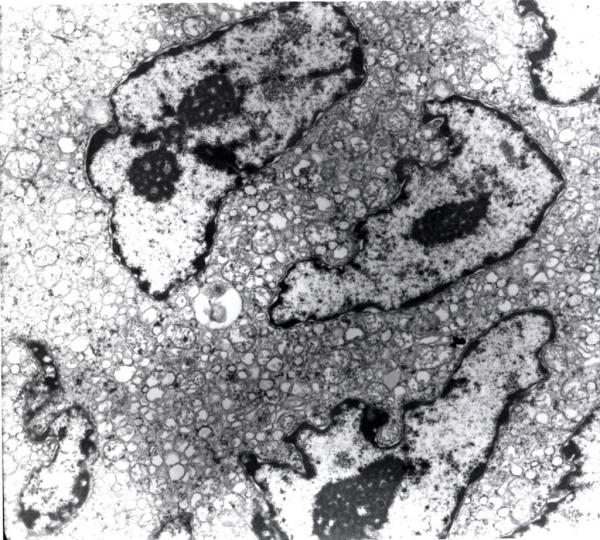
Background: Osteoclast-like giant cell tumors are usually observed in osseous tissue or as tumors of tendon sheath, characterized by the presence of multinucleated giant cells and mononuclear stromal cells. It has been reported in various extraosseous sites including breast, skin, soft tissue, salivary glands, lung, pancreas, female genital tract, thyroid, larynx and heart. However, extraosseous occurrence of such giant cell tumors in the kidney is extremely rare and is usually found in combination with a conventional malignancy. De-novo primary malignant giant cell tumors of the kidney are unusual lesions and to our knowledge this is the second such case.
Case presentation: We report a rare case of extraosseous primary denovo malignant giant cell tumor of the renal parenchyma in a 39-year-old Caucasian female to determine the histogenesis of this neoplasm with a detailed literature review.
Conclusion: Primary denovo malignant giant cell tumor of the kidney is extremely rare. The cellular origin of this tumor is favored to be a pluripotential mesenchymal stromal cell of the mononuclear/phagocytic cellular lineage. Awareness of this neoplasm is important in the pathological interpretation of unusual findings at either fine needle aspiration or frozen section of solid renal masses.
Figures


Similar articles
-
Giant cell tumor of the nasal cavity: case report.Eur Arch Otorhinolaryngol. 2007 Feb;264(2):205-8. doi: 10.1007/s00405-006-0143-6. Epub 2006 Sep 15. Eur Arch Otorhinolaryngol. 2007. PMID: 16977452
-
Solitary osteoclast-like giant cell tumor of the kidney: a case report.Urology. 2012 Nov;80(5):e67-8. doi: 10.1016/j.urology.2012.06.047. Urology. 2012. PMID: 23107416
-
Osteoclast-Like Giant Cell Tumor of the Parotid Gland: Report of a Case Diagnosed on Fine-Needle Aspiration Cytology With Histological and Immunohistochemical Findings.Diagn Cytopathol. 2016 Jun;44(6):548-51. doi: 10.1002/dc.23482. Epub 2016 Apr 15. Diagn Cytopathol. 2016. PMID: 27079183
-
Malignant osteoclast-like giant cell tumor of the kidney.Urology. 1998 Mar;51(3):495-8. doi: 10.1016/s0090-4295(97)00649-3. Urology. 1998. PMID: 9510362 Review.
-
Osteoclast-like giant cell tumor of the pancreas.Int J Clin Oncol. 2002 Dec;7(6):376-80. doi: 10.1007/s101470200059. Int J Clin Oncol. 2002. PMID: 12494256 Review.
Cited by
-
Primer malignant giant cell tumour of kidney: a case report.Ann R Coll Surg Engl. 2021 Oct;103(9):e288-e291. doi: 10.1308/rcsann.2020.7113. Epub 2021 Apr 14. Ann R Coll Surg Engl. 2021. PMID: 33851880 Free PMC article.
-
Osteoclast-like giant cell tumor arising in the soft tissue of the breast: report of a case.Surg Today. 2009;39(1):48-51. doi: 10.1007/s00595-008-3774-y. Epub 2009 Jan 8. Surg Today. 2009. PMID: 19132468
-
A Primary Kidney Giant Cell Tumor of Soft Tissue Caused Peritoneal Dissemination, Considered to Be Malignant Transformation: A Case Report.Diagnostics (Basel). 2023 Feb 16;13(4):752. doi: 10.3390/diagnostics13040752. Diagnostics (Basel). 2023. PMID: 36832239 Free PMC article.
-
Giant cell tumor-like lesion of the urinary bladder: a report of two cases and literature review; giant cell tumor or undifferentiated carcinoma?Diagn Pathol. 2009 Dec 31;4:48. doi: 10.1186/1746-1596-4-48. Diagn Pathol. 2009. PMID: 20043822 Free PMC article.
-
Osteoclasts in Tumor Biology: Metastasis and Epithelial-Mesenchymal-Myeloid Transition.Pathol Oncol Res. 2021 Apr 30;27:609472. doi: 10.3389/pore.2021.609472. eCollection 2021. Pathol Oncol Res. 2021. PMID: 34257573 Free PMC article. Review.
References
-
- Meis JM, Dorfman HD, Nathanson SD, Haggar AM, Wu KK. Primary malignant giant cell tumor of bone: "dedifferentiated" giant cell tumor. Mod Pathol. 1989;2:541–546. - PubMed
Publication types
MeSH terms
LinkOut - more resources
Full Text Sources
Medical
