Comparative Study
. 2004 Jun 24;42(6):961-72.
doi: 10.1016/j.neuron.2004.06.002.

CBP histone acetyltransferase activity is a critical component of memory consolidation

Affiliations
Comparative Study

CBP histone acetyltransferase activity is a critical component of memory consolidation

Edward Korzus et al. Neuron. .

Abstract

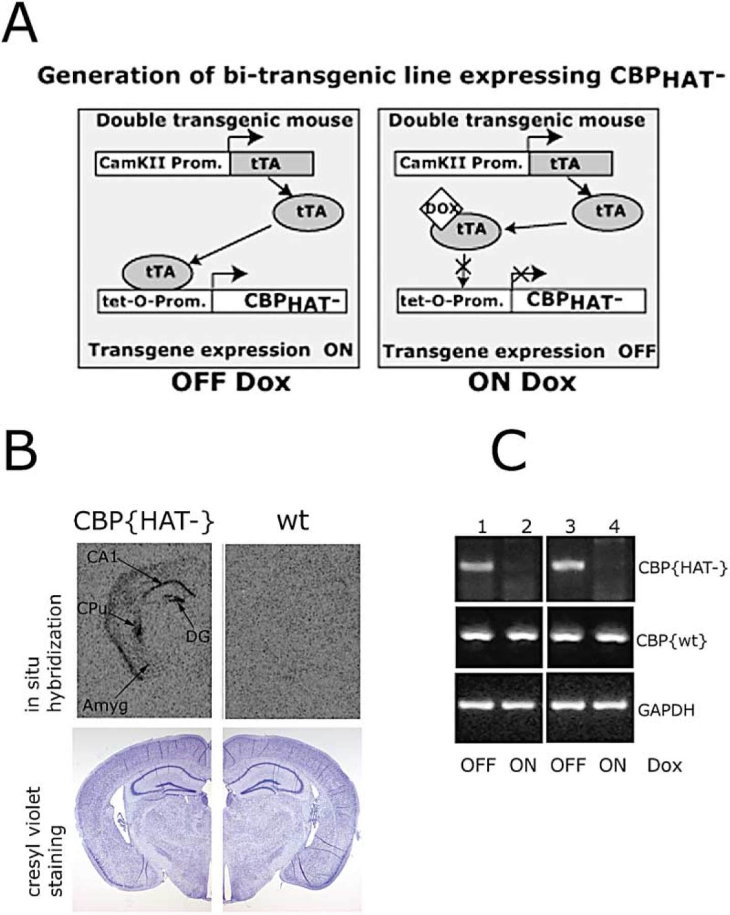
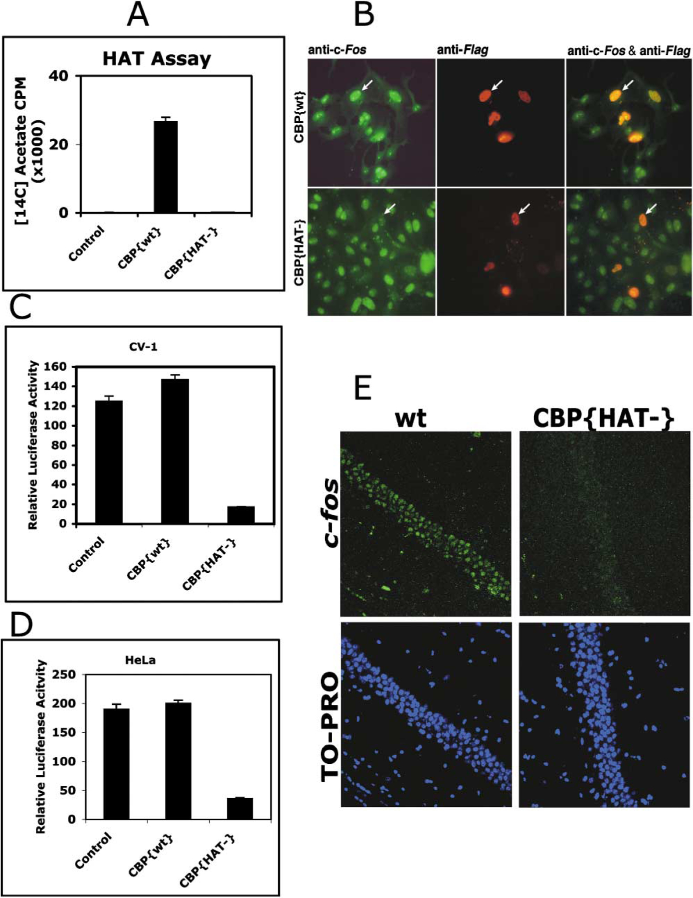
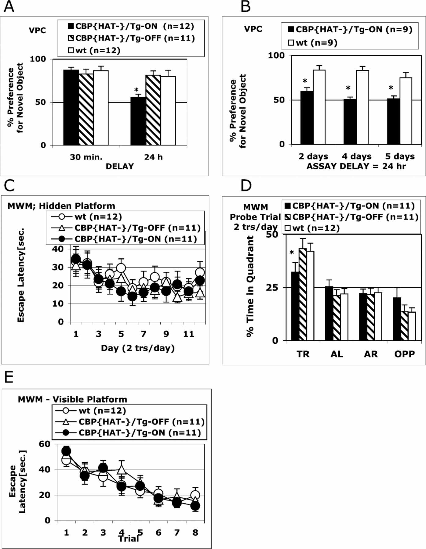
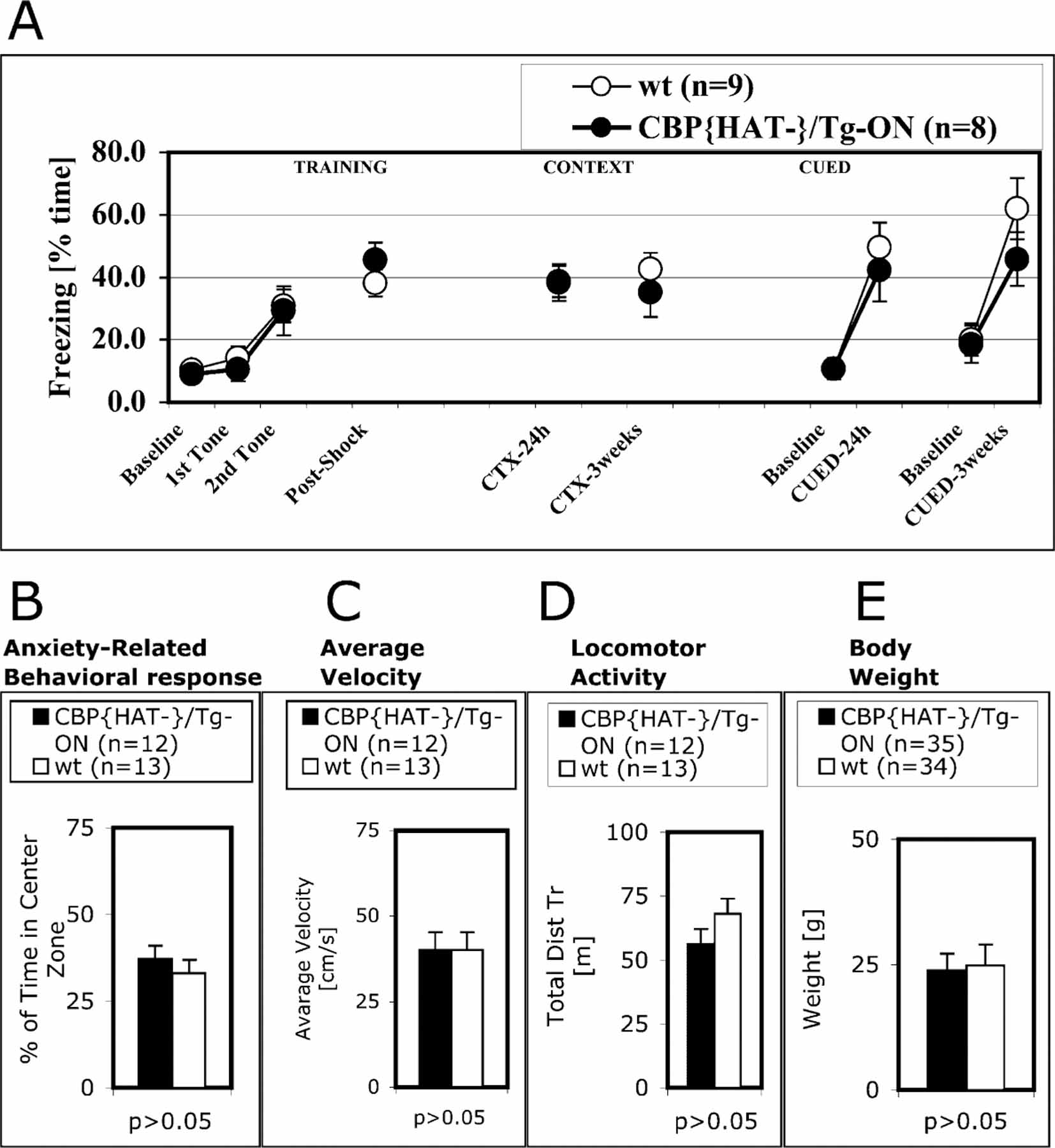
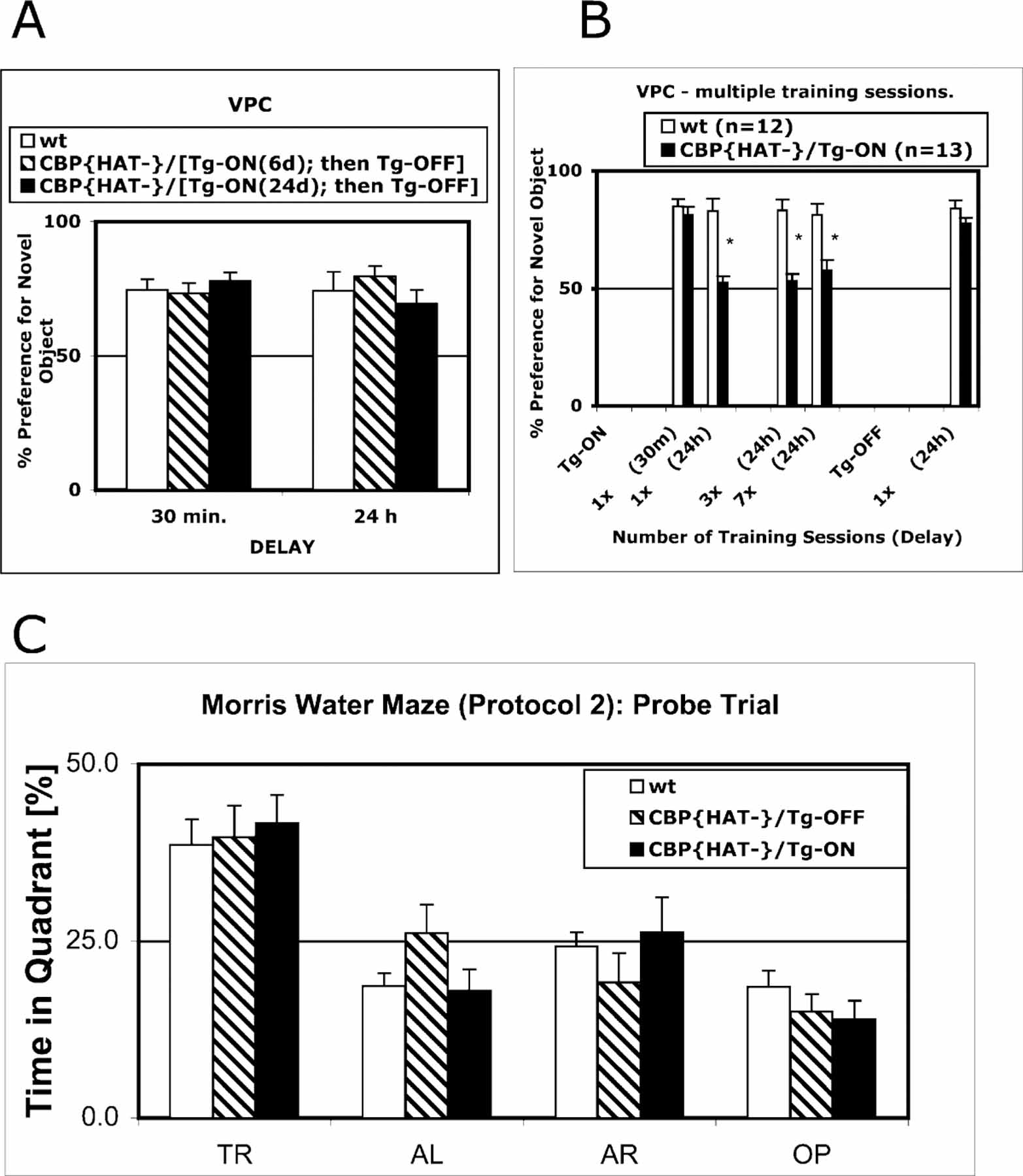
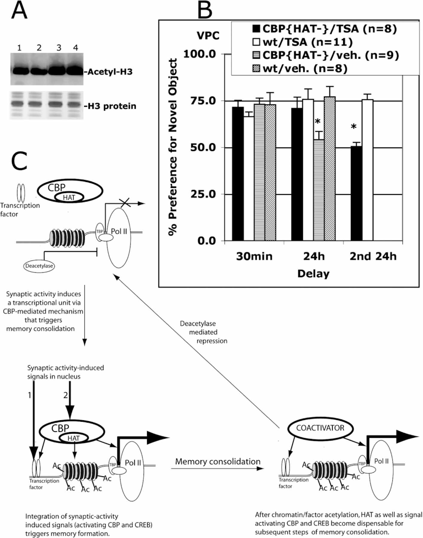
The stabilization of learned information into long-term memories requires new gene expression. CREB binding protein (CBP) is a coactivator of transcription that can be independently regulated in neurons. CBP functions both as a platform for recruiting other required components of the transcriptional machinery and as a histone acetyltransferase (HAT) that alters chromatin structure. To dissect the chromatin remodeling versus platform function of CBP or the developmental versus adult role of this gene, we generated transgenic mice that express CBP in which HAT activity is eliminated. Acquisition of new information and short-term memory is spared in these mice, while the stabilization of short-term memory into long-term memory is impaired. The behavioral phenotype is due to an acute requirement for CBP HAT activity in the adult as it is rescued by both suppression of transgene expression or by administration of the histone deacetylase inhibitor Trichostatin A (TSA) in adult animals.


Figures

Figure 1.
Figure 1.. Generation of CBP Mutant Mice
(A) Schematic of the generation of bitransgenic mice expressing CBP{HAT}. (B) Coronal brain sections of CBP{HAT} mice and control wild-type (wt) littermates. Top: in situ analysis of CBP{HAT} transgene expression using 35S-labeled oligo probe. The transgene expression has been detected in CA1 and dentate gyrus fields of the hippocampus, caudate putamen, and at a low level throughout the neocortex. Bottom: coronal brain sections (10 µm) of CBP{HAT} and wt mice corresponding to top panels and stained with cresyl violet showed no alterations in gross neuroanatomy. Abbreviations: Amyg, amygdala; CA1, pyramidal cell field of hippocampus; DG, dentate gyrus; CPu, caudate putamen. (C) RT-PCR analysis showed that the expression of CBP{HAT} transgene is tightly controlled by doxycycline. First strand cDNA was generated by reverse transcription followed by PCR analysis of CBP{HAT}, CBP{wt}, and GAPDH (internal control) expression. 50 ng of total RNA was used in reactions 1 and 2 with oligoes hybridizing with CBP{HAT} mRNA. For all other reactions, 10 ng of total RNA was used. 1, RNA from hippocampus, CBP{HAT} mice OFF-Dox for 4 days; 2, RNA from hippocampus, CBP{HAT} mice ON-Dox; 3, RNA from CA1, CBP{HAT} mice OFF-Dox for 7 days; 4, RNA from CA1, CBP{HAT} mice ON-Dox.
Figure 2.
Figure 2.. CBP Acetyltransferase Activity Is Required for c-fos Gene Expression
(A) Generation of substitution mutation in the mouse CBP (Y1540/F1541 to 1540/1541) abolished detectable acetylation of histones using[14C]acetyl-CoA as a substrate. Histone acetyltransferase activity was determined by liquid HAT assay (see Experimental Procedures) using purified CBP{wt} or CBP{HAT} proteins. (B) CBP{HAT} expression blocks c-fos gene expression in vivo. CV-1 cells were transfected with Flag-tagged CBP wild-type or Flag-tagged CBP{HAT} expressing vectors. After 24 hr, cells were immunostained for c-fos protein (green) and for wild-type or mutant CBP proteins, both tagged with the Flag epitope (red). Left panels show immunostaining for endogenous c-fos protein expression. Middle panels show cells expressing transfected CBP{wt} or CBP{HAT}. Right panels show overlap. (C and D) Transiently expressed CBP{HAT} in CV-1 (C) or HeLa (D) cells efficiently reduced transcription of the c-fos promoter-driven luciferase. (E) Physiological levels of c-fos expression is reduced in CBP{HAT}/Tg-ON. Fresh brain sections were prepared from wt and CBP{HAT}/Tg-ON animals expressing the transgene for 7 days. Rabbit anti-c-fos antibodies and Alexa488-goat anti-rabbit IgG were used to detect the physiological level of c-fos expression. Specimens were counterstained with selective nuclei fluorescent marker TO-PRO3 Iodine and analyzed using laser scanning confocal microscope Olympus BX61 (40× oil). Specimens are from animals exhibiting representative levels of c-fos expression for the CBP{HAT}/Tg-ON and wild-type littermate mice.
Figure 3.
Figure 3.. Two Forms of Declarative Memory: Spatial and Recognition Memory Are Impaired in CBP{HAT} Mice
(A) CBP{HAT} mutant mice exhibit a severe impairment in recognition memory. Three groups were tested on the VPC task at 30 min and 24 hr delays: CBP{HAT}/Tg-ON (n = 12) that had expressed the transgene for 18 days before the assay, CBP{HAT}/Tg-OFF (n = 11) with blocked transgene expression, and wt (n = 13). CBP{HAT}/Tg-ON mice exhibited normal acquisition, short-term memory, and recall when tested on the VPC task at the 30 min delay. However, CBP{HAT}/Tg-ON mice were severely impaired on the VPC task at the 24 hr delay (*p < 0.05). We found no effect of Dox treatment in wild-type mice and therefore combined the data for wild-type ON/OFF Dox. t test for the % preference for novel object after 30 min delay: wt/OFF-Dox versus wt/ON-Dox, p > 0.85; wt/OFF-Dox, average = 86%, n = 7, SEM ± 0.8; wt/ON-Dox, average = 88%, n = 6, SEM ± 0.5. t test for the % preference for novel object after 24 hr delay: wt/OFF-Dox versus wt/ON-Dox, p > 0.89; wt/OFF-Dox, average = 79%, n = 7, SEM ± 0.8; wt/ON-Dox, average = 81%, n = 6, SEM ± 0.8. (B) The performance of CBP{HAT}/Tg-ON and wt mice was tested on the VPC task at the 24 hr delay after CBP{HAT} transgene expression was activated for 2, 4, or 5 days before training. Expression of the CBP{HAT} transgene for 2 days before training was sufficient to block long-term memory formation assessed at the 24 hr delay. Both groups of animals, CBP{HAT}/Tg-ON and control wt littermates, received exactly the same Dox treatment before and during the experiment. (C) Performance on the MWM task during training on Protocol 1 (see Experimental Procedures). CBP{HAT}/Tg-ON mice were trained together with two control groups: CBP{HAT}/Tg-OFF and wt mice for 12 days (2 trials/day, 1 hr ITI). No differences in escape latencies were found between CBP{HAT}/Tg-ON mice and two control groups. (D) CBP{HAT}/Tg-ON mice show a deficiency in spatial memory on the MWM task. A probe trial performed 1 day after training was complete revealed a significant impairment in spatial localization in the CBP{HAT}/Tg-ON group compared to controls (t test for time in target quadrant: *CBP{HAT}/Tg-ON versus wt, p < 0.02; CBP{HAT}/Tg-ON versus CBP{HAT}/Tg-OFF, p < 0.05). We found no effect of Dox treatment in wild-type mice and therefore combined the data for wild-type ON/OFF Dox. t test for the time in target quadrant: wt/OFF-Dox versus wt/ ON-Dox, p > 0.9; wt/OFF-Dox: average = 43.2%, n = 7, SEM ± 4.8; wt/ON-Dox, average = 44.1%, n = 5, SEM ± 6.7. (E) CBP{HAT}/Tg-ON mice showed normal acquisition on the visual platform version of the MWM task indicating normal vision, motivation, and swimming ability. The design of the experiments in this figure (except B) includes two types of animals (wild-types and double transgenics) and two treatment groups (Dox or no Dox). We performed the behavioral analysis on these four groups. However, we found no effect of Dox treatment in wildtype mice and therefore combined the data for wild-type ON/OFF Dox.
Figure 4.
Figure 4.. Fear Conditioning, Anxiety-Related Response, Weight, and Locomotor Activity Are Normal in CBP{HAT} Mice
(A) CBP{HAT}/Tg-ON mice and control wt littermates showed the same freezing in tests of contextual and cued fear conditioning after 24 hr and 3 week delays. (B–D) An open field activity test (see Experimental Procedures) showed that time in center (B), average velocity (C), and locomotor activity (D) were normal in CBP{HAT}/TgOn mice. (E) Weights of CBP{HAT}/Tg-ON mice were the same as wt littermates.
Figure 5.
Figure 5.. Deficit in Recognition Memory in CBP{HAT} Mice Can Be Reversed by Elimination of CBP{HAT} Transgene Expression but not by Multiple Training Sessions
(A) Performance in CBP{HAT} mice was rescued when the expression of CBP{HAT} was suppressed. Three groups were used for the experiment: CBP{HAT}/Tg-ON(24d)-OFF (n = 12) that had expressed the transgene for 24 days, CBP{HAT}/Tg-ON(6d)-OFF that had expressed the transgene for 6 days (n = 11), and wt (n = 13). Following transgene expression for 6 or 24 days, we suppressed expression and tested the animals on the VPC task at 30 min and 24 hr delays using a new set of objects. CBP{HAT} mice with suppressed transgene expression exhibited normal performance on the VPC task at the 30 min delay [ANOVA, F(2,33) = 0.45, p > 0.05; CBP{HAT}/Tg-ON(24d)-OFF = 78.1 ± 3.0, CBP{HAT}/Tg-ON(6d)-OFF = 73.2 ± 3.9, wt = 74.5 ± 4.0] as well as at the 24 hr delay [ANOVA, F(2,33) = 0.75, p > 0.05; CBP{HAT}/Tg-ON(24d)-OFF = 69.5 ± 5.2, CBP{HAT}/Tg-ON(6d)-OFF = 79.6 ± 3.8, wt = 74.3 ± 7.1]. (B) Multiple training sessions did not rescue the recognition memory deficit in CBP{HAT} mice. Two groups (wt [n = 12] and CBP{HAT}/ Tg-ON [n = 13]) were trained in multiple training sessions (40 min exposure to the same object every 24 hr) and tested on the VPC task at 24 hr delays except for the first test, which was performed at the 30 min delay (t test for performance, *p > 0.05). (C) Performance on the hidden platform version of the MWM task was rescued by intense training (Protocol 2, see Experimental Procedures).
Figure 6.
Figure 6.. Administration of the Histone Deacetylase Inhibitor TSA Reverses the Recognition Memory Deficit in CBP{HAT} Mice
(A) I.p. TSA injection increases histone acetylation levels in the hippocampus. Nuclear extracts were prepared from hippocampi isolated from wt mice sacrificed 6 hr after i.p. injection of vehicle (lanes 1 and 2) or TSA (lanes 3 and 4). Acid-extracted histones were visualized with Coomassie blue staining. The level of histone H3 acetylation was assessed in Western blot assay using anti-acetylated H3 antibodies (Upstate). (B) I.p. TSA administration before training rescued a recognition memory deficit in CBP{HAT} mice. Four groups of mice were tested: CBP{HAT}/TSA (Group 1), wt/TSA (Group 2), CBP{HAT}/veh (Group 3), and wt/veh (Group 4). TSA or DMSO alone (veh) was administered 2 hr before training on the VPC task as described in Experimental Procedures. Then, we measured performance of these four groups on the VPC task at 30 min and 24 hr delays. 4 days later, the recognition memory was assessed again (Groups 1 and 2) on the VPC task at the 24 hr delay using a new set of objects (2nd 24 hr). This assay provided a control showing that Group 1 has strong memory impairment if TSA was not administered. (C) Model of acetylation-dependent memory consolidation. The proposed model takes into account five observations: (1) NMDAR-dependent neuronal activation is required for memory formation (Davis et al., 1992); (2) neuronal activity-dependent transcription is required for memory consolidation (Andrew, 1980; Davis and Squire, 1984); (3) NMDA-dependent transcription requires a signaling pathway to activate transcription factors such as CREB and a separate signaling pathway to activate CBP (Chawla et al., 1998; Impey et al., 2002); (4) behaviorally induced CREB phosphorylation is transient and does not correlate with peak Fos induction (Stanciu et al., 2001); and (5) CBP acetyltransferase activity is required for long-term memory consolidation but not for short-term memory (our data). This model suggests that CBP-mediated histone acetylation during transcriptional activation is a limiting step in the molecular mechanism controlling memory stabilization. Initial steps include induction of transient CREB phosphorylation, CBP activation, and CBP-mediated histone acetylation at a specific transcriptional unit in response to the initial learning event. Subsequently, prolonged elevated transcription required for memory consolidation could be maintained by CBP- and CREB phosphorylation-independent nuclear mechanisms even after signals to CREB and CBP are no longer present. This ongoing transcription would remain active until the competing deacetylase-dependent repression mechanism shut off transcription.

Comment in

Similar articles

Cited by

References

    1. Andrew RJ (1980). The functional organization of phases of memory consolidation. In Advances in the Study of Behaviour, Hinde RA, Beer C, and Bunsel M, eds. (New York: Academic Press; ), pp. 337–367.
    1. Bachevalier J (1990). Ontogenetic development of habit and memory formation in primates. Ann. N Y Acad. Sci 608, 457–484. - PubMed
    1. Bading H, Ginty DD, and Greenberg ME (1993). Regulation of gene expression in hippocampal neurons by distinct calcium signaling pathways. Science 260, 181–186. - PubMed
    1. Bito H, Deisseroth K, and Tsien RW (1996). CREB phosphorylation and dephosphorylation: a Ca(2+)- and stimulus duration-dependent switch for hippocampal gene expression. Cell 87, 1203–1214. - PubMed
    1. Bourtchuladze R, Frenguelli B, Blendy J, Cioffi D, Schutz G, and Silva AJ (1994). Deficient long-term memory in mice with a targeted mutation of the cAMP-responsive element-binding protein. Cell 79, 59–68. - PubMed

Publication types

MeSH terms