Review
. 2004 Jul;25(7):360-7.
doi: 10.1016/j.it.2004.05.001.

Mitochondrial hyperpolarization: a checkpoint of T-cell life, death and autoimmunity

Affiliations
Review

Mitochondrial hyperpolarization: a checkpoint of T-cell life, death and autoimmunity

Andras Perl et al. Trends Immunol. 2004 Jul.

Abstract

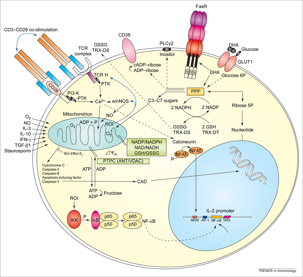
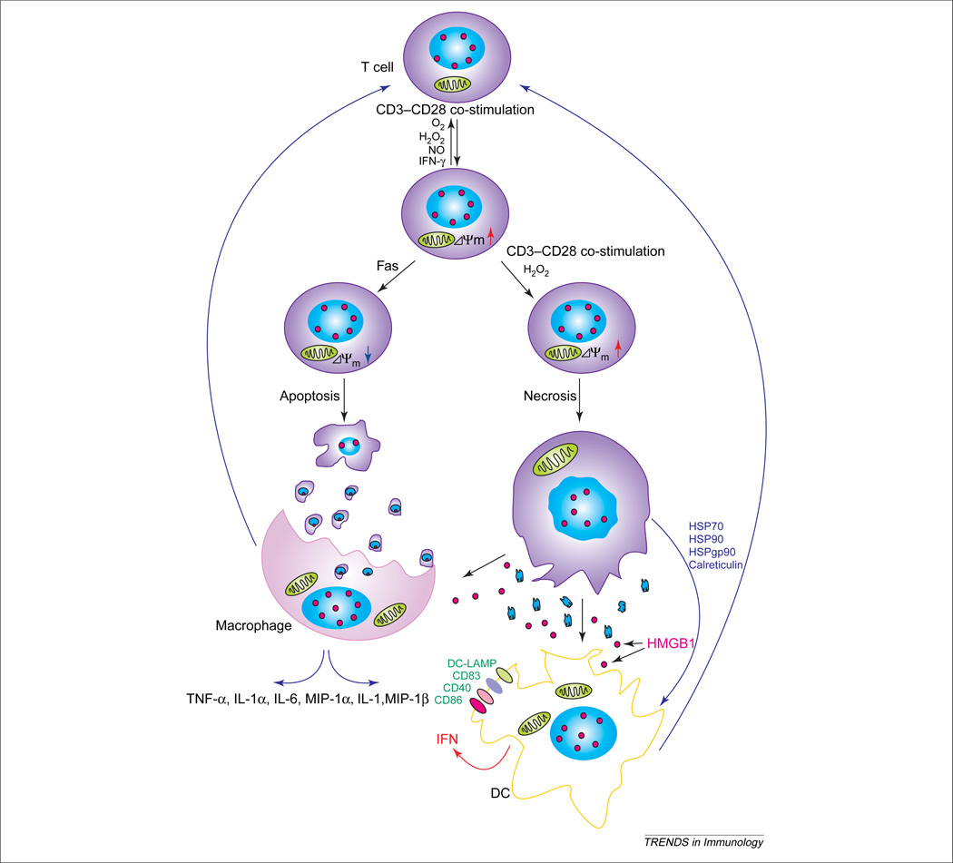
T-cell activation, proliferation and selection of the cell death pathway depend on the production of reactive oxygen intermediates (ROIs) and ATP synthesis, which are tightly regulated by the mitochondrial transmembrane potential (ΔΨm). Mitochondrial hyperpolarization (MHP) and ATP depletion represent early and reversible steps in T-cell activation and apoptosis. By contrast, T cells of patients with systemic lupus erythematosus (SLE) exhibit persistent MHP, cytoplasmic alkalinization, increased ROI production and depleted ATP, which mediate enhanced spontaneous and diminished activation-induced apoptosis and sensitize lupus T cells to necrosis. Necrotic, but not apoptotic, cell lysates activate dendritic cells and might account for increased interferon a production and inflammation in lupus patients. MHP is proposed as a key mechanism of SLE pathogenesis and is therefore a target for pharmacological intervention.


Figures

Figure 1
Figure 1
Mitochondrial and metabolic checkpoints of T-cell activation and apoptosis signals. Antigen binding-initiated signaling through the T-cell receptor (TCR)–CD3–TCRζ-chain complex and the CD28 co-stimulatory molecule activate phosphoinositide 3-kinase (PI3-K) and protein tyrosine kinase (PTK). Increased cytosolic Ca2+; concentration activates the serine/threonine phosphatase calcineurin, which dephosphorylates the NFAT. Dephosphorylated NFAT can translocate to the nucleus, where it promotes transcription of interleukin-2 (IL-2) in concert with AP-1 and NF-κB. Ca2+ flux into mitochondria increases production of reactive oxygen intermediates (ROIs) and NF-κB activation [22]. Excess ROI production and disruption of ΔΨm lead to activation-induced cell death executed by caspase-3 (digesting vitally important proteins PARP, 70K U1RNP, lamin and actin) and caspase-activated DNase (CAD). Cleavage by caspase-3 is thought to expose cryptic epitopes and cause autoantigenicity of self-antigens [70]. Dehydroascorbate (DHA) is imported through glucose transporter 1 (GLUT1) and is metabolized through the pentose phosphate pathway (PPP), thereby enhancing reduced glutathione (GSH) levels. DHA also increases surface expression of Fas receptor (FasR) [64]. Glutathione reductase and TRX reductase synthesize GSH and TRX-DT at the expense of NADPH. Formulation of the PPP and its efficiency to provide NADPH is dependent on the expression of G6PD (glucose 6-phosphate dehydrogenase) and transaldose (TAL) [34,43]. Dcm is controlled by intracellular GSH/NADH/NADPH levels, integrity of the permeability transition pore complex (PTPC), largely composed of the adenine nucleotie translocator (ANT) (inner membrane) and voltage-dependent anion channel (VDAC) (outer membrane), and translocation and dimerization of proand anti-apoptotic Bcl-2 family members in the intermembrane space [21]. TCR-activation-induced mitochondrial hyperpolarization is mediated through nitric oxide (NO) production by endothelial/neuronal NO synthase (e/nNOS), which, in turn, is regulated by ROIs and Ca2+; [23]. Abbreviations: GSSG, oxidized glutathione; IFN-γ, interferon γ; P, phosphate; PLCγ2, phospholipase Cγ2; TGF-β1, transforming growth factor β1; TRX-DT, thioredoxin.
Figure 2
Figure 2
Impact of ΔΨm on cell death pathway selection and activation of T cells by macrophages and dendritic cells (DCs). CD3–CD28 co-stimulation, reactive oxygen intermediate (ROI), H2O2, nitric oxide (NO) and cytokines, such as interferon-γ (IFN-γ), might elicit reversible elevation of ΔΨm [i.e. mitochondrial hyperpolarization (MHP)]. Transient MHP is an early event in Fas-mediated apoptosis, resulting in cellular shrinkage and formation of membrane-enclosed apoptotic bodies that are phagocytosed by macrophages. Repeated T-cell receptor (TCR) activation or exposure to ROIs induces necrosis instead of MHP, which, in turn, leads to cellular swelling, membrane breakdown and release of proinflammatory cell lysate. While high-mobility group B1 (HMGB1) protein remains immobilized on chromatin of apoptotic bodies, it is released from necrotic cells. HMGB1, heat-shock protein 70 (HSP70), HSP90, HSPgp95 and calreticulin induce differentiation of DCs and activate macrophages. DCs produce IFN-γ, whereas macrophages produce a series of proinflammatory cytokines. Both cell types increase expression of co-stimulation molecules, thus contributing to T-cell activation. These positive-feedback loops might play key roles in aberrant T-cell activation in patients with systemic lupus erythematosus (SLE). Abbreviations: DC-LAMP, lysosome-associated membrane glycoprotein; MIP-1γ, macrophage inflammatory protein 1γ; TNF-γ, tumor necrosis factor γ.

Similar articles

Cited by

References

    1. Perl A, et al. Metabolic switches of T-cell activation and apoptosis. Antioxid. Redox Signal. 2002;4:427–443. - PubMed
    1. Susin SA, et al. The central executioner of apoptosis: multiple connections between protease activation and mitochondria in Fas/Apo-1/CD95- and ceramide-induced apoptosis. J. Exp. Med. 1997;186:25–37. - PMC - PubMed
    1. Banki K, et al. Elevation of mitochondrial transmembrane potential and reactive oxygen intermediate levels are early events and occur independently from activation of caspases in Fas signaling. J. Immunol. 1999;162:1466–1479. - PMC - PubMed
    1. Puskas F, et al. Stimulation of the pentose phosphate pathway and glutathione levels by dehydroascorbate, the oxidized form of vitamin C. FASEB J. 2000;14:1352–1361. - PubMed
    1. Li P-F, et al. p53 regulates mitochondrial membrane potential through reactive oxygen species and induces cytochrome c-independent apoptosis blocked by bcl-2. EMBO J. 1999;18:6027–6036. - PMC - PubMed

Publication types

MeSH terms

LinkOut - more resources